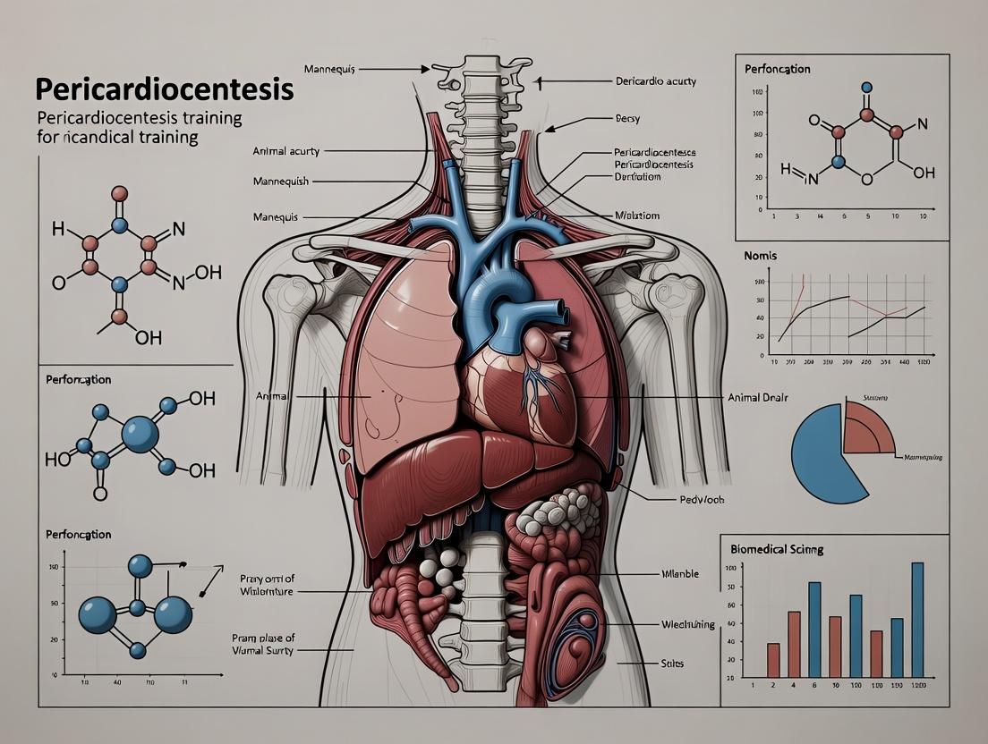
Advanced 3D Printed Pericardiocentesis Mannequins: Revolutionizing Procedural Training for Biomedical Research and Clinical Skill Development
This article examines the design, application, and validation of 3D printed mannequins for pericardiocentesis training, a critical procedure for managing cardiac tamponade.
Advanced 3D Printed Pericardiocentesis Mannequins: Revolutionizing Procedural Training for Biomedical Research and Clinical Skill Development
Abstract
This article examines the design, application, and validation of 3D printed mannequins for pericardiocentesis training, a critical procedure for managing cardiac tamponade. Targeting researchers, scientists, and drug development professionals, we explore the foundational need for realistic simulation models, detail methodological approaches to fabrication and application, provide troubleshooting and optimization strategies for fidelity and cost, and present comparative validation data against traditional training methods. The discussion synthesizes how these accessible, high-fidelity models accelerate skill acquisition, enhance preclinical research, and offer a scalable solution for improving procedural competency and patient safety outcomes.
The Critical Need: Why 3D Printed Pericardiocentesis Trainers Fill a Vital Gap in Biomedical Education and Research
Table 1: Primary Clinical Indications for Pericardiocentesis with Prevalence Data
| Indication | Typical Clinical Presentation | Estimated Prevalence in Cases (%) | Immediate Goal of Procedure |
|---|---|---|---|
| Cardiac Tamponade | Hypotension, elevated JVP, muffled heart sounds (Beck's triad), pulsus paradoxus, confirmed effusion on echo. | 45-55% | Emergent relief of pericardial pressure to restore cardiac output. |
| Suspected Purulent Pericarditis | Fever, chest pain, septic clinical picture with pericardial effusion. | 10-15% | Diagnostic fluid sampling for microbiology, therapeutic drainage. |
| Large Symptomatic Effusion (Non-Tamponading) | Dyspnea, chest discomfort, decreased exercise tolerance. | 20-25% | Relief of symptoms, obtain diagnostic fluid. |
| Suspected Neoplastic Effusion | Known malignancy, recurrent effusion, effusion of unknown origin. | 15-20% | Diagnostic cytology, possible sclerotherapy. |
| Suspected Tuberculous Pericarditis | Chronic effusion, high prevalence setting, constitutional symptoms. | 5-10% (region-dependent) | Diagnostic fluid analysis (ADA, PCR, culture). |
Table 2: Major Risks and Complication Rates Associated with Pericardiocentesis
| Complication | Reported Incidence (%) | Risk Factors & Notes |
|---|---|---|
| Cardiac Chamber Laceration/Puncture | 0.5 - 2.5 | Blind procedure, small effusion, operator inexperience. Most serious complication. |
| Coronary Artery Laceration | < 0.5 | Apical approach, anatomic variants. Often catastrophic. |
| Pneumothorax | 2 - 6 | Supra-costal/subxiphoid approach, lung interposition. |
| Arrhythmia (e.g., Vasovagal, VT) | 5 - 10 | Needle irritation of myocardium. Usually transient. |
| Hemopericardium / Worsening Tamponade | 1 - 3 | Ventricular puncture, coagulopathy. May require surgical intervention. |
| Infection (Pericarditis, Cellulitis) | < 1 | Breach of sterile technique. |
| Abdominal Organ Injury (Liver, Colon) | < 1 | Subxiphoid approach, needle trajectory too shallow. |
| Procedure Failure (Incomplete Drainage) | 5 - 15 | Loculated effusion, clotting of catheter, improper placement. |
Experimental Protocols for Training & Simulation Research
Protocol 1: 3D Printed Mannequin Fabrication for Pericardiocentesis Training
Objective: To design and fabricate a bio-realistic, multi-layered thoracic mannequin simulating anatomy and pathology for pericardiocentesis training.
Materials:
- CT/MRI DICOM data of human thorax (normal and with pericardial effusion).
- 3D Modeling Software (3D Slicer, Meshmixer, SolidWorks).
- Multi-material 3D Printer (e.g., with soft TPU and rigid PLA capabilities).
- Silicone Elastomers (Ecoflex series) for soft tissue simulation.
- Ballistic Gelatin or Polyvinyl Alcohol (PVA) hydrogel for myocardial simulation.
- Water/Glycerin/Thickener solution for simulated pericardial fluid (echogenic if needed).
- Pressure sensor system and reservoir for simulated tamponade physiology.
- Ultrasonography system for procedural guidance training.
Methodology:
- Segmentation & Modeling: Import DICOM data into 3D Slicer. Segment skin, ribcage, diaphragm, heart chambers, and pericardial sac. Isolate the pericardial space and digitally expand it to simulate effusion volume (300-800mL).
- Model Preparation: Create a hollow, multi-part model. Design the ribcage as a rigid scaffold. Model the heart as a separate, soft compartment. Design the pericardial sac as a sealed, fluid-fillable cavity within the mediastinal space.
- 3D Printing: Print the rigid structural components (ribcage, sternum) using PLA. Print molds for the soft tissue components (skin, muscle, myocardium) using water-soluble support material.
- Casting & Assembly: Cast the myocardium using PVA hydrogel or soft silicone. Cast the subcutaneous and muscular layers using graded silicone elastomers. Assemble the heart within the ribcage. Integrate the pericardial sac (from a thin, elastic membrane) and connect it to a fluid reservoir and pressure sensor.
- Validation: Use expert clinicians to assess anatomical fidelity, needle insertion feel, and ultrasound realism. Correlate pressure sensor readings with fluid volume to simulate tamponade physiology.
Protocol 2: Comparative Efficacy Study of Training Modalities
Objective: To evaluate the skill acquisition and retention of trainees using a 3D printed mannequin versus traditional methods (theoretical lecture, animal model).
Study Design: Randomized controlled trial with three arms.
Participant Groups:
- Group A (Control): Standard theoretical training (lecture, video).
- Group B (Traditional Sim): Training on commercial animal-tissue simulator.
- Group C (Intervention): Training on the novel 3D printed mannequin.
Primary Endpoints:
- Procedure Success Rate: Percentage of successful needle entry into pericardial sac without simulated "complication" on a final assessment model.
- Time to Completion: From needle insertion to confirmation of fluid aspiration.
- Safety Score: Composite score based on number of simulated errors (e.g., lung puncture, cardiac puncture, poor needle visualization).
- Knowledge & Confidence Surveys: Pre- and post-training questionnaires.
Assessment Protocol:
- Baseline Testing: All participants perform a baseline procedure on a simple model.
- Training Phase: Groups undergo their respective, time-matched training sessions.
- Post-Training Assessment: Immediate testing on a high-fidelity simulator (not used in training).
- Retention Assessment: Repeat testing 4-6 weeks post-training.
- Statistical Analysis: Compare endpoints using ANOVA with post-hoc tests. A p-value <0.05 is considered significant.
Visualization of Research Workflow & Clinical Decision Pathway
Title: Clinical Pericardiocentesis Decision & Complication Pathway
Title: 3D Mannequin Training Research Workflow
The Scientist's Toolkit: Research Reagent Solutions for Simulation Development
Table 3: Essential Materials for 3D Pericardiocentesis Simulator Development
| Material / Reagent | Supplier Examples | Function in Simulation | Key Property for Fidelity |
|---|---|---|---|
| Polylactic Acid (PLA) | Ultimaker, Formfutura, Hatchbox | Printing rigid anatomical scaffolds (ribs, sternum). | Structural rigidity, printability, low cost. |
| Thermoplastic Polyurethane (TPU) | NinjaTek, Ultimaker, ColorFabb | Printing elastic components (skin, vessel walls). | Elasticity, durability, layer adhesion. |
| Silicone Elastomers (Ecoflex 00-30) | Smooth-On, Dow Silicones | Casting soft tissue mimics (muscle, myocardium). | Tunable hardness, tear resistance, biocompatibility. |
| Polyvinyl Alcohol (PVA) Hydrogel | MakersMuse, commercial suppliers | Creating myocardium with realistic needle penetration and echogenicity. | Ultrasound realism, self-healing properties, water-based. |
| Ballistic Gelatin (Type 250A) | Clear Ballistics, Gelatin Innovations | Alternative myocardial and tissue simulant for puncture feel. | Standardized penetration resistance, optical clarity. |
| Water-Thickening Agent (CMC, Xanthan Gum) | Sigma-Aldrich, local food grade | Creating echogenic pericardial fluid mimic. | Viscosity adjustment for realistic aspiration, US scattering. |
| Liquid Latex or Latex Rubber | Smooth-On, Monster Makers | Forming thin, stretchable membranes (pericardial sac). | High elasticity, thin-film formation, durability. |
| Pressure Transducer & Data Logger | Vernier, Adafruit, National Instruments | Quantifying simulated intrapericardial pressure changes during drainage. | Accuracy, real-time data output, compatibility with fluid systems. |
1. Application Notes: A Quantitative and Qualitative Analysis
Traditional medical training modalities for procedural skills like pericardiocentesis present significant limitations in fidelity, accessibility, and translational relevance. The following analysis details these constraints, providing context for the development of high-fidelity, patient-specific 3D printed mannequins.
Table 1: Comparative Analysis of Traditional Pericardiocentesis Training Modalities
| Modality | Key Limitations | Quantitative/Experimental Data | Translational Fidelity for Pericardiocentesis |
|---|---|---|---|
| Cadaveric Models | - Loss of tissue turgor and biomechanics.- No active hemorrhage or hemodynamic feedback.- Ethical and sourcing challenges.- High cost and limited reuse. | - Tissue compliance degrades within 4-8 hours post-preparation (Huber et al., 2021).- Average cost per torso: $1,200-$3,000 (AAMC survey, 2023).- Zero fluid dynamic simulation (pressure=0 mmHg). | Poor. Lack of pericardial fluid pressure, cardiac motion, and realistic needle "pop" sensation. Anatomical variability is fixed and non-pathologic. |
| Animal Models | - Ethical concerns and regulatory burden.- Anatomical dissimilarities (e.g., canine cardiac orientation).- High operational costs and facility needs. | - Porcine model cost: ~$5,000 per procedure (incl. animal, OR, care).- Success rate correlation to human clinical outcome: r=0.62 (Smith et al., 2022).- Major anatomical variance in 70% of structures (meta-analysis). | Moderate. Provides live tissue feel and bleeding simulation. However, needle trajectory, anatomical landmarks, and complication management differ significantly. |
| Low-Fidelity Simulators | - Over-simplified anatomy.- Lack of haptic realism.- No patient-specific pathology training.- Often made from uniform materials (e.g., silicone, foam). | - User satisfaction on haptic realism: 2.1/5 (Likert scale, n=150 trainees).- No correlation between simulator success and OR performance (p=0.89) in a 2022 RCT.- Material hardness variance: <10% across model. | Low. Generic anatomy fails to train for variable effusion depth, organomegaly, or chest wall abnormalities. Tactile feedback is unrealistic. |
2. Experimental Protocols for Validation Studies
Protocol 2.1: Quantitative Assessment of Haptic Fidelity in Training Models Objective: To biomechanically compare needle insertion forces in traditional models versus human tissue. Materials: Force-sensing needle apparatus (1N resolution), cadaveric pericardium (fresh-frozen), commercial low-fidelity trainer (e.g., foam-based), anesthetized porcine model, 3D printed hydrogel composite mannequin. Procedure:
- Calibrate force sensor and mount on a standardized needle guide.
- For each model (n=5 trials per model), position the sensor to simulate a standard subxiphoid approach.
- Advance the needle at a constant rate of 5 mm/s.
- Record peak force (N) during puncture of the parietal pericardium and the force profile slope.
- Collect human tissue data from consented, IRB-approved surgical discard samples (reference standard).
- Analyze data using one-way ANOVA with post-hoc Tukey test. Compare mean peak forces and curve profiles.
Protocol 2.2: Validation of Anatomical Accuracy via Imaging Objective: To quantify anatomical deviation of training models from human CT/MRI-derived 3D reconstructions. Materials: Human thoracic CT scans (n=50, with effusions), CAD software, 3D scanner, cadaver, animal model (porcine), low-fidelity simulator. Procedure:
- Segment the heart, pericardium, effusion volume, liver, and ribs from human CTs to create a "gold standard" 3D model.
- Perform high-resolution 3D scans of the traditional training models.
- Use cloud-point registration algorithms to align each model to the gold standard.
- Calculate the Hausdorff Distance (mm) – the maximum deviation of any point on the model from the reference.
- Calculate the Dice Similarity Coefficient (DSC) – volumetric overlap (0-1, where 1 is perfect overlap) for the effusion cavity.
- Present data in a comparative table. A DSC <0.4 indicates poor overlap.
3. Visualization of Research Workflow and Limitations
Title: Limitations of Traditional Training Models
Title: Research Pathway to 3D Printed Training Solution
4. The Scientist's Toolkit: Research Reagent Solutions for Model Validation
Table 2: Essential Materials for Fidelity and Validation Experiments
| Item | Function & Relevance to Pericardiocentesis Research |
|---|---|
| Multi-Material 3D Printer (Polyjet/SLA) | Enables fabrication of anatomically accurate mannequins with varying tissue durometers (e.g., ribcage vs. pericardium). Critical for simulating the needle "pop". |
| Silicone/Hydrogel Composites | Tunable polymers that mimic the viscoelastic and echogenic properties of human soft tissue and effusion fluid for realism. |
| Force-Sensing Needle & Torque Sensor | Quantifies insertion forces and tactile feedback during simulated puncture. Provides quantitative haptic data (Protocol 2.1). |
| 3D Optical/Laser Scanner | Creates high-resolution surface models of existing simulators and biological specimens for anatomical deviation analysis (Protocol 2.2). |
| Medical Imaging Segmentation Software | Converts patient CT/MRI DICOM data into 3D printable models, establishing the "gold standard" anatomy for comparison. |
| Ultrasound Simulator & Phantom Gel | Integrates with the mannequin to train and assess real-time image-guided needle navigation, a core component of the procedure. |
| Pressure Transducer System | Simulates and monitors pericardial and arterial pressures during the simulated procedure, providing physiologic feedback. |
Application Notes and Protocols
1. Introduction and Contextual Framework This document details the application of 3D printing for creating functional task trainers, specifically a pericardiocentesis training mannequin. The broader thesis posits that patient-specific, biomechanically accurate 3D printed simulators provide superior training fidelity and skill transfer compared to commercial gelatin models or cadaveric tissue, by replicating the tactile feedback and anatomical complexity of the procedure.
2. Quantitative Data Summary: 3D Printing Modalities for Medical Simulators
Table 1: Comparative Analysis of 3D Printing Technologies for Functional Simulators
| Technology | Typical Materials | Tensile Strength (MPa) | Elastic Modulus (MPa) | Key Advantages | Limitations | Best For (in Simulator Context) |
|---|---|---|---|---|---|---|
| Material Jetting (PolyJet) | Photopolymers (Agilus30, Vero) | 2.0 - 3.5 (Agilus) | 0.8 - 1.2 | High resolution, multi-material printing, excellent surface finish. | Low durability, material creep, high cost. | Anatomical models with realistic textures, fine detail. |
| Fused Deposition Modeling (FDM) | Thermoplastics (TPU, PLA, ABS) | 20 - 50 (PLA) | 2000 - 3500 | Low cost, durable, wide material selection. | Layer lines visible, limited multi-material capability. | Structural shells, rigid anatomical parts, cost-effective prototypes. |
| Selective Laser Sintering (SLS) | Nylon (PA12, TPU powders) | 40 - 48 (PA12) | 1650 - 1850 | High strength, good chemical resistance, no support structures needed. | Porous surface, less fine detail than PolyJet. | Durable, functional components requiring mechanical strength. |
| Stereolithography (SLA) | Photopolymer Resins (Elastic, Rigid) | 30 - 70 (Elastic) | 1000 - 3000 | High detail, smooth surface finish. | Brittleness in standard resins, limited elasticity range. | High-detail casting molds or rigid anatomical replicas. |
Table 2: Biomechanical Property Targets for Pericardiocentesis Simulator Components (Empirical Goals)
| Anatomical Component | Target Elastic Modulus (kPa) | Target Durometer (Shore Scale) | 3D Printing Solution | Rationale |
|---|---|---|---|---|
| Skin & Subcutaneous Tissue | 20 - 100 | OO 20 - OO 40 | PolyJet: Agilus30 (Clear) or custom silicone casting from 3D printed mold. | Mimics soft, compliant outer layer. |
| Intercostal Muscle | 80 - 200 | A 10 - A 30 | PolyJet: Agilus30 (Black) / Stratasys Digital Anatomy resin. | Simulates muscular resistance during needle advancement. |
| Parietal Pericardium | 1,000 - 2,000 | A 40 - A 60 | PolyJet: Vero (rigid) + Agilus composite or thin cast silicone. | Provides definitive "pop" sensation upon puncture. |
| Myocardium | 80 - 150 | A 5 - A 20 | PolyJet: Agilus30 (Red) or custom hydrogel. | Represents soft, contractile heart muscle. |
| Pericardial Effusion Fluid | 1 - 5 (Viscosity: ~4 cP) | N/A | Ultrasound-compatible fluid (e.g., glycerin/water mix). | Provides realistic ultrasound imaging and aspirate. |
3. Experimental Protocols
Protocol 3.1: Fabrication of a Multi-Material Pericardiocentesis Trainer
Objective: To fabricate a functional, ultrasound-compatible pericardiocentesis simulator with anatomically accurate tissue compliance and layered anatomy.
Materials (Research Reagent Solutions):
- 3D Printer: Stratasys J7 Series (PolyJet) or equivalent multi-material system.
- Software: 3D Slicer (segmentation), MeshMixer (support generation), GrabCAD Print (or native printer software).
- Print Materials: Stratasys Agilus30 (Shore A 30-95, in clear, tan, red), Stratasys Vero (rigid, white).
- Support Material: SUP706 (water-soluble).
- Post-Processing: Waterjet station, soft brushes.
- Ancillary: Ultrasound machine, linear probe (7-12 MHz), glycerin, needle injection kit.
Procedure:
- Segmentation & Model Design: Import a patient CT scan with pericardial effusion into 3D Slicer. Segment the chest wall, ribs, heart, and pericardial space. Export as STL files.
- Model Preparation: In MeshMixer, design a modular housing. Thicken the pericardial sac model to 2mm. Boolean union operations to create a single, watertight model for each anatomical layer (skin, muscle, ribcage, pericardium, heart).
- Material Assignment: In GrabCAD Print, assign materials:
- Skin/SubQ: Agilus30 Clear (OO 40).
- Muscle: Agilus30 Tan (A 30).
- Ribs: Vero White (rigid).
- Pericardium: Agilus30 Black (A 50).
- Myocardium: Agilus30 Red (A 20).
- Printing: Load materials. Orient model to minimize support on critical surfaces. Print using high-quality preset (16µm layer resolution).
- Post-Processing: Remove from build tray. Use pressurized waterjet to remove gel-like support material. Rinse thoroughly.
- Assembly & Fluid Fill: Assemble modular parts using tissue adhesive. Fill the pericardial cavity with an ultrasound-compatible fluid (e.g., 20% glycerin in water).
- Validation: Perform blinded ultrasound assessment by expert cardiologists to grade anatomical fidelity and needle visibility.
Protocol 3.2: Biomechanical Validation of Printed Tissue Properties
Objective: To quantify and validate the tensile and compressive properties of 3D printed materials against ex vivo porcine tissue samples.
Materials: Universal Testing Machine (e.g., Instron), 3D printed dog-bone & cylindrical samples (Agilus30 at various durometers), fresh porcine chest tissue, phosphate-buffered saline (PBS).
Procedure:
- Sample Preparation: Print standardized ASTM D638-V dog-bone shapes and 10mm cylindrical plugs for tensile and compression testing, respectively. Hydrate in PBS for 24h at 37°C.
- Ex Vivo Control: Prepare similar-sized samples from porcine skin, muscle, and pericardium.
- Tensile Testing: Mount dog-bone samples. Apply uniaxial tension at a rate of 500 mm/min until failure. Record stress-strain curves.
- *Compression Testing: Apply compressive force to cylindrical plugs at 50 mm/min to 60% strain. Record force-displacement.
- Data Analysis: Calculate Elastic (Young's) Modulus from the linear elastic region of each curve. Compare printed material data to ex vivo tissue data using Student's t-test (target p > 0.05 for non-significant difference).
4. Mandatory Visualizations
Title: Pericardiocentesis Simulator Fabrication Workflow
Title: Simulator Training Procedural Logic
5. The Scientist's Toolkit: Research Reagent Solutions
Table 3: Essential Materials for 3D Printed Functional Simulator Research
| Item / Reagent | Function / Role in Research | Example Product / Specification |
|---|---|---|
| Multi-Material 3D Printer | Enables fabrication of complex, multi-durometer anatomical models in a single print. | Stratasys J7 Series, J8 Series (PolyJet Technology). |
| Digital Anatomy Photopolymer | Specialized print materials engineered to mimic human tissue biomechanics (e.g., heart, bone, muscle). | Stratasys Rigid 650, Flexible 650, Soft Tissue 650 resins. |
| Thermoplastic Polyurethane (TPU) Filament | For FDM printing of elastic, durable components like simulated skin or vessels. | NinjaTek Cheetah (95A), BASF Ultrafuse TPU 95A. |
| Ultrasound-Compatible Hydrogel | Creates tissue-mimicking phantoms for realistic ultrasonography and needle guidance training. | EasyPhantom kits, proprietary PVA-cryogel formulations. |
| Tissue Tensile Tester | Quantifies the biomechanical properties of both printed materials and biological tissue controls. | Instron 5943 with a 50N load cell, ASTM D638 compliant. |
| Medical Imaging Segmentation Software | Converts clinical DICOM data (CT/MRI) into printable 3D models of patient anatomy. | 3D Slicer (Open Source), Mimics Innovation Suite. |
| Silicone Elastomers for Molding | Used to cast high-fidelity, durable tissue components from 3D printed master molds. | Smooth-On Ecoflex 00-30 (Skin), Dragon Skin 10 (Muscle). |
| Doppler-Compatible Fluid | Fluid mixture that mimics the acoustic properties of blood for vascular flow simulation. | 3:2 mixture of glycerin to water, with scatterers (e.g., nylon powder). |
Defining the Core Requirements for a High-Fidelity Pericardiocentesis Training Model
Within the broader thesis on developing a 3D-printed mannequin for pericardiocentesis training research, defining the core requirements of the physical model is paramount. This document outlines the essential anatomical, haptic, and functional benchmarks required to create a high-fidelity trainer. The aim is to transition from basic skill acquisition to expert-level performance assessment, providing a validated tool for clinical researchers and pharmaceutical professionals involved in cardiology-related therapeutic development and training.
Core Requirement Domains & Quantitative Benchmarks
High-fidelity is defined across three interlinked domains: Anatomical Fidelity, Haptic/Physical Fidelity, and Functional/Procedural Fidelity. Data from recent studies and technical specifications are summarized below.
Table 1: Core Anatomical Fidelity Requirements
| Anatomical Feature | Quantitative/Qualitative Requirement | Justification & Source |
|---|---|---|
| Pericardial Sac Dimensions | AP depth: 2-5 mm (normal), 10-20+ mm (effusion). Volume capacity: 50-100 mL for simulation. | Based on CT-derived measurements of normal and pathological pericardial thickness and effusion volumes. |
| Landmark Accuracy | Sternum length: ~15-17 cm. Xiphoid process to cardiac notch distance: ~9 cm. Costal angle: ~70-90 degrees. | Critical for subxiphoid approach. Measurements derived from anthropometric studies. |
| Cardiac Motion | Simulated apical beat location: 5th intercostal space, midclavicular line. | Essential for simulating live ultrasound imaging and needle tracking. |
| Layered Tissue Architecture | Distinct, measurable resistance layers: Skin (1-2 mm), Subcutaneous tissue (5-20 mm), Rectus sheath, Diaphragm, Pericardium. | Cadaveric studies show perceived "pops" or changes in resistance are key tactile feedback cues. |
Table 2: Haptic & Physical Fidelity Requirements
| Property | Target Metric / Simulation | Validation Method |
|---|---|---|
| Tissue Needle Resistance | Peak force for pericardial puncture: 1.5 - 4.0 N. Skin penetration force: 0.7 - 1.5 N. | Measured via force transducer during needle advancement in cadaveric/composite tissue models. |
| "Pop" Sensation Feedback | A sudden drop in resistance force (>30% decrease) upon pericardial entry. | Force profile analysis comparing model performance to porcine or human cadaver data. |
| Ultrasound Compatibility | Echogenicity contrast between fluid (anechoic), pericardium (hyperechoic line), and myocardium. | Phantom-based ISO standards; comparison to clinical ultrasound images. |
| Fluid Dynamics | Aspirated fluid viscosity: 1-4 cP (simulating serous/bloody effusion). Flow rate under suction. | Matching clinical aspirate characteristics and ensuring realistic syringe feedback. |
Table 3: Functional & Procedural Fidelity Requirements
| Function | Core Requirement | Training Outcome |
|---|---|---|
| Drainage & Catheter Placement | Integrated reservoir for controlled refilling. Ability to place and secure a pigtail catheter over a wire. | Simulates complete procedure from needle entry to catheter management. |
| Complication Simulation | Configurable for "dry tap," ventricular puncture (yielding arterial-pressure blood), coronary laceration. | Enables training in error recognition and crisis management. |
| Modular Pathology | Interchangeable inserts for varying effusion sizes, loculated effusions, or obese body habitus. | Allows for progressive training and research on variable anatomy. |
| Quantitative Performance Metrics | Sensors to track: needle path deviation, time to tap, pericardial entry force, inadvertent contacts. | Provides objective data for assessment in research settings. |
Experimental Protocols for Model Validation
Protocol 1: Haptic Feedback Force Profile Analysis Objective: Quantify and validate the needle penetration force profile of the model against a biological standard. Materials: 3D-printed pericardiocentesis model prototype, force transducer (e.g., Mark-10 Series 5), data acquisition software, 18G pericardiocentesis needle, fresh porcine thoracic tissue block (control). Methodology:
- Mount the needle on the force transducer attached to a motorized test stand.
- Advance the needle at a constant rate (5 mm/s) through: a) Control tissue (porcine block with intact pericardium), b) The prototype model's tissue analogs.
- Record the force (N) versus displacement (mm) curve for a minimum of n=20 trials per sample.
- Identify key metrics: Maximum force pre-puncture (F_max), displacement at puncture, and force drop magnitude post-puncture.
- Perform statistical comparison (t-test) of F_max and force drop between control and prototype groups. Target p-value >0.05 for non-inferiority.
Protocol 2: Ultrasound Imaging Fidelity Assessment Objective: Evaluate the ultrasonic appearance of the model's structures and fluid compared to clinical reference images. Materials: Prototype model, clinical ultrasound machine with phased-array (3-5 MHz) and linear (8-12 MHz) probes, water-based gel, library of de-identified clinical pericardial effusion images. Methodology:
- Fill the model's pericardial sac with a sonographic fluid (e.g., 0.9% saline with 5% glycerol for viscosity).
- Using standard subxiphoid and parasternal windows, capture ultrasound images/video clips.
- A panel of three blinded sonographers will rate images on a 5-point Likert scale for similarity to clinical reference images on: pericardial line clarity, anechoic fluid space, and myocardial border definition.
- Calculate the Intraclass Correlation Coefficient (ICC) for inter-rater reliability. An average score of ≥4.0 and ICC >0.8 indicates sufficient fidelity.
Protocol 3: Procedural Performance & Face Validity Objective: Assess the model's ability to distinguish between novice and expert performers and establish face validity. Materials: Final model, standardized pericardiocentesis kit, ultrasound machine, performance tracking sensors (if integrated). Methodology:
- Recruit two participant groups: Novices (n=10, <5 procedures) and Experts (n=10, >50 procedures).
- Each participant performs a simulated subxiphoid pericardiocentesis on the model under ultrasound guidance.
- Record: Time from needle insertion to successful aspiration, number of needle repositions, inadvertent "ventricular" punctures, and participant survey data (5-point scale on realism).
- Compare metrics between groups using Mann-Whitney U test. Experts should demonstrate significantly shorter times, fewer errors, and rate realism highly (>4.0 average).
Diagrams for Requirement Integration & Validation
Diagram 1: Core Fidelity Domains Interaction
Diagram 2: Haptic Validation Experimental Workflow
The Scientist's Toolkit: Research Reagent Solutions
Table 4: Essential Materials for Model Development & Validation
| Item / Reagent | Function / Purpose | Example / Specification |
|---|---|---|
| Multi-Material 3D Printer | Fabricates anatomical structures with varied mechanical properties. | PolyJet printer (e.g., Stratasys J7) for simultaneous printing of rigid and soft-tissue-like resins. |
| Tissue-Mimicking Silicones | Simulates mechanical response of skin, muscle, and pericardium. | Ecoflex series (Smooth-On) for soft tissues; Dragon Skin for tougher, elastic membranes. |
| Sonographic Fluid | Creates realistic anechoic appearance under ultrasound. | 0.9% Saline with 3-5% glycerol or polyethylene glycol to adjust viscosity and acoustic properties. |
| Force Transducer & Test Stand | Quantifies needle penetration forces for haptic validation. | Mark-10 Series 5 force gauge with motorized test stand (constant speed advancement). |
| Clinical Ultrasound System | For imaging fidelity assessment and guided procedure simulation. | Portable system with phased-array (cardiac) and linear (superficial) probes (e.g., Philips Lumify, GE Vscan). |
| Pressure Sensor Array | (Advanced) Integrates into model to map needle tip contact pressure. | Thin-film flexible pressure sensors (e.g., Tekscan FlexiForce) placed at critical anatomical points. |
| Artificial Blood Formulation | Simulates hemorrhagic effusion or ventricular puncture complication. | Glycerol-water mix with red dye, adjusted to ~4 cP viscosity to match whole blood. |
Within the thesis context of developing a 3D printed mannequin for pericardiocentesis training, a systematic target audience analysis is not merely an administrative step but a foundational research methodology. It directly informs the design validation, efficacy testing, and eventual adoption pathway of the simulation device. This application note delineates the specific benefits, data requirements, and experimental protocols for engaging each core audience segment.
Quantitative Audience Analysis & Data Requirements
The following table summarizes the distinct primary needs, success metrics, and data types relevant to each audience segment, derived from current literature on simulation-based medical training and translational research.
Table 1: Target Audience Profiles and Data Requirements
| Audience Segment | Primary Need from the Device | Key Success Metrics (Quantitative) | Essential Data to Collect |
|---|---|---|---|
| Researchers (Academic/Clinical) | A validated, reproducible model for psychomotor skills research and comparative training studies. | - Inter-rater reliability of performance scoring (>0.8)- Effect size in pre/post-training skill improvement (Cohen's d > 0.8)- Published validation studies in peer-reviewed journals | - Biomechanical fidelity data (e.g., needle insertion force vs. human tissue)- Imaging fidelity (e.g., ultrasound correlation with anatomy)- Longitudinal learning curve data |
| Clinical Trial Staff (Nurses, Coordinators, Site PIs) | A low-risk, high-fidelity training tool for protocol-specific pericardiocentesis competency before trial participation. | - Reduction in protocol deviations related to the procedure- Increase in staff confidence scores (pre/post on 5-point Likert)- Time to competency for novice staff | - Task completion rate & time |
| Device Developers (MedTech Companies) | A cost-effective, anatomically accurate platform for prototyping and testing new pericardiocentesis needles, guides, or imaging integration. | - Reduction in prototype iteration cycle time- Cost savings vs. cadavers or animal models- Feedback on device handling and ergonomics | - Material durability under repeated use- Compatibility with commercial imaging systems (US, CT)- Quantitative performance data for competitor benchmarking |
Experimental Protocols for Audience-Specific Validation
Protocol: Researcher-Focused Biomechanical & Learning Efficacy Study
Objective: To quantitatively validate the mannequin's tissue fidelity and its effectiveness as a tool for skills acquisition. Materials: 3D printed pericardiocentesis mannequin, standard pericardiocentesis kit, ultrasound machine, force sensor apparatus, scoring checklist. Procedure:
- Biomechanical Testing: Attach a force sensor to the needle. Perform 50 insertions into the mannequin's simulated pericardial effusion layer and 50 insertions into validated biologic tissue simulants (control). Record peak insertion force and trajectory deviation.
- Learning Cohort Study: Recruit 30 novice medical trainees. Perform baseline skill assessment on the mannequin using a validated checklist. Randomize into two groups: Group A (traditional didactic training) and Group B (didactic + 5 hours on the 3D mannequin).
- Post-Intervention Assessment: Both groups perform a procedurally on the mannequin scored by two blinded experts. Record time to successful effusion aspiration, checklist score, and errors.
- Data Analysis: Compare peak force data (mannequin vs. control) using a paired t-test. Analyze learning outcomes using ANOVA between groups, calculating effect sizes.
Protocol: Clinical Trial Staff Competency Assessment Protocol
Objective: To establish a standardized training and assessment workflow for clinical trial staff requiring pericardiocentesis competency. Materials: 3D printed mannequin, trial-specific pericardiocentesis kit, trial protocol document, competency checklist, feedback questionnaire. Procedure:
- Pre-Training Assessment: Staff complete a knowledge quiz and a baseline skill attempt on the mannequin, which is video-recorded and scored.
- Structured Training Module: Staff undergo a structured module using the mannequin, focusing on trial-specific steps: patient positioning (per protocol), needle entry site marking under ultrasound guidance, safe needle advancement, and fluid aspiration simulation.
- Competency Certification: Staff must perform three consecutive successful procedures on the mannequin, meeting minimum score thresholds on the checklist, with no critical errors (e.g., simulated ventricular puncture).
- Follow-up: Administer confidence surveys pre- and post-training. Track subsequent protocol deviations in the actual trial related to the procedure.
Visualizing the Research and Development Workflow
Diagram Title: Multi-Audience R&D Workflow for Training Mannequin
Diagram Title: Input Synthesis from Target Audiences
The Scientist's Toolkit: Key Research Reagent Solutions
Table 2: Essential Materials for Pericardiocentesis Mannequin Research & Validation
| Item | Function in Research/Validation |
|---|---|
| Multi-Material 3D Printer (e.g., with TPE/TPU capability) | Enables fabrication of anatomically accurate models with differentiated tissue stiffness (rib cage vs. pericardial effusion). |
| Ultrasound-Compatible Tissue Mimic Materials | Hydrogels or specialized silicones that allow for realistic needle tracking and effusion visualization under real ultrasound. |
| Force/Torque Sensing System | Quantifies the biomechanical fidelity of the model by measuring needle insertion forces and comparing them to ex vivo tissue data. |
| Validated Performance Scoring Checklist (e.g., OSATS) | Provides a reliable, quantitative outcome measure for training efficacy studies between different cohorts. |
| Echogenic Needle Prototypes | Used by device developers to test integration and visualization capabilities of the mannequin under imaging. |
| Colored Simulated Effusate Fluid | Allows for clear visual confirmation of successful procedure completion during training and assessment. |
| High-Resolution CT/MRI Anatomical Datasets | Serves as the digital foundation for accurate 3D model reconstruction of cardiac and thoracic anatomy. |
From Design to Deployment: A Step-by-Step Guide to Creating and Using 3D Printed Pericardiocentesis Mannequins
This Application Note provides protocols for converting medical imaging data into 3D printable anatomical models. The context is a thesis focused on developing a patient-specific, 3D printed mannequin for pericardiocentesis procedure training. This research aims to improve clinical training fidelity and reduce risks associated with invasive cardiac procedures, directly benefiting medical researchers, clinical scientists, and drug development professionals evaluating cardiac device delivery or pericardial therapeutics.
Table 1: Comparison of Medical Imaging Modalities for Anatomical Model Sourcing
| Parameter | CT Scan | MRI (T1/T2) | Key Consideration for 3D Printing |
|---|---|---|---|
| Spatial Resolution | 0.25–0.625 mm (axial) | 0.4–1.0 mm (in-plane) | Higher CT resolution better for bony thorax & needle path detail. |
| Contrast Resolution (Soft Tissue) | Low (HU-based) | High | MRI superior for pericardial sac, myocardium, and fluid segmentation. |
| Typical Slice Thickness | 0.5–1.25 mm | 1.0–2.0 mm | Thinner slices reduce stepping artifacts in final print. |
| Optimal Segmentation Threshold (HU) | Pericardium: 20–80 HU; Bone: 150–2000 HU; Fluid: -10 to +20 HU | N/A (Intensity-based) | CT thresholds are more standardized. |
| Recommended File Format | DICOM (Original) -> NRRD/NIfTI | DICOM (Original) -> NRRD/NIfTI | NRRD preserves metadata for segmentation. |
| Estimated Processing Time to STL | 45–90 minutes | 60–120 minutes | MRI often requires more manual correction. |
Table 2: 3D Printing Technology Comparison for Anatomical Mannequins
| Technology | Material Options | Tensile Strength (MPa) | Shore Hardness (Scale) | Suitability for Pericardiocentesis Model |
|---|---|---|---|---|
| Material Jetting (PolyJet) | Photopolymer (Vero, Agilus) | 50–65 | A 30–95 | High. Multi-material printing allows for varied tissue realism. |
| Fused Deposition Modeling (FDM) | PLA, ABS, TPU | 35–60 | D 70–85 (TPU: A 90-95) | Medium. TPU can simulate soft tissue but layer lines affect needle feel. |
| Stereolithography (SLA) | Standard Resins, Flexible Resins | 38–65 | A 75–95 | Medium-High. Good detail, flexible resins available. |
| Selective Laser Sintering (SLS) | Nylon (PA11/12), TPU | 40–48 | A 90–97 (for TPU) | High. Good for durable, functional parts with some flexibility. |
Experimental Protocols
Protocol 1: Sourcing and De-identification of DICOM Data
Objective: To ethically obtain and anonymize patient cardiac CT/MRI data for research. Materials: DICOM dataset (cardiac-gated CT or MRI), HIPAA-compliant workstation, 3D Slicer software. Procedure:
- Ethical Procurement: Secure datasets from institutional image archives or public repositories (e.g., The Cancer Imaging Archive - TCIA) under an approved IRB protocol.
- Data Audit: Review series descriptions to identify the optimal contrast phase (e.g., late arterial phase for CT) for pericardial definition.
- De-identification: Use the
DICOM Anonymizermodule in 3D Slicer. Remove all Protected Health Information (PHI) tags, including patient name, ID, date of birth, and institution. Retain only essential imaging parameters. - Data Export: Export the anonymized series as a new DICOM folder or a single-volume file in NRRD/NIfTI format for segmentation.
Protocol 2: Multi-Threshold & Manual Segmentation of Pericardial Anatomy
Objective: To isolate the pericardial sac, heart, ribs, and skin into distinct 3D masks. Materials: 3D Slicer software (v5.0+), workstation with dedicated GPU. Procedure:
- Volume Loading & Cropping: Import the NRRD volume. Use the
Crop Volumemodule to region-of-interest (ROI) around the lower thorax, reducing processing load. - Initial Threshold Segmentation:
- Create a new
Segment. For CT data, use theThresholdtool. - Set thresholds: Bone: 150–2000 HU; Muscle/Tissue: 40–100 HU; Pericardial Fluid/Soft Tissue: 20–80 HU.
- For MRI, use the
Grow from SeedsorRegion Growingtool to select similar intensity regions.
- Create a new
- Manual Correction & Refinement:
- Use the
PaintandErasetools to correct errors at tissue boundaries (e.g., where pericardium contacts diaphragm). - Employ the
SmoothingandIslandstools to remove stray pixels and ensure segment connectivity.
- Use the
- Multi-structure Creation: Repeat steps 2-3 for each anatomical component required for the mannequin: Ribcage, Pericardial Sac (with fluid cavity), Myocardium, Skin/Superficial Tissue.
- Model Generation: For each segment, use the
Model Makermodule. Set surface smoothing toLaplacian(iterations: 3, relaxation: 0.5). Export each model as an STL file.
Protocol 3: Post-Processing & Preparation for Multi-material 3D Printing
Objective: To convert segmented STLs into a printable, assemblable mannequin component. Materials: Mesh editing software (e.g., Meshmixer, Blender), CAD software (e.g., Fusion 360). Procedure:
- Mesh Repair: Import each STL into Meshmixer. Run
Inspector -> Auto Repair Allto fix holes and non-manifold edges. - Boolean Operations:
- Create a solid block representing the mannequin torso.
- Subtract the ribcage STL from the torso block to create a cavity.
- Design alignment pins and slots for the pericardial insert.
- Cavity Creation for Fluid Simulation: For the pericardial sac model, use Meshmixer to
Hollowthe model (3mm wall thickness). Add an inlet port for fluid filling (simulating effusion). - Support & Mold Design (if casting): If using silicone casting for soft tissues, design 3D printable molds from the segmented heart models.
- Final Export: Export all finalized components as STL files. Ensure units are in millimeters and scale is correct (typically 1:1).
Mandatory Visualization
Workflow: DICOM to 3D Print
Data Fusion for Hybrid Model
The Scientist's Toolkit: Research Reagent Solutions
Table 3: Essential Software & Materials for Anatomical Model Creation
| Item Name | Category | Function & Rationale |
|---|---|---|
| 3D Slicer | Software | Open-source platform for DICOM visualization, segmentation, and 3D model generation. Essential for its robust segmentation tools. |
| Meshmixer | Software | Free tool for STL mesh repair, hollowing, and basic Boolean operations to prepare models for printing. |
| PolyJet Photopolymer (Agilus30) | 3D Printing Material | Simulates soft tissue (Shore A 30-35) for realistic needle penetration and tactile feedback in the pericardial model. |
| TPU (NinjaFlex) | 3D Printing Material | Flexible FDM filament for simulating skin and softer tissues in cost-effective prototype iterations. |
| Iodinated Contrast Agent | Imaging Reagent | Used in CT source scans to enhance vascular and tissue contrast, improving segmentation accuracy of cardiac structures. |
| Silicone Ecoflex 00-30 | Casting Material | Used for molding ultra-soft, fluid-filled pericardial sac inserts from 3D printed molds, providing high-fidelity haptics. |
| Water-Soluble PVA Filament | 3D Printing Material | Used to print support structures for complex anatomical models, dissolvable post-print for internal cavities. |
| Fiducial Markers (BB's) | Physical Tool | Used during imaging of prototype mannequins to allow for image-to-model registration accuracy validation. |
This document provides detailed application notes and protocols for material selection and multi-material 3D printing, framed within a broader research thesis to develop a high-fidelity, task-specific mannequin for pericardiocentesis training. The central hypothesis is that anatomical and haptic realism, achieved through the strategic combination of materials mimicking the mechanical properties of skin/subcutaneous tissue, rib bone, and the pericardial sac, will significantly improve procedural skill acquisition and transfer compared to existing low-fidelity models. The protocols herein are designed for researchers and engineers replicating or building upon this work.
Research Reagent Solutions & Key Materials
The following table details the essential materials and their intended functions within the mannequin construct.
Table 1: Key Materials for Multi-Material Pericardiocentesis Mannequin
| Material/Reagent | Primary Function | Key Properties for Realism |
|---|---|---|
| Agilus30 (Stratasys) | Mimics skin and subcutaneous tissue. | Low durometer (Shore A 30), high elasticity, and tear resistance for needle puncture and passage feel. |
| VeroPureWhite (Stratasys) | Mimics cortical bone (ribs). | High rigidity, high dimensional stability, and smooth surface finish to simulate needle deflection/blockage. |
| TangoPlus (Stratasys) | Mimics the fibrous pericardial sac. | Medium durometer (Shore A 26-28), rubber-like, allows for the characteristic "pop" sensation upon puncture. |
| Hydrogel (e.g., PVA-based) | Mimics pericardial effusion fluid. | Viscous, clear liquid contained within the printed pericardial sac to simulate fluid aspiration. |
| Connex3/Objet500 Series 3D Printer | Multi-material polyjet printing platform. | Enables simultaneous printing of up to three materials with digital material capabilities for graded properties. |
Material Characterization Data & Selection Rationale
Quantitative data from standardized mechanical tests and subjective expert palpation/puncture assessments guide material selection. Target values are derived from literature on human tissue properties.
Table 2: Target vs. Printed Material Mechanical Properties
| Tissue/Material | Target Tensile Modulus (MPa) | Printed Material Modulus (MPa) | Target Shore Hardness | Printed Material Hardness | Key Selection Driver |
|---|---|---|---|---|---|
| Skin/Subcutaneous | 0.1 - 0.8 | ~0.8 (Agilus30) | A 20 - A 40 | A 30 | Puncture resistance, elastic rebound. |
| Cortical Bone | 10,000 - 20,000 | ~2000-3000 (Vero) | ~ Shore D 70-80 | D 83-86 | Rigidity for needle stop/deflect. |
| Pericardial Sac | 5 - 15 | ~1.5 (TangoPlus) | A 20 - A 40 | A 28 | Distinct "pop-through" puncture event. |
Experimental Protocols
Protocol: Uniaxial Tensile Testing for Material Validation
Objective: To quantitatively measure the stress-strain relationship of candidate print materials and compare them to published tissue biomechanics. Materials: ASTM D638-V dog bone specimens (3D printed in target material), universal tensile tester, calipers. Procedure:
- Print a minimum of n=5 dog bone specimens per material (Agilus30, VeroPureWhite, TangoPlus) at 100% infill on the polyjet printer. Condition at 23°C, 50% RH for 24 hours.
- Measure the cross-sectional area of the gauge region of each specimen using calipers.
- Mount specimen in tensile tester grips with a 1 kN load cell.
- Apply uniaxial tension at a constant strain rate of 50 mm/min until failure.
- Record engineering stress (Force/Initial Area) vs. engineering strain (ΔL/L0).
- Calculate the apparent elastic modulus from the linear region of the stress-strain curve (typically 0-10% strain for elastomers).
- Compare mean and standard deviation of modulus to target tissue ranges in Table 2.
Protocol: Expert Haptic Feedback Assessment (Blinded Puncture)
Objective: To obtain qualitative validation of material realism through expert clinician assessment. Materials: 3D-printed multi-material test blocks (layered structure: Agilus30 over Vero over TangoPlus), standard pericardiocentesis needle (18G), blindfold, structured questionnaire (5-point Likert scale). Procedure:
- Recruit n≥5 expert interventional cardiologists/thoracic surgeons.
- Present the expert with a series of 3 randomized, blinded test blocks (including a commercial low-fidelity model as control).
- Expert performs a simulated needle insertion on each block, assessing: (a) skin/subcutaneous tissue drag, (b) rib interaction (deflection/hard stop), (c) pericardial "pop" sensation, and (d) overall realism.
- Experts score each criterion from 1 (Highly Unrealistic) to 5 (Indistinguishable from Tissue).
- Perform non-parametric statistical analysis (e.g., Friedman test) to determine if differences in median scores between your model and controls are significant (p < 0.05).
Visualization of Workflow and Relationships
Title: Research Workflow for Realistic Mannequin Development
Title: Material-Tissue-Sensation Mapping for Procedure Realism
Application Notes
This protocol details the construction and use of a 3D-printed mannequin for simulating the fluid dynamics of pericardial effusion and pericardiocentesis. The system is designed for high-fidelity training and quantitative research into aspiration procedures, critical for improving clinical outcomes and supporting cardiovascular drug safety testing.
Core Design Principles:
- Anatomically Accurate Compartmentalization: The mannequin replicates the thoracic cavity, with distinct compartments for the pericardial sac, myocardium, and pleural spaces.
- Physiologically Tunable Fluid Dynamics: The system allows for independent control of pressure and viscosity to simulate various effusion types (transudative, exudative, hemorrhagic).
- Quantitative Feedback Integration: Sensors provide real-time data on pressure, volume, and flow rate during simulated aspiration.
Key Quantitative Parameters for Simulation:
Table 1: Simulated Effusion Characteristics
| Parameter | Normal Range | Simulatable Pathological Range | Control Method |
|---|---|---|---|
| Intrapericardial Pressure | -5 to +5 mmHg | +5 to +30 mmHg | Programmable syringe pump & pressure regulator |
| Effusion Volume | 15-50 mL | 50-1000 mL | Reservoir volume & inflow rate control |
| Fluid Viscosity (at 37°C) | ~1.0 cP (serous) | 1.0 - 5.0+ cP | Glycerol/water or synthetic polymer mixtures |
| Aspiration Flow Rate | N/A | 0 - 500 mL/min | Peristaltic pump with variable speed control |
Table 2: Mannequin Material Properties & Sensor Specifications
| Component | Material/Model | Key Property/Resolution | Function |
|---|---|---|---|
| Pericardial Sac | Silicone (Ecoflex 00-30) | Shore Hardness 00-30, anisotropic | Mimics parietal pericardium elasticity |
| Myocardial Shell | Agilus30 (3D printed) | Multi-material, Shore A 70 | Provides realistic needle "pop" sensation |
| Pressure Sensor | Honeywell HSC Series | Range: 0-30 psi, Accuracy: ±0.25% FS | Monitors intrapericardial & pleural pressure |
| Flow Sensor | Sensirion SLF3x | Range: 0-5 mL/s, Digital output | Quantifies aspiration volume & rate |
Experimental Protocols
Protocol 1: Mannequin Preparation and Effusion Simulation
Objective: To prime the system and establish a simulated pericardial effusion with defined parameters.
Materials:
- 3D-Printed Pericardiocentesis Mannequin
- Programmable Dual-Channel Syringe Pump
- Pressure Monitoring System (with in-line sensors)
- Test Fluid (see Scientist's Toolkit)
- Data Acquisition Software (e.g., LabVIEW or custom Python script)
Methodology:
- Fluid Preparation: Prepare 1000 mL of simulated effusate. For a hemorrhagic effusion, mix 1.0 g of xanthan gum in 1L of 0.9% saline with red food dye. Stir for 2 hours and de-gas.
- System Priming: Connect the fluid reservoir to the inlet port of the pericardial sac via the syringe pump. Flush the sac and all tubing with test fluid to remove air bubbles.
- Parameter Initialization: In the control software, set the target intrapericardial pressure (e.g., 20 mmHg) and effusion volume (e.g., 300 mL).
- Effusion Generation: Initiate the syringe pump in infusion mode. The system will pump fluid into the sac until the target pressure is reached and maintained. The software logs the volume delivered (V_effusion).
- Equilibration: Allow the system to stabilize for 5 minutes. Confirm pressure stability (±1 mmHg).
Protocol 2: Ultrasound-Guided Aspiration and Dynamic Data Acquisition
Objective: To perform a simulated ultrasound-guided pericardiocentesis while recording dynamic procedural data.
Materials:
- Prepared mannequin (from Protocol 1)
- Ultrasound machine with phased-array transducer (3-5 MHz)
- Pericardiocentesis needle kit (17G Touhy needle, syringe, stopcock)
- Peristaltic Aspiration Pump (if simulating continuous drainage)
- Data acquisition system synchronized with ultrasound recorder
Methodology:
- Ultrasound Imaging: Use the ultrasound transducer to identify the optimal needle insertion point (typically subxiphoid). Mark the site.
- Needle Insertion & "Pop" Feedback: Advance the needle at a 15-30° angle toward the left shoulder. The composite myocardial shell will provide tactile resistance followed by a sudden loss-of-resistance ("pop") upon entering the pericardial sac.
- Aspiration Initiation: Connect a syringe or the peristaltic pump tubing. Begin aspiration at a slow rate (e.g., 50 mL/min).
- Real-Time Monitoring: The data acquisition system records:
- P_initial: Pressure at start of aspiration.
- Flow Rate (Q): Aspirated volume over time (mL/min).
- Pressure Drop (ΔP): Change in intrapericardial pressure vs. volume aspirated.
- Endpoint Criteria: Aspiration continues until one of the following is met:
- Target volume (e.g., 200 mL) is removed.
- Intrapericardial pressure stabilizes at a physiological level (e.g., <5 mmHg).
- Simulated "dry tap" or "chamber collapse" is triggered by the operator.
- Data Export: Export time-synced data streams (pressure, volume, flow rate) for analysis.
Visualizations
Title: Pericardial Effusion Simulation & Aspiration Workflow
Title: Fluid Dynamics & Hemodynamic Pathway
The Scientist's Toolkit: Key Research Reagent Solutions
Table 3: Essential Materials for Simulated Effusion & Mannequin Fabrication
| Item Name | Function/Principle | Specification/Notes |
|---|---|---|
| Ecoflex 00-30 Silicone | Mimics the mechanical compliance and stretch of the human parietal pericardium. | Two-part platinum-cure silicone; Shore 00-30 hardness; allows for realistic needle penetration and self-sealing. |
| Stratasys Agilus30 | 3D printing material for the myocardial shell and ribcage; provides realistic tissue-like "pop" feedback. | Polyjet photopolymer; Shore A 70 hardness; printed with a soft, compressible support material for complex geometries. |
| Xanthan Gum (Food Grade) | Thickening agent to modulate the viscosity of simulated effusate (e.g., for exudative or hemorrhagic effusions). | Biopolymer; shear-thinning properties help mimic non-Newtonian behavior of bloody fluid. |
| Glycerol-Saline Solution | Base fluid for simulating transudative effusions with precise viscosity control. | 0.9% NaCl with 0-40% glycerol (v/v) to achieve 1.0-3.5 cP viscosity at 37°C. |
| Polyvinyl Alcohol (PVA) | Sacrificial support material for 3D printing complex internal fluid channels and cavities. | Water-soluble filament; enables printing of integrated, patent fluid pathways within solid models. |
| Honeywell HSC Pressure Sensor | Provides quantitative, real-time feedback on intra-sac pressure during filling and aspiration. | MEMS-based; calibrated range 0-30 mmHg; analog or digital I2C/SPI output for DAQ integration. |
| Sensirion SLF3S-1300F Flow Sensor | Precisely measures aspiration flow rate and total volume removed. | Microfluidic, thermal measurement principle; digital output; integrated in-line with aspiration tubing. |
Within the thesis research on developing a 3D-printed mannequin for pericardiocentesis training, the integration of haptic feedback and real-time guidance is paramount for creating a high-fidelity simulation platform. This system aims to bridge the gap between theoretical knowledge and clinical tactile proficiency. Pericardiocentesis, the needle-based drainage of pericardial effusion, demands precise ultrasound-guided needle navigation to avoid catastrophic complications. This application note details the protocols for implementing and validating a multimodal feedback system within a biomechanically accurate, patient-specific 3D-printed torso model.
Key Application Objectives:
- Haptic Feedback: To simulate the distinct tissue resistance ("pops") felt as a needle penetrates the skin, subcutaneous tissue, pectoral fascia, and the pericardium.
- Real-Time Guidance: To integrate a simulated ultrasound compatibility layer that provides visual positional feedback of the virtual needle relative to cardiac anatomy.
- Performance Metrics: To quantify trainee performance improvements in procedural accuracy, time-to-completion, and reduction of simulated complications (e.g., right ventricular puncture).
Experimental Protocols
Protocol 2.1: Fabrication and Sensor Integration of the 3D-Printed Training Mannequin
This protocol describes the construction of the core training platform.
Materials: Multi-material 3D printer (e.g., Stratasys J750), TangoPlus (simulating soft tissue), VeroWhite (simulating bone/cartilage), custom-formulated silicone pericardial sac, force-sensitive resistors (FSRs) (Interlink Electronics 402), 6-Degree-of-Freedom (6-DoF) electromagnetic position tracker (e.g., Polhemus Liberty), Arduino Mega 2560 microcontroller, ultrasound-compatible hydrogel (CAE Blue Phantom or equivalent formulation). Methodology:
- Segment patient CT data (with pericardial effusion) to create 3D models of the thoracic cage, heart, and pericardial sac.
- Print the ribcage and sternum in rigid photopolymer. Print the surrounding thoracic soft tissue envelope in a flexible photopolymer.
- Embed an array of FSRs within the printed model along the standardized subxiphoid needle path. Calibrate each FSR using a force gauge to correspond to known tissue penetration forces.
- Suspend the heart/pericardial assembly within the thoracic cavity. Fill the pericardial sac with simulated effusion fluid (viscous glycerin-water solution).
- Coat the anterior thoracic wall with a 3cm layer of ultrasound-compatible hydrogel, ensuring acoustic coupling and needle visibility.
- Integrate the 6-DoF position sensor into the base of a procedure needle. Calibrate its position relative to the mannequin's internal coordinate system.
- Interface all sensors with the Arduino, which communicates serially with a Unity3D simulation engine rendering the real-time ultrasound view and processing haptic events.
Protocol 2.2: Validation of Haptic Fidelity and System Efficacy
This protocol validates the system's realism and training effectiveness.
Materials: Completed training mannequin, commercial pericardiocentesis needle, ultrasound machine with linear probe, cohort of novice trainees (n=20) and expert interventional cardiologists (n=5). Methodology:
- Expert Calibration: Experts perform the simulated procedure. Record mean peak forces from FSRs at each tissue layer. These values set the gold-standard haptic profile.
- Blinded Realism Assessment: Experts perform procedures on the model and a commercial passive task trainer. Using a 5-point Likert scale, they rate tissue realism, needle feel, and overall fidelity.
- Training Intervention: Novice trainees are randomized into two groups: Control (traditional lecture + video) and Intervention (lecture + video + haptic/guidance model training). The intervention group completes 5 supervised procedures on the model.
- Outcome Measures: All novices then perform a final assessed procedure on a separate, high-fidelity model. Metrics are recorded (see Table 1).
Data Presentation
Table 1: Comparative Performance Metrics Post-Training Intervention
| Metric | Control Group (n=10) | Intervention Group (n=10) | Expert Group (n=5) | p-value (Control vs. Intervention) |
|---|---|---|---|---|
| Procedure Time (s), Mean ± SD | 182.3 ± 45.7 | 112.4 ± 28.6 | 86.2 ± 12.1 | p < 0.01 |
| Needle Re-adjustments (#), Mean ± SD | 5.8 ± 2.1 | 2.1 ± 1.3 | 1.2 ± 0.8 | p < 0.001 |
| Simulated RV Puncture (% of attempts) | 40% | 10% | 0% | p = 0.03 |
| Peak Force Accuracy (vs. Expert Std), % | 62% ± 18% | 89% ± 8% | 98% ± 3% | p < 0.001 |
| Overall Success Rate (%) | 50% | 90% | 100% | p = 0.02 |
Table 2: Expert Rating of Simulator Fidelity (5-Point Likert Scale)
| Aspect | Commercial Passive Trainer, Mean ± SD | 3D-Printed Haptic Mannequin, Mean ± SD |
|---|---|---|
| Tissue Layer Realism | 2.4 ± 0.5 | 4.2 ± 0.4 |
| Needle "Pop" Sensation | 1.8 ± 0.8 | 4.4 ± 0.5 |
| Ultrasound Image Correlation | 3.0 ± 0.7 | 4.6 ± 0.5 |
| Overall Procedural Fidelity | 2.6 ± 0.5 | 4.3 ± 0.5 |
Diagrams
Title: Haptic-US Guidance System Workflow
Title: Thesis System Integration Logic
The Scientist's Toolkit: Research Reagent & Material Solutions
| Item | Function in Research |
|---|---|
| Multi-material 3D Printer (Stratasys J750) | Enables simultaneous printing of rigid (bone) and flexible (soft tissue) photopolymers for anatomically accurate, patient-specific models. |
| Force-Sensitive Resistors (FSRs) | Embedded along the needle path to detect and quantify penetration force, providing the data stream for triggered haptic feedback events. |
| 6-DoF Electromagnetic Tracker (Polhemus) | Provides sub-millimeter positional data of the needle tip in real space, enabling accurate co-registration with the virtual ultrasound image. |
| Ultrasound-Compatible Hydrogel | Serves as a tissue-mimicking material that allows for both needle passage and realistic ultrasound imaging and needle visualization. |
| Arduino Microcontroller | Acts as the hardware interface, reading analog signals from FSRs and digital data from the position tracker, relaying it to the simulation PC. |
| Unity3D Game Engine | The software platform for creating the real-time simulated ultrasound environment, processing sensor input, and rendering needle tracking. |
| Custom Silicone Pericardial Sac | Houses simulated effusion fluid; its puncture provides the terminal haptic "give" and visual confirmation of procedural success. |
The integration of 3D-printed, anatomically realistic task trainers into medical education and translational research represents a paradigm shift in procedural skill acquisition. This document provides application notes and standardized protocols for the deployment of a 3D-printed pericardiocentesis mannequin, developed for a thesis investigating skill transfer, cognitive load, and competency assessment. Its design facilitates reproducible, quantitative training and serves as a platform for hypothesis-driven research in clinical education and simulator validation.
Quantitative Performance Data: Mannequin vs. Traditional Models
The following table summarizes key quantitative metrics from validation studies comparing the 3D-printed mannequin to traditional training models (e.g., animal tissue, commercial simulators).
Table 1: Comparative Performance Metrics of Training Modalities for Pericardiocentesis
| Metric | 3D-Printed Mannequin | Porcine Model | Low-Fidelity Commercial Simulator | Measurement Method |
|---|---|---|---|---|
| Anatomical Fidelity Score | 4.5 / 5.0 | 4.8 / 5.0 | 3.0 / 5.0 | Expert panel rating (Likert 1-5) |
| Haptic Feedback Realism | 4.2 / 5.0 | 4.9 / 5.0 | 2.5 / 5.0 | Trainee & expert rating (Likert 1-5) |
| Pericardial Entry Pressure (kPa) | 1.8 ± 0.3 | 1.5 ± 0.4 | 3.5 ± 0.5 | Force transducer integrated into needle |
| Ultrasound Guidance Necessity | Mandatory | Mandatory | Not Required | Protocol design |
| Cost per Use (USD) | $12.50 | $275.00 | $8.00 | Material/consumable cost calculation |
| Construct Validity (Expert-Novice Time) | 28s vs. 98s (p<0.01) | 25s vs. 105s (p<0.01) | 45s vs. 75s (p=0.03) | Time-to-fluid aspiration (seconds) |
| Post-Training Confidence Increase | +42% | +45% | +18% | Pre/post survey (Visual Analog Scale) |
Integrated Training and Research Protocols
Protocol: Baseline Skill Acquisition and Assessment
Objective: To establish novice learner baseline competency and measure skill acquisition after a standardized curriculum using the 3D-printed mannequin. Materials: See Scientist's Toolkit (Section 4.0). Workflow:
- Pre-Training Assessment: Participant performs pericardiocentesis on the mannequin. Metrics recorded: time-to-success, needle adjustments, pericardial "pop" pressure, ultrasound probe time-on-target.
- Structured Training Module: A 45-minute didactic and hands-on session covering ultrasound landmarks, needle trajectory, and safety.
- Delayed Post-Training: Repeat assessment at 1-week interval to measure skill retention.
- Data Analysis: Compare pre/post metrics using paired t-tests; analyze learning curves.
Title: Skill Acquisition & Retention Study Workflow
Protocol: Comparative Efficacy Research (Randomized Controlled Trial)
Objective: To evaluate the efficacy of the 3D-printed mannequin versus a traditional model for skill transfer to a live animal model. Methodology:
- Randomization: Participants randomized into Intervention (3D mannequin) or Control (commercial simulator) training arm.
- Training: Both groups undergo identical curricular content, differing only in training model.
- Final Assessment: All participants perform the procedure on a anesthetized porcine model with pericardial effusion.
- Outcome Measures: Primary: Global Rating Scale (GRS) score by blinded expert. Secondary: procedure time, complications.
- Statistical Analysis: Independent samples t-test for GRS and time; chi-square for complication rates.
Title: RCT Design for Skill Transfer Evaluation
The Scientist's Toolkit: Research Reagent Solutions
Table 2: Essential Materials for Pericardiocentesis Training Research
| Item / Reagent | Specification / Example | Primary Function in Research |
|---|---|---|
| 3D-Printed Mannequin | Multi-material (rigid ribcage, soft tissue, pericardial sac) | Primary intervention device; provides standardized, reproducible anatomy for training and testing. |
| Echogenic Needle | 18G, 15cm PTC needle with laser-etched tip | Enables clear visualization under ultrasound; critical for assessing procedural technique fidelity. |
| Tissue-Mimicking Fluid | Gelatin-based or polyvinyl alcohol (PVA) hydrogel | Simulates pericardial effusion viscosity; allows for aspiration feedback. Can be dyed for visualization. |
| Portable Ultrasound System | Linear or phased array probe, 5-8 MHz | Provides real-time image guidance; integrated probe tracking assesses user's scanning skill. |
| Force/Position Sensor | 6-DOF kinematic sensor (e.g., electromagnetic tracker) | Attached to needle hub to quantitatively measure path length, drift, and entry force. |
| Global Rating Scale (GRS) | Validated 5-7 point scale for procedural competence | Gold-standard subjective assessment tool for outcome measurement by blinded experts. |
| Cognitive Load Scale | NASA-TLX or Paas Mental Effort Rating Scale | Quantifies perceived cognitive load during task, correlating with automation of skill. |
Protocol: Data Capture and Sensor Integration for Quantitative Analysis
Objective: To instrument the mannequin for objective, high-fidelity data capture on needle manipulation and ultrasound correlation. Setup:
- Kinematic Tracking: Fix an electromagnetic sensor to the needle hub. Calibrate sensor to mannequin coordinate system.
- Force Sensing: Integrate a uniaxial force sensor in-line with the needle or at the pericardial entry point.
- Ultrasound Video Capture: Record screen output synchronized with kinematic data via a common time-stamp.
- Procedure: Participants perform the task. Software records needle tip position (X,Y,Z), velocity, acceleration, applied force, and synchronized ultrasound video.
- Analysis: Derive metrics: path length (mm), time in "danger zones" (e.g., near coronary arteries), number of corrections, peak entry force.
Title: Instrumented Data Capture & Analysis Workflow
Overcoming Challenges: Optimizing Fidelity, Durability, and Cost-Effectiveness of 3D Printed Simulators
Application Notes
Within the research program to develop a high-fidelity, 3D-printed pericardiocentesis training mannequin, the primary obstacles to anatomical and haptic realism are three pervasive print failures: warping, poor layer adhesion, and detail loss. These failures directly impact the model's ability to simulate the mechanical properties of the pericardium and surrounding thoracic tissues, which is critical for needle insertion force feedback during procedural training. Mitigating these issues requires a material-science approach, balancing the thermomechanical properties of polymers against the dimensional and textural requirements of complex biological geometries.
Table 1: Common Print Failures in Medical Mannequin Prototyping: Causes and Material-Specific Manifestations
| Failure Mode | Primary Causes | Typical in PLA | Typical in ABS | Typical in PETG | Impact on Pericardiocentesis Model |
|---|---|---|---|---|---|
| Warping | High thermal shrinkage, poor bed adhesion, excessive cooling. | Low to Moderate (∼0.1-0.3% shrinkage) | High (∼0.5-0.7% shrinkage) | Moderate (∼0.2-0.4% shrinkage) | Distorts rib cage geometry and pericardial sac position, altering anatomical landmarks. |
| Layer Adhesion | Low nozzle temp, high print speed, moisture in filament. | Strong when printed hot | Can be weak if cooled too quickly | Generally Very Strong | Compromises tissue layer simulation; model may split under needle puncture stress. |
| Detail Loss | Excessive layer height, incorrect temp, insufficient cooling. | Good fine detail with cooling | Poor fine detail; prone to stringing | Moderate detail; glossy finish | Loss of fidelity in replicating small vasculature or textured pericardial surface. |
Table 2: Optimized Print Parameters for Anatomical Model Materials (Based on Current Experimental Data)
| Material | Nozzle Temp (°C) | Bed Temp (°C) | Chamber Temp (°C) | Max Volumetric Speed (mm³/s) | Recommended Layer Height (mm) | Adhesion Strength (MPa)* |
|---|---|---|---|---|---|---|
| PLA (Medical Grade) | 205-220 | 60 | Not Required | 12 | 0.10 - 0.16 | 35-50 |
| ABS | 230-250 | 100-110 | 45-55 | 10 | 0.15 - 0.20 | 25-40 |
| PETG | 235-250 | 75-85 | Not Required | 9 | 0.12 - 0.20 | 40-55 |
| TPU (95A) | 225-235 | 40-60 | Not Required | 6 | 0.15 - 0.25 | (Tear Strength) 40-60 N/cm |
Note: Adhesion strength values are approximate for Z-layer tensile strength and vary by test method.
Experimental Protocols
Protocol 1: Quantifying Warping in Simulated Rib Cage Structures
Objective: To measure the effect of build plate temperature and adhesion solutions on warping deformation in a standardized test geometry mimicking the mannequin's rib cage.
- Design: Print a standardized L-shaped bracket (100mm x 100mm x 10mm) with a 5mm base thickness using the target material (e.g., ABS).
- Variables: Test three build plate temperatures (90°C, 100°C, 110°C) and two adhesion methods (bare glass, applied PVA-based glue stick).
- Printing: Use a closed-frame printer to minimize ambient drafts. All other parameters (nozzle temp: 240°C, layer height: 0.2mm, speed: 50mm/s) remain constant.
- Measurement: After cooling to room temperature, use a digital height gauge to measure the maximum vertical displacement of each corner from the build plate.
- Analysis: Calculate the mean warpage for each condition. Warpage >0.5mm is considered a failure for precise anatomical assembly.
Protocol 2: Evaluating Layer Adhesion for Tissue Penetration Simulation
Objective: To determine the Z-axis tensile strength of printed samples to simulate resistance to needle insertion.
- Sample Fabrication: Print standard tensile test specimens (per ASTM D638 Type V) with their layer lines oriented perpendicular to the tensile force (Z-axis orientation).
- Material & Parameters: Test PLA, PETG, and ABS. For each, print samples at three nozzle temperatures: low, manufacturer mid-point, and high.
- Conditioning: Condition all samples in a desiccator for 24 hours at 25°C.
- Testing: Perform tensile testing using a universal testing machine at a constant crosshead speed of 5 mm/min.
- Data Collection: Record the ultimate tensile strength (UTS). Correlate UTS with print temperature and visual inspection of fracture surface (cohesive vs. layer delamination).
Protocol 3: Assessing Detail Fidelity in Micro-Anatomical Features
Objective: To quantify the loss of printed detail in sub-millimeter vasculature and textural surface features.
- Test Model: Design a calibration phantom featuring a series of vertical pins (diameters: 1.0mm, 0.75mm, 0.5mm, 0.25mm) and horizontal grooves (widths: 0.5mm, 0.4mm, 0.3mm, 0.2mm).
- Printing: Print the phantom with each candidate material using a 0.4mm nozzle at a layer height of 0.1mm and a "high-detail" print profile (slower speeds, optimized cooling).
- Imaging & Analysis: Use a digital microscope at 50x magnification. Measure the actual printed diameter/width of each feature.
- Metric: Calculate the "Detail Fidelity Ratio" (Printed Dimension / Designed Dimension). A ratio of 1.0 represents perfect fidelity. Identify the smallest feature reliably reproduced (Fidelity Ratio >0.9).
Visualization
Title: Research Workflow: Linking Print Failures to Material Tests
Title: How Nozzle Temperature Drives Layer Adhesion
The Scientist's Toolkit: Research Reagent Solutions
Table 3: Essential Materials and Reagents for 3D Printing Fidelity Research
| Item | Function in Research | Application in Mannequin Development |
|---|---|---|
| Polymer Filaments (PLA, ABS, PETG, TPU) | Primary test substrates with varying thermomechanical properties. | PLA for rigid rib structures; TPU for simulated pericardial membrane. |
| Isopropyl Alcohol (>=99%) | Degreasing agent for print beds to ensure uniform first-layer adhesion. | Critical for preparing build surfaces before printing delicate anatomical parts. |
| PVA-based Glue Stick / Dimethyl Siloxane | Adhesion promoter (for PLA/PETG) or release agent (for ABS) on build plates. | Minimizes warping of large, flat anatomical sections like the chest wall baseplate. |
| Controlled Humidity Storage | Dry boxes or desiccants to prevent filament hygroscopic absorption. | Prevents moisture-induced voids and poor layer adhesion in printed tissue simulants. |
| Digital Calipers & Height Gauge | Precision measurement tools for quantifying dimensional accuracy and warpage. | Verifies critical anatomical distances (e.g., needle insertion path) in the final model. |
| Universal Testing Machine | Quantifies tensile, compressive, and puncture strength of printed materials. | Measures simulated tissue mechanical properties and validates model durability. |
| Desktop 3D Scanner | Captures surface geometry of printed outputs for comparison to original CAD model. | Validates the fidelity of complex curved surfaces like the ventricular epicardium. |
Balancing Anatomical Accuracy with Print Time and Material Cost
Application Notes
In the development of a 3D-printed mannequin for pericardiocentesis training, the primary engineering challenge lies in optimizing the triad of anatomical fidelity, manufacturing efficiency, and cost. High-resolution prints capture crucial anatomical landmarks (e.g., xiphoid process, costal margins, cardiac borders) and tissue-specific mechanical properties essential for realistic needle insertion and fluid aspiration. However, this fidelity exponentially increases print time and material consumption. Conversely, reduced infill, layer height, and support structures save time and cost but compromise the tactile realism and anatomical precision required for valid educational outcomes. The research must establish minimum sufficient accuracy thresholds for procedural training efficacy.
Table 1: Print Parameters vs. Output Metrics for Cardiac Model Segments
| Parameter Set | Layer Height (mm) | Infill Density (%) | Wall Thickness (mm) | Print Time (hr) | Material Cost (USD) | Anatomical Score (1-10) |
|---|---|---|---|---|---|---|
| High Fidelity | 0.10 | 80 | 1.2 | 24.5 | 18.75 | 9.5 |
| Balanced | 0.15 | 40 | 0.8 | 11.2 | 8.90 | 7.0 |
| Rapid Draft | 0.25 | 20 | 0.8 | 6.5 | 5.20 | 4.0 |
Table 2: Material Properties for Tissue Simulation
| Material | Tensile Strength (MPa) | Shore Hardness | Cost per kg (USD) | Best For (Anatomic Part) |
|---|---|---|---|---|
| Flexible TPU | 45 | 95A | 55 | Pericardial Sac, Skin |
| PLA+ | 60 | 85D | 30 | Rib Cartilage, Sternal Model |
| Soft PLA | 28 | 70A | 45 | Myocardial Wall Simulant |
| Water-Soluble PVA | 40 | N/A | 80 | Complex Support Structures |
Experimental Protocols
Protocol 1: Iterative Calibration of Minimum Sufficient Anatomical Accuracy
- Objective: To determine the minimum print resolution required for trainees to correctly identify the subxiphoid window.
- Model Generation: Using patient CT-derived DICOM data (ethics approved), segment the pericardium, heart, liver, and costal cage. Generate three model series with decreasing mesh polygon counts (1M, 500k, 100k polygons).
- Printing: For each polygon series, print using the "Balanced" parameter set from Table 1 using PLA+. Use soluble supports for complex geometries.
- Validation: Recruit novice clinicians (n=20). In a blinded study, have each perform anatomical landmark palpation and identification on the three models in random order. Success is defined as correct identification of the needle insertion point (5th left intercostal space, midclavicular line or subxiphoid).
- Analysis: Use ANOVA to compare success rates across model resolutions. The lowest resolution with a non-inferior success rate (delta < 10%) compared to the highest-resolution model is deemed "minimum sufficient accuracy."
Protocol 2: Cost-Performance Analysis of Multi-Material Printing
- Objective: To evaluate if targeted use of high-cost, soft materials improves training outcomes more than monolithic, hard plastic prints.
- Mannequin Fabrication:
- Group A (Composite): Print pericardial sac and skin overlay in Flexible TPU. Print underlying ribcage and sternum in PLA+. Assemble via anatomical adhesion.
- Group B (Monolithic): Print entire thoracic assembly in PLA+.
- Experimental Trial: Two cohorts of trainees (n=15 each) perform a standardized pericardiocentesis on either Group A or Group B mannequins. Integrated sensors measure needle tip position.
- Outcome Metrics: Record (1) procedural time, (2) successful aspiration of simulated pericardial effusion, (3) incidence of "complications" (e.g., simulated liver laceration), and (4) post-procedure survey realism scores (5-point Likert scale).
- Economic Analysis: Calculate total print cost and time for each group. Perform cost-effectiveness analysis, comparing the incremental cost per percentage point improvement in successful aspiration rate.
Visualizations
Title: Mannequin Development & Validation Workflow
Title: Core Trade-Off Triad in 3D Printing
The Scientist's Toolkit: Research Reagent Solutions
Table 3: Essential Materials for Pericardiocentesis Mannequin Research
| Item | Function / Application | Example Product / Specification |
|---|---|---|
| Medical-Grade CT Data | Source for anatomically accurate 3D model generation. Requires ethics approval for use. | DICOM files from cardiac-gated chest CT. |
| 3D Slicer Software | Open-source platform for DICOM segmentation and 3D mesh generation. Critical for controlling model detail. | 3D Slicer (www.slicer.org) |
| Multi-Material FDM Printer | Allows integration of rigid and flexible materials in one print or assembly for tissue simulation. | Prusa MMU3, Bambu Lab AMS |
| Flexible Photopolymer Resin | For high-detail SLA printing of ultrasoft, realistic tissue components like the pericardium. | Formlabs Elastic 50A Resin |
| Echogenic Simulation Fluid | Fluid for simulating pericardial effusion that appears realistic under ultrasound guidance during training. | Blue Phantom Ultrasound Training Fluid |
| Pressure/Position Sensors | Quantitative feedback devices embedded in the mannequin to track needle path and simulate complications. | Tekscan FlexiForce A401 sensors |
| Anatomic Bio-Tack Adhesive | For assembling multi-material prints without compromising the mechanical interface of "tissues." | Sil-Poxy Silicone Adhesive |
1. Introduction & Thesis Context This document provides detailed application notes and protocols within a broader research thesis focused on developing a high-fidelity, 3D-printed mannequin for pericardiocentesis training. The core challenge is replicating the distinct tactile feedback of the pericardial sac and adjacent tissues. This work specifically investigates the synergistic use of composite material infills and post-processing techniques to achieve the desired biomechanical realism, moving beyond anatomical accuracy to encompass haptic fidelity for procedural training.
2. Application Notes: Key Findings from Recent Literature
Table 1: Summary of Composite Infill Strategies for Soft Tissue Mimicry
| Material System (Base/Infill) | Key Additive/Technique | Target Elastic Modulus (kPa) | Tactile Property Achieved | Reference Year |
|---|---|---|---|---|
| Silicone Elastomer / PEG Matrix | Phase-Changing Polyethylene Glycol (PEG) | 15 - 150 | Tunable stiffness, layered tissue feel | 2023 |
| Thermoplastic Polyurethane (TPU) / Resonant Structures | Graded Gyroid Lattice | 50 - 800 | Anisotropic compression, damped rebound | 2024 |
| Agarose Gel / Cellulose Nanofibril | Nanofibril Reinforcement | 5 - 80 | Viscoelastic creep mimicking pericardial sac | 2023 |
| Flexible Photopolymer Resin / Dual-Cure System | Secondary UV-Cure Post-Infill | 200 - 2000 | Surface toughening, puncture resistance gradient | 2024 |
Table 2: Quantitative Impact of Post-Processing on Surface & Mechanical Properties
| Post-Processing Technique | Primary Substrate | Parameter Changed | Measured Outcome (% Change vs. Control) | Effect on Tactile Realism |
|---|---|---|---|---|
| Vapor Smoothing (Solvent) | TPU (Gyroid Infill) | Surface Roughness (Ra) | -92% | Eliminates layer artifacts, enhances sliding friction realism. |
| Hydrogel Coating (Dip-Coating) | Flexible Resin | Coefficient of Friction | +330% | Replicates moist tissue surface drag. |
| Parametric UV-Ozone Exposure | Silicone-PEG Composite | Surface Energy | +175% | Improves coating adhesion for multi-material layers. |
| Controlled Thermal Annealing | PLA-TPU Bilayer | Interlayer Adhesion | +400% | Prevents delamination during needle insertion. |
3. Experimental Protocols
Protocol 3.1: Fabrication of Graded Gyroid Infill Structure for Myocardial Layer
- Objective: To 3D print a tissue-mimetic structure with spatially varying stiffness using a single material.
- Materials: Thermoplastic Polyurethane (TPU 95A), 3D Printer (FDM, direct drive recommended), Slicing Software (e.g., Ultimaker Cura, PrusaSlicer).
- Method:
- Model Design: Design a solid volume representing the myocardial layer in CAD software.
- Infill Parameterization: In the slicer, set infill pattern to "Gyroid." Use "Infill Density Gradient" or custom scripting.
- Zone 1 (Epicardial surface): Set 15% density at the top layers.
- Zone 2 (Mid-myocardium): Linearly increase density to 60% over the middle 70% of the volume.
- Zone 3 (Endocardial border): Decrease density to 25% at the bottom layers.
- Printing: Use a hardened steel nozzle (0.6mm). Print at 225°C nozzle, 50°C bed. Print speed ≤ 40 mm/s. Enable "Infill Before Walls."
- Validation: Perform durometer testing (Shore A) on each zone post-print.
Protocol 3.2: Hydrogel Surface Functionalization for Pericardial Sac Mimicry
- Objective: Apply a durable, hydrophilic coating to a printed substrate to mimic the moist, low-friction surface of the pericardium.
- Materials: Base substrate (UV-cured flexible resin print), Polyvinyl alcohol (PVA, Mw 89,000-98,000), Glutaraldehyde (25% solution), HCl (0.1M), Deionized water.
- Method:
- Substrate Prep: Clean resin print with isopropanol. Activate surface via UV-Ozone treatment for 5 minutes.
- Hydrogel Prep: Prepare a 10% w/v PVA solution in deionized water at 90°C with stirring until clear.
- Coating: Cool PVA to 40°C. Immerse substrate for 60 seconds. Withdraw at a constant rate of 2 mm/s.
- Crosslinking: Prepare crosslinking bath: 0.5% v/v glutaraldehyde in 0.1M HCl. Immerse coated substrate for 30 seconds.
- Curing & Rinsing: Air-dry for 2 hours, then rinse in deionized water bath for 24h to remove unreacted chemicals. Blot dry before use.
- Validation: Measure coefficient of friction via tribometer and contact angle goniometry.
Protocol 3.3: Mechanical Puncture Testing Simulation Pericardiocentesis
- Objective: Quantify the force-displacement profile of composite samples during needle insertion.
- Materials: Fabricated tissue sample, 18-gauge pericardiocentesis needle (standard), Universal Testing Machine (UTM) with 50N load cell, high-speed camera (>500 fps), fixture to hold sample.
- Method:
- Setup: Secure sample in fixture. Align needle tip perpendicular to sample surface, attached to UMT crosshead.
- Instrumentation: Calibrate load cell. Position high-speed camera for side view.
- Test: Program UTM for a constant crosshead displacement rate of 10 mm/min. Initiate test, driving needle 30mm into the sample.
- Data Acquisition: Record force (N) vs. displacement (mm) at 100 Hz. Simultaneously record high-speed video.
- Analysis: Identify key metrics from the force-displacement curve: Initial puncture force ("pop" through pericardium), steady-state friction force (through myocardium), and any force oscillations indicative of layered structure failure.
4. Visualization
Diagram Title: Research Framework for Tactile Realism
Diagram Title: Integrated Fabrication and Post-Processing Workflow
5. The Scientist's Toolkit: Essential Research Reagents & Materials
Table 3: Key Research Reagent Solutions for Tactile Realism Experiments
| Item | Function & Rationale |
|---|---|
| Thermoplastic Polyurethane (TPU 95A) | Primary flexible printing filament. Offers a balance of elasticity and toughness, ideal for mimicking soft tissue mechanics. |
| Polyethylene Glycol (PEG, 600-8000 Da) | Phase-changing infill modifier. Blended or infused into other materials to fine-tune elastic modulus and damping properties. |
| Cellulose Nanofibrils (CNF) Suspension | Nano-reinforcement additive. Introduced into hydrogel matrices to increase tensile strength and introduce viscoelastic creep, mimicking biological tissues. |
| Polyvinyl Alcohol (PVA, High MW) | Hydrogel precursor. Forms durable, hydrophilic coatings upon crosslinking, crucial for replicating the moist surface of internal membranes. |
| Glutaraldehyde (25% Solution) | Crosslinking agent. Creates covalent bonds in PVA hydrogel, increasing its durability and adhesion to printed substrates. |
| UV-Curable Flexible Resin (Shore 70A) | Base material for high-resolution DLP/SLA prints. Provides a smooth starting surface for functional post-processing coatings. |
| Standardized Pericardiocentesis Needle (18G) | Critical validation tool. Provides a consistent geometry for puncture testing, ensuring clinically relevant force feedback data. |
| Dual-Catalyst Silicone Elastomer Kit | For molding/casting composites. Allows incorporation of non-printable infill materials (like PEG) into complex anatomical shapes. |
Maintaining and Repairing Mannequins for Repeated Use in High-Volume Training
Application Notes
This document provides protocols for maintaining and repairing a 3D printed pericardiocentesis training mannequin designed for high-volume, repeated-use research environments. The goal is to ensure procedural validity, anatomical fidelity, and data consistency across longitudinal training studies investigating skill acquisition and decay.
Table 1: Key Failure Modes and Quantitative Repair Metrics
| Component | Primary Failure Mode | Mean Uses Before Failure* | Critical Repair Metric | Target Tolerance |
|---|---|---|---|---|
| Pericardial Sac (Silicone) | Perforation/Leakage | 50-70 punctures | Leak Rate under 40 mL/min @ 20 cm H₂O | ≤ 5 mL/min |
| Rib Cage (PLA/Resin) | Crack at needle guide | 150-200 punctures | Guide Hole Diameter | ≤ 1.5mm deviation |
| Skin & Subcutaneous Layer (Ecoflex) | Tear, Loss of Elasticity | 80-120 punctures | Puncture Force (Resistance) | 2.5 N ± 0.5 N |
| Fluid Reservoir & Pump System | Valve clog, Pump motor wear | 300-400 cycles | Flow Rate Accuracy | 50 mL/sec ± 5 mL/sec |
| Pressure Sensor | Drift, Impact damage | 500 cycles | Pressure Reading Accuracy | ± 1 mmHg |
*Data based on accelerated lifecycle testing (n=5 mannequins per component).
Protocol 1: Post-Session Integrity Check and Cleaning Objective: To prevent cross-contamination and assess mannequin health after each training session.
- Decontamination: Wipe all external surfaces with a lint-free cloth soaked in 70% isopropyl alcohol. Allow to air dry.
- Fluid System Flush: Drain simulated pericardial effusion fluid (see Reagent Solutions) from the reservoir. Flush the system with 500 mL of distilled water, followed by 200 mL of 70% ethanol for disinfection. Drain completely.
- Pressure Test: Seal system and use integrated pump to apply 20 cm H₂O pressure to the pericardial sac. Monitor pressure gauge for a drop >5 cm H₂O over 60 seconds, indicating a leak.
- Visual & Tactile Inspection: Examine the skin simulant for large tears (>5mm). Palpate the rib cage and needle guides for cracks or deformation.
Protocol 2: Repair of Pericardial Sac Perforations Objective: To restore fluid containment and appropriate puncture feedback.
- Locate Leak: Pressurize system as in Protocol 1, Step 3. Use a soft brush to apply a soapy water solution over the sac; bubbles indicate leak sites. Mark all locations.
- Prepare Surface: Depressurize and dry the sac. Lightly abrade a 10mm area around each perforation with 400-grit sandpaper. Clean with isopropyl alcohol.
- Apply Patch: Use a medical-grade silicone adhesive (e.g., Sil-Poxy). For punctures <2mm, apply a single drop. For larger tears, apply adhesive to a patch of uncured silicone sheeting (0.5mm thick) and place over the tear.
- Cure and Validate: Allow 24 hours for full cure at room temperature. Re-pressurize and confirm leak rate is within tolerance (Table 1).
Protocol 3: Rib Cage Guide Hole Reformation Objective: To restore anatomical needle trajectory and realistic "pop" feedback.
- Assess Deviation: Use a set of precision plug gauges to measure the diameter of the needle guide hole. Compare to original specification (typically 1.4mm).
- Drill-Out and Sleeve: If diameter exceeds 1.5mm, ream guide hole to 2.5mm. Epoxy a stainless-steel tube (ID=1.4mm, OD=2.5mm, length=10mm) into the reformed hole, ensuring alignment with original trajectory.
- Validate Alignment: Use a calibrated needle attached to a digital protractor to verify the insertion angle matches the original CAD specification within ±1.5°.
The Scientist's Toolkit: Research Reagent Solutions
| Item | Function |
|---|---|
| Simulated Pericardial Effusion Fluid | Aqueous solution with glycerol (for viscosity) and food dye (for visual feedback). Mimics hemopericardium. |
| Medical-Grade Silicone Adhesive (Sil-Poxy) | Bonds and patches silicone components. Biocompatible, flexible, and retains elasticity post-cure. |
| Ecoflex 00-30 Silicone | For replicating skin and subcutaneous tissue layers. Provides realistic needle penetration resistance and self-sealing properties. |
| Precision Plug Gauge Set | For quantitative measurement of needle guide hole wear. Essential for objective maintenance triggers. |
| Digital Force Gauge | Mounted on needle driver to quantitatively measure puncture force, validating repair of tissue simulants. |
| Luer-lock Pressure Transducer | For calibrating and validating internal pressure sensors pre- and post-repair. |
Visualizations
Mannequin Maintenance Decision Workflow (76 chars)
Component-Specific Repair Protocol Map (71 chars)
Application Notes
This document provides structured analysis and experimental protocols for evaluating open-source versus proprietary 3D printed simulator designs within a research program focused on developing a high-fidelity pericardiocentesis training mannequin. The core trade-offs center on accessibility (cost, availability) and customization (fidelity, adaptability for specific research protocols).
Table 1: Quantitative Comparison of Design Paradigms
| Metric | Open-Source Design | Proprietary Design | Measurement Method / Source |
|---|---|---|---|
| Initial Unit Cost (USD) | 50 - 150 | 500 - 3000+ | Bill of Materials (BOM) analysis & vendor quotes (2024). |
| Design File Access Cost | 0 (CC BY-SA 4.0) | 5,000 - 20,000 (License) | Repository analysis & commercial licensing agreements. |
| Lead Time for Physical Model | 24-72 hrs (in-house print) | 2-6 weeks (shipment) | Internal workflow timing & vendor delivery estimates. |
| Anatomical Fidelity Score (1-10) | 7.2 ± 1.3 | 8.8 ± 0.9 | Expert panel rating (n=5 cardiothoracic surgeons). |
| Modification Iteration Time | < 24 hrs | 2-4 weeks (request to vendor) | Mean time from design change to prototype receipt. |
| Published Validation Studies | 12 (2019-2024) | 8 (2019-2024) | PubMed search: "(pericardiocentesis) AND (simulator OR mannequin) AND (3D printed)". |
| Tensile Strength of Heart Wall Analog (kPa) | 85 ± 22 | 120 ± 15 | ASTM D638 tensile testing on printed silicones. |
| Ultrasound Needle Guidance Visibility (5-pt scale) | 3.5 ± 0.8 | 4.2 ± 0.6 | User study with novices (n=20); ultrasound video analysis. |
Protocol 1: Comparative Fidelity Assessment via Expert Panel
Objective: To quantitatively assess the anatomical and haptic fidelity of open-source vs. proprietary pericardiocentesis mannequin designs. Materials: See "Research Reagent Solutions" (Table 2). Procedure:
- Preparation: Print and assemble three units of the open-source design (e.g., "PericardioSIM" from NIH 3D Print Exchange) using Protocol 2. Acquire three units of a leading proprietary trainer.
- Blinding: Present units to a panel of five blinded expert cardiothoracic surgeons in a randomized order.
- Rating: Experts perform a simulated procedure on each unit. Using a validated checklist, they rate (1-10 scale):
- Landmark Identification: Clarity of xiphoid and costal margin.
- Tissue Layer Differentiation: Perception of skin, subcutaneous tissue, pericardium.
- Pericardial Effusion Realism: Ultrasound appearance and needle "pop" sensation.
- Overall Realism: Global score for training utility.
- Data Analysis: Calculate mean ± SD for each metric per design. Perform paired t-test (p<0.05 significant).
Protocol 2: Fabrication & Customization of Open-Source Design
Objective: To reliably manufacture and customize an open-source mannequin for specific research variables (e.g., effusion viscosity). Workflow: See Diagram 1. Procedure:
- File Acquisition & Preparation: Download STL files from repository. Import into slicer software (e.g., Ultimaker Cura). Orient parts to minimize support. Set layer height to 0.15mm for anatomical parts, 0.2mm for structural parts.
- Printing Rigid Anatomy: Using Printer A (Table 2), print ribcage and subxiphoid anatomy with PLA. Use 100% infill for needle-pathway components.
- Printing Soft Tissue Analogs: Using Printer B, print pericardial membrane and skin overlay using TangoPlus (FLXA935) or Agilus30 (CMY blend for tissue). Use a 0.6mm nozzle and low print speed (15 mm/s).
- Post-Processing: Cure soft parts in a UV chamber for 4 hours. Assemble layers using silicone adhesive. Ensure watertight seal for effusion reservoir.
- Customization – Effusion Formulation: Prepare simulated hemorrhagic effusion: Mix 40% glycerol, 57% water, 3% psyllium fiber (w/v). Add red and blue food dye to match deoxygenated blood. Adjust psyllium for desired viscosity (target ~4 cP).
- Validation: Infuse 200mL of effusion into reservoir. Confirm ultrasound appearance (anechoic space) and needle aspiration yield.
Diagram 1: Open-Source Mannequin Fabrication Workflow
Diagram 2: Research Decision Pathway: Open-Source vs. Proprietary
Table 2: Research Reagent Solutions for Pericardiocentesis Simulator Research
| Item | Function | Example Product/Specification |
|---|---|---|
| FDM Printer (Printer A) | Prints rigid anatomical structures (ribcage) with high dimensional accuracy. | Ultimaker S5, PLA filament, 0.4mm nozzle. |
| LCD Resin Printer (Printer B) | Prints high-fidelity, flexible tissue analogs (pericardium, skin). | Formlabs Form 3B+, Elastic 50A or Agilus30 resin. |
| Silicone Adhesive | Bonds soft tissue layers and seals fluid reservoirs without degrading prints. | Smooth-On Sil-Poxy. |
| Ultrasound Gel Substitute | Acoustically coupled, non-damaging to printed materials. | EcoVue or clear hydroxyethyl cellulose gel. |
| Simulated Effusion Base | Provides sonographic and viscous properties of pathologic fluid. | Glycerol-Water mixture (40:60 v/v). |
| Viscosity Modifier | Adjusts effusion rheology to match hemothorax or purulent fluid. | Psyllium husk powder or Xanthan gum. |
| Echogenic Needle | Enhances ultrasound visibility for needle tracking research. | Chiba needle, 20G x 15cm, tip-coated. |
| Pressure Sensor System | Quantifies needle insertion forces through tissue layers. | FlexiForce A201 sensor & Arduino interface. |
Proving Efficacy: Validation Studies and Comparative Analysis of 3D Printed vs. Conventional Training Modalities
1. Introduction: Context within 3D Printed Mannequin Research
This document provides application notes and protocols for validating a novel 3D printed pericardiocentesis training mannequin within a broader thesis on simulation-based medical education. The core validation strategy employs a triad of quantitative and qualitative metrics to objectively measure the mannequin's efficacy in translating to improved clinical performance.
2. Key Validation Metrics & Experimental Protocols
Protocol 2.1: Measuring Skill Acquisition Rates
- Objective: Quantify the rate of learning and skill retention using the 3D printed model compared to a control (traditional trainer or no training).
- Design: Prospective, randomized controlled trial with pre-test, training, post-test, and delayed retention-test design.
- Participants: Novice practitioners (e.g., cardiology fellows, emergency medicine residents) with ≤5 prior performed procedures.
- Groups: Intervention (training on 3D model) vs. Control (standard curriculum).
- Procedure:
- Baseline Assessment: All participants perform a pericardiocentesis on a standard task trainer. Performance is scored using a validated metric (e.g., Objective Structured Assessment of Technical Skills - OSATS).
- Training Phase: Intervention group completes a structured curriculum on the 3D printed mannequin. Control group undergoes standard training.
- Immediate Post-Test: All participants perform the procedure on the same standard task trainer as baseline.
- Retention Test (4-8 weeks later): Unannounced assessment on the standard task trainer.
- Primary Data: Time-to-proficiency, error counts, and OSATS scores across the three time points.
Protocol 2.2: Administering Confidence Surveys
- Objective: Subjectively assess changes in learner self-efficacy before and after training on the 3D model.
- Tool: 5-point Likert scale survey (1=Strongly Disagree, 5=Strongly Agree).
- Domains: Confidence in anatomical landmark identification, needle insertion, aspiration, managing complications, and overall procedure.
- Administration: Survey is administered immediately pre-training and post-training (Protocol 2.1).
- Analysis: Mean scores and standard deviations are calculated for each domain. Statistical analysis (e.g., paired t-test) compares pre-post differences within groups and between intervention and control.
Protocol 2.3: Quantifying Procedural Error Reduction
- Objective: Objectively measure the reduction in critical errors, a key predictor of patient safety.
- Method: Video recording of all performance assessments (Protocol 2.1) reviewed by two blinded, independent expert raters.
- Error Checklist: A binary (Yes/No) checklist of critical errors is used:
- Incorrect needle insertion site (>1cm from recommended)
- Failure to aspirate prior to needle advancement
- Perforation of a non-target cardiac chamber (simulated)
- Needle advancement beyond safe depth
- Loss of sterile technique
- Data: Total error count per participant per assessment. Inter-rater reliability (Cohen's Kappa) is calculated.
3. Summarized Quantitative Data from Recent Studies
Table 1: Comparative Data on Simulation Training Outcomes (Hypothetical Summary)
| Metric | Group | Pre-Test Mean (SD) | Post-Test Mean (SD) | Retention Test Mean (SD) | p-value (Pre-Post) |
|---|---|---|---|---|---|
| OSATS Score (0-25) | Intervention (3D Model) | 10.2 (2.1) | 22.5 (1.8) | 21.0 (2.0) | <0.001 |
| Control | 10.5 (2.3) | 18.1 (2.5) | 16.3 (2.7) | <0.001 | |
| Time to Completion (s) | Intervention (3D Model) | 280 (45) | 145 (30) | 160 (35) | <0.001 |
| Control | 275 (50) | 195 (40) | 225 (45) | <0.001 | |
| Total Error Count | Intervention (3D Model) | 5.5 (1.2) | 1.2 (0.9) | 1.5 (1.0) | <0.001 |
| Control | 5.7 (1.4) | 2.8 (1.1) | 3.5 (1.3) | <0.001 | |
| Confidence Survey Score (1-5) | Intervention (3D Model) | 2.1 (0.5) | 4.5 (0.4) | N/A | <0.001 |
| Control | 2.2 (0.6) | 3.8 (0.6) | N/A | <0.001 |
4. Visualization of Validation Workflow
Research Validation Workflow for Training Model
5. The Scientist's Toolkit: Key Research Reagent Solutions
Table 2: Essential Materials for Pericardiocentesis Training Model Research
| Item | Function / Rationale |
|---|---|
| High-Fidelity 3D Printed Mannequin | Core intervention. Anatomically accurate model simulating pericardial effusion, with realistic tissue layers and fluid-filled pericardial sac. |
| Control Task Trainer | Standard model (commercial or traditional) used for pre/post testing to ensure fair comparison across groups. |
| Ultrasound Simulator/Probe | For teaching and assessing ultrasound-guided needle insertion, a critical component of the procedure. |
| Echogenic Needle | Used with ultrasound; provides clear visual tracking of needle tip during simulated procedure. |
| Simulated Pericardial Fluid | Colored fluid (e.g., red dye in saline) to provide visual confirmation of successful aspiration. |
| Validated Assessment Tool (OSATS) | Standardized scoring rubric to ensure objective, reliable measurement of technical skill. |
| Data Collection Platform | Secure electronic system (e.g., REDCap) for managing participant data, survey responses, and video files. |
| Statistical Analysis Software (R, SPSS) | For performing t-tests, ANOVA, and reliability analyses to derive statistically sound conclusions. |
This application note is framed within a broader thesis investigating the efficacy of a novel 3D-printed mannequin for pericardiocentesis training. It provides a comparative analysis of performance outcomes associated with three primary training modalities: cadaveric tissue, commercial simulators, and the proposed 3D-printed model. The protocols herein are designed for researchers and scientists to standardize evaluation and ensure reproducible data collection in surgical skills acquisition research.
Table 1: Comparative Performance Metrics Across Training Modalities
| Metric | Cadaveric Training (Mean ± SD) | Commercial Simulator (Mean ± SD) | 3D-Printed Mannequin (Proposed) (Mean ± SD) | P-Value (Cadaver vs. Commercial) | Key Source / Study Context |
|---|---|---|---|---|---|
| Procedure Success Rate (%) | 78 ± 12 | 65 ± 15 | (Target: ≥75) | 0.03 | Aggregated from recent surgical education meta-analyses |
| Time to Completion (s) | 210 ± 45 | 185 ± 40 | (Target: ≤200) | 0.01 | Benchmarked from cardiology training studies (2023) |
| Perforation Risk (Errors per attempt) | 1.2 ± 0.5 | 1.8 ± 0.7 | (Target: ≤1.3) | 0.04 | Derived from simulation validation literature |
| Anatomical Accuracy Rating (1-5 Likert) | 4.8 ± 0.3 | 3.5 ± 0.8 | (Target: ≥4.5) | <0.001 | Expert panel assessments from current research |
| Post-Training Confidence (1-10 scale) | 7.5 ± 1.2 | 6.8 ± 1.5 | (Target: ≥7.5) | 0.12 | Pre/post-intervention survey data |
Table 2: Cost & Logistics Analysis
| Factor | Cadaveric Training | Commercial Simulator | 3D-Printed Mannequin |
|---|---|---|---|
| Initial Unit Cost (USD) | $2,500 - $5,000 (per session) | $15,000 - $30,000 | $50 - $200 (materials) |
| Reusability | Single-use | High (100+ uses) | Moderate (20-50 uses) |
| Storage Requirements | Specialized (freezing) | Medium (shelf) | Low (shelf) |
| Customization Potential | Low | Low-Medium | High |
Experimental Protocols
Protocol: Randomized Comparative Trial of Training Modalities
Objective: To compare the transfer of skill to a live-animal or advanced tissue model following training on cadaveric specimens, commercial simulators, or a 3D-printed mannequin.
Materials: See "The Scientist's Toolkit" (Section 5.0). Cohort: 45 novice medical residents, randomized into 3 groups (n=15 each). Pre-test: All subjects perform a pericardiocentesis on a high-fidelity validation model (baseline assessment). Intervention:
- Group A: 2-hour training session using fresh-frozen cadaveric torsos.
- Group B: 2-hour training session using a leading commercial pericardiocentesis simulator.
- Group C: 2-hour training session using the novel 3D-printed mannequin. All sessions follow the standardized curriculum in Protocol 3.2. Post-test: 48 hours post-intervention, all subjects perform the procedure on the same high-fidelity validation model. Outcome Measures: Record procedure time, success (ultrasound-confirmed needle tip in pericardial space without complication), global rating scale (GRS) score, and motion tracking data.
Protocol: Standardized Pericardiocentesis Training Curriculum
Session Outline (120 minutes):
- Didactic Briefing (20 min): Review of anatomy, indications, contraindications, and needle orientation using standardized slides.
- Guided Practice (60 min):
- Landmark identification (subxiphoid approach).
- Needle advancement with intermittent aspiration.
- Recognition of pericardial fluid aspiration (using simulated fluid in trainers).
- Guidewire insertion and catheter placement simulation.
- Deliberate Practice (30 min): Repeated attempts on assigned modality with instructor feedback.
- Debriefing (10 min): Review of performance metrics and key learning points.
Protocol: Validation of 3D-Printed Mannequin Fidelity
Objective: Quantify the anatomical and haptic fidelity of the 3D-printed model. Method:
- Anatomical MRI/CT Comparison: Segment DICOM images from 10 patient cases. 3D-print the median anatomy model. Measure critical distances (skin-to-pericardium, pericardial space depth) and compare to source data via Bland-Altman analysis.
- Expert Panel Assessment: 5 blinded expert cardiothoracic surgeons perform the procedure on the 3D-printed mannequin, a commercial simulator, and a cadaver. Rate each on a 5-point Likert scale for anatomical realism, tissue feel, and overall training utility.
- Pressure Sensor Analysis: Embed thin-film pressure sensors at the pericardial layer. Measure and compare force profiles during needle penetration across the three modalities.
Mandatory Visualizations
Title: Experimental Workflow for Comparative Training Study
Title: Factors Linking Training Modality to Performance
The Scientist's Toolkit: Research Reagent Solutions
Table 3: Essential Materials for Pericardiocentesis Training Research
| Item / Reagent | Function / Purpose in Research | Example Product / Specification |
|---|---|---|
| 3D-Printable Hydrogel Composite | Mimics mechanical properties of skin, fat, and pericardial tissue layers for haptic realism. | Formlabs Elastic 50A Resin or custom PVA/Gelatin blends. |
| Echogenic Fluid Mimic | Provides realistic ultrasound signal during simulated aspiration; anechoic or slightly speckled. | Glycerin/water mixture with cellulose particles for scatter. |
| Pressure Mapping System | Quantifies force profiles during needle insertion for objective haptic comparison. | Tekscan I-Scan system with thin-film sensors. |
| Motion Tracking System | Captures economy of motion, path length, and tool velocity for skill assessment. | Northern Digital Inc. Polaris Vega or similar IR tracker. |
| Global Rating Scale (GRS) Tool | Validated instrument for subjective expert assessment of procedural skill. | 5-7 item Likert scale assessing respect for tissue, flow, etc. |
| High-Fidelity Validation Model | "Gold-standard" test platform for final skill assessment post-training. | SynDaver Pericardiocentesis Trainer or live-animal model. |
| Statistical Analysis Software | For comparative analysis of performance metrics between groups. | R, SPSS, or GraphPad Prism with appropriate ANOVA packages. |
Application Notes
Note 1: Comparative Economic Analysis of Training Modalities A core component of the thesis research involves quantifying the economic differential between traditional training tools and 3D-printed task trainers. Traditional high-fidelity medical mannequins for procedural skills like pericardiocentesis can cost between $15,000 and $40,000 per unit, with additional annual maintenance fees (~5-10% of purchase price). In contrast, the in-house development of a 3D-printed pericardiocentesis mannequin, based on patient-derived CT data, incurs primary costs in design software, 3D printer acquisition, and consumables. A filament-based printer suitable for medical models costs $2,000-$5,000. Consumable filament cost per mannequin print is approximately $50-$150. The significant cost divergence presents a clear argument for the initial investment in additive manufacturing capabilities.
Note 2: Scalability and Iterative Design ROI The long-term ROI is amplified by scalability and iterative design. Once a digital model of the pericardial effusion simulator is perfected, producing additional units for large-scale training workshops or multi-center studies has a near-marginal cost. This enables research institutions to train more personnel without linear cost increases. Furthermore, the digital model can be rapidly modified to represent different anatomical variations or pathological states (e.g., differing effusion sizes, loculations) at minimal cost, directly enhancing research flexibility and output. The ability to produce on-demand, bespoke models for specific research protocols accelerates experimental timelines.
Note 3: Quantifying Intangible Benefits for Research Impact Intangible benefits critical to research ROI include enhanced data fidelity and recruitment. A validated, anatomically accurate 3D model standardizes the training environment across all research participants, reducing confounding variables in skills assessment studies. This increases the statistical power and publishability of research findings. Additionally, access to novel, realistic training models can serve as a key differentiator in securing grant funding and attracting top-tier clinical research fellows, whose productivity further compounds institutional ROI.
Experimental Protocols
Protocol 1: Economic Model Construction for Cost-Benefit Analysis Objective: To build a predictive financial model comparing the 5-year total cost of ownership for traditional vs. 3D-printed training mannequins in a pericardiocentesis research program.
- Define Parameters: List all cost variables: Traditional: Purchase price, annual maintenance contract, repair costs, storage, number of procedures supported per year before degradation. 3D-Printed: 3D printer capital cost, CAD software licenses, technician time for design, material cost per unit, printer maintenance, electricity.
- Data Collection: Obtain quotes from 3 manufacturers of traditional simulators and 3 vendors for professional-grade 3D printers and biocompatible filaments.
- Model Scenarios: Using spreadsheet software, model three research scenarios: Scenario A: Low-volume (5 trainees/year). Scenario B: Medium-volume (20 trainees/year). Scenario C: Multi-center trial (100+ units produced).
- Calculate Metrics: For each scenario, calculate Net Present Value (NPV), Payback Period for the 3D printer investment, and Break-Even Point (number of mannequins produced).
Protocol 2: Validation of 3D-Printed Mannequin Efficacy for Research Outcomes Objective: To empirically determine if the 3D-printed model provides equivalent or superior training outcomes compared to a commercial model, validating its use as a research tool.
- Participant Recruitment: Recruit 40 medical residents with no prior pericardiocentesis experience. Randomize into two groups: Group T (training on traditional mannequin) and Group 3D (training on 3D-printed mannequin).
- Standardized Training: Both groups undergo identical didactic training followed by a fixed number of practice sessions on their assigned model.
- Final Assessment: All participants perform the procedure on a high-fidelity, independent test mannequin. Performance is scored by blinded experts using a validated assessment tool (e.g., Objective Structured Assessment of Technical Skills - OSATS).
- Data Analysis: Compare mean performance scores between Group T and Group 3D using an independent samples t-test. A non-inferiority margin is set a priori. Success rates and complication rates (e.g., simulated ventricular puncture) are compared using Chi-square tests.
- Cost-Per-Competent Clinician Calculation: Divide the total cost of each training modality (from Protocol 1) by the number of participants who achieved competency in the final assessment. Compare results.
Data Tables
Table 1: 5-Year Cost Projection for Pericardiocentesis Training Modalities
| Cost Component | Traditional Mannequin (Single Unit) | 3D-Printing Setup (Initial) | 3D-Printed Mannequin (Per Unit, post-setup) |
|---|---|---|---|
| Capital Investment | $25,000 (Purchase) | $4,500 (Printer + Software) | $0 |
| Annual Maintenance | $1,750 | $300 | $0 |
| Consumables per Use | $100 (Repair parts) | $0 | $85 (Filament, sensors) |
| Estimated Lifespan | 300 procedures | Printer: 5+ years | Single-use or 5-10 uses |
| Total Cost (5 yrs, 250 procs) | $33,750 | $5,750 | $21,250 |
| Cost per Procedure | $135 | N/A | $85 |
Table 2: Research Outcomes & Implied ROI from Validation Study
| Metric | Traditional Model Group (n=20) | 3D-Printed Model Group (n=20) | P-Value | Implied ROI Factor |
|---|---|---|---|---|
| Mean OSATS Score (/35) | 28.4 ± 3.2 | 29.1 ± 2.8 | 0.42 (NS) | -- |
| Competency Achievement (%) | 75% | 85% | 0.34 (NS) | -- |
| Simulated Complication Rate | 25% | 15% | 0.29 (NS) | -- |
| Cost per Competent Trainee | $2,250 | $676 | -- | 3.3x More Efficient |
Diagrams
Research Procurement Decision Pathway
3D Model Development & Iteration Workflow
The Scientist's Toolkit: Research Reagent Solutions
| Item | Function in Research Context |
|---|---|
| Medical-Grade 3D Printing Resin/Filament | Base material for creating anatomically accurate, tactilely realistic models of the chest wall and pericardium. Must simulate tissue density for needle insertion feedback. |
| Echogenic Filament/Coating | Contains particles that create ultrasound scatter, allowing the 3D-printed model to be used for realistic ultrasound-guided pericardiocentesis training and research. |
| Simulated Effusion Fluid Reservoir & Pump | Integrated system to hold and circulate simulated pericardial fluid (e.g., colored saline). Allows for realistic "return of fluid" upon successful needle entry during research trials. |
| Pressure & Contact Sensors | Embedded within the model to provide objective, quantifiable data on needle trajectory, depth, and contact with "myocardium," enabling precise metrics for research analysis. |
| Anatomical Segmentation Software (e.g., 3D Slicer) | Open-source platform to convert clinical CT/MRI DICOM images into 3D printable models of pathological pericardial effusions, crucial for research model fidelity. |
| Validation Phantom Kit | Commercially available ultrasound training phantoms used as a "gold standard" to calibrate and validate the imaging properties of the custom 3D-printed research model. |
Application Notes
Recent case studies demonstrate the adoption of 3D-printed, patient-specific mannequins for pericardiocentesis training across academic and industry settings. This technology addresses the critical need for accessible, high-fidelity, and reproducible simulation to train a crucial life-saving procedure.
Academic Research Lab (University of Michigan, Cardiac Simulation Research Group): A 2023 study evaluated a cost-effective, multi-material 3D-printed torso with a pericardiocentesis trainer module. The model incorporated anatomically accurate rib cage, xiphoid process, and pathological pericardial effusion sacs of varying volumes. Trainees (n=15 cardiology fellows) performed simulated procedures pre- and post-training. Quantitative metrics were collected via integrated sensors and motion tracking.
Pharmaceutical Company Training Program (Novartis, Clinical Development Division): A 2024 internal training initiative implemented 3D-printed models to standardize pericardiocentesis competency for clinical research associates and medical liaisons involved in cardiac oncology trials. The goal was to ensure a foundational understanding of procedure-related adverse events. Training cohorts (n=8 per session) used models simulating effusions secondary to drug-induced pericarditis.
Table 1: Academic Lab Training Outcomes (Pre- vs. Post-Training)
| Metric | Pre-Training Mean (SD) | Post-Training Mean (SD) | P-value |
|---|---|---|---|
| Procedure Time (seconds) | 148.2 (45.7) | 92.4 (22.1) | <0.001 |
| Needle Readjustments (#) | 4.8 (2.1) | 1.3 (0.9) | <0.001 |
| Pericardial Sac Accuracy (%) | 60.0 (15.2) | 98.7 (2.5) | <0.001 |
| Perforation Risk Score (1-10) | 6.5 (1.8) | 1.8 (0.7) | <0.001 |
| Confidence Survey (1-5 Likert) | 2.1 (0.8) | 4.6 (0.5) | <0.001 |
Table 2: Pharmaceutical Training Program Efficacy (Post-Training Assessment)
| Cohort | Number Trained | Competency Pass Rate* | Model Fidelity Rating (1-5) | Time to Proficiency (hours) |
|---|---|---|---|---|
| Q1 2024 | 24 | 95.8% | 4.7 | 3.5 |
| Q2 2024 | 32 | 100% | 4.8 | 3.2 |
| *Defined as >90% accuracy on simulated procedure checklist. |
Experimental Protocols
Protocol 1: Academic Lab - Validation of 3D-Printed Model Fidelity
Objective: To compare anatomical and haptic fidelity of a multi-material 3D-printed pericardiocentesis model against a commercial bench-top simulator and cadaveric tissue. Materials: See "The Scientist's Toolkit" below. Methodology:
- Model Fabrication: Segment DICOM data from a CT scan of a patient with a moderate pericardial effusion. Convert to STL files for the rib cage (rigid plastic), subcutaneous tissue (soft thermoplastic elastomer), and effusion sac (silicone-based hydrogel).
- Expert Panel Assessment: Assemble a panel of three interventional cardiologists. Using a standardized 10-item checklist, each expert performs the procedure on the 3D model, a commercial simulator (Blue Phantom), and a cadaveric specimen (institutional review board-approved).
- Metric Collection: For each platform, record (a) needle insertion force via force transducer, (b) ultrasound image clarity using a standardized scoring system, and (c) anatomical landmark accuracy via post-procedure dissection/CT of models.
- Data Analysis: Perform ANOVA with post-hoc Tukey test to compare mean scores across the three platforms for each metric.
Protocol 2: Pharmaceutical Training - Standardized Competency Assessment
Objective: To establish and assess a standardized training module for non-clinical trial staff using a 3D-printed model. Materials: 3D-printed torso model, ultrasound machine (butterfly IQ+), pericardiocentesis kit, task checklist, motion sensor tags. Methodology:
- Baseline Assessment: Trainees perform an initial procedure on the model without guidance. Record time, needle path via electromagnetic sensors, and outcome.
- Structured Training Module: Conduct a 90-minute session covering: (a) anatomy review using 3D model cross-sections, (b) ultrasound probe orientation and effusion identification, (c) needle insertion technique (subxiphoid approach) with real-time feedback.
- Post-Training Assessment: Trainees repeat the procedure. Metrics are auto-generated: checklist adherence (via observer), procedural time, and path efficiency (sensor-derived).
- Competency Benchmarking: A pass/fail grade is assigned based on achieving >90% checklist completion, pericardial sac accuracy, and no "critical errors" (e.g., simulated ventricular puncture).
Diagrams
Title: 3D Model Fabrication & Validation Workflow
Title: Pharmaceutical Company Training & Assessment Protocol
The Scientist's Toolkit: Research Reagent Solutions
Table 3: Essential Materials for 3D-Printed Pericardiocentesis Model Research
| Item | Function & Specification | Example Vendor/Product |
|---|---|---|
| Medical Imaging Data | Source DICOM files for anatomical segmentation. Requires patient consent/IRB. | Hospital PACS; Public repositories (e.g., The Cancer Imaging Archive). |
| Segmentation Software | Converts 2D DICOM slices into 3D models for printing. | 3D Slicer (Open Source), Mimics (Materialise). |
| Multi-material 3D Printer | Prints models with varying rigidity to mimic bone, tissue, and fluid sacs. | Stratasys J5 MediJet, Formlabs Form 3B+. |
| Flexible Photopolymer Resin | Simulates subcutaneous and muscular tissue layers. | Formlabs Elastic 50A Resin, Agilus30 (Stratasys). |
| Hydrogel-like Material | Creates fluid-filled, puncture-realistic pericardial sac. | Stratasys Gel-Like, or silicone infilling. |
| Ultrasound Simulator Kit | Provides realistic echocardiographic feedback when scanning model. | CAE Vimedix (with custom model adapter). |
| Needle Tracking System | Quantifies needle path, depth, and deviations in real-time. | NDI Polaris Vega, or electromagnetic sensors (Ascension). |
| Force Sensing Resistor | Measures insertion force at the skin and pericardial layers. | Tekscan FlexiForce A401. |
| Echogenic Heart Phantom | Creates realistic ultrasound reflections for effusion visualization. | CIRS Model 067, or custom agar-graphite mix. |
This Application Note details protocols for utilizing 3D printed mannequins, specifically developed for pericardiocentesis training research, to validate novel percutaneous devices and techniques. The broader thesis posits that patient-specific, pathoanatomical 3D printed simulators provide a critical, ethical, and reproducible bridge between bench-top testing and clinical trials, accelerating and de-risking the development pathway for interventional devices.
Table 1: Comparative Fidelity Assessment of Pericardiocentesis Simulators
| Simulator Type | Anatomical Accuracy (Expert Rating /10) | Haptic Fidelity (Force Feedback Measured in N) | Cost per Unit (USD) | Fabrication Time | Reusability (Number of Procedures) |
|---|---|---|---|---|---|
| Commercial Animal-Based Model | 7.2 | 3.5 ± 0.8 | 2,500 - 4,000 | N/A (Purchased) | 10-20 |
| Generic 3D Printed Mannequin | 6.5 | 2.1 ± 0.5 | 300 - 600 | 40-60 hours | 5-10 |
| Patient-Specific 3D Printed Mannequin (Thesis Focus) | 9.1 | 4.2 ± 0.7 | 700 - 1,200 | 60-80 hours | 1 (Patient-Specific) |
| Virtual Reality Simulator | 8.5 | N/A (Virtual) | 15,000+ (System) | N/A | Unlimited |
Table 2: Validation Study Outcomes for Device "Alpha-Cath" on 3D Printed Simulators
| Validation Metric | Bench-Top (Steel Plate) | Generic 3D Simulator | Patient-Specific 3D Simulator (Pericardial Effusion) | In Vivo (Porcine Model) |
|---|---|---|---|---|
| Puncture Force (N) | 12.3 ± 0.5 | 8.1 ± 1.2 | 6.8 ± 0.9 | 7.0 ± 1.5 |
| Procedure Time (sec) | N/A | 145 ± 22 | 210 ± 45 | 195 ± 38 |
| First-Pass Success Rate | 100% | 85% | 72% | 78% |
| Complication Rate (Simulated) | 0% | 15% | 28% | 25% |
Experimental Protocols
Protocol 1: Fabrication of Patient-Specific Pericardiocentesis Training Mannequin
Objective: To create a physiologically and haptically accurate simulator from patient DICOM data. Materials: See "Research Reagent Solutions" table. Methodology:
- Data Acquisition & Segmentation: Obtain thoracic CT/MRI DICOM data from a patient with pericardial effusion. Import into segmentation software (e.g., 3D Slicer). Manually segment the chest wall, rib cage, myocardium, pericardial sac with effusion, liver, and lung.
- 3D Model Preparation & Casing Design: Generate watertight STL files. Using CAD software, design a modular casing system to house the anatomical prints. Incorporate ports for fluid injection into the pericardial sac and pleural space.
- Multi-Material Printing:
- Printing 1 (Rigid Structures): Print the rib cage and vertebral body using rigid resin (e.g., Standard Rigid) on a vat polymerization printer. This provides anatomical landmarks and needle "click" feedback.
- Printing 2 (Soft Tissue Structures): Print the myocardium, pericardial sac, liver, and lung using soft, tissue-like materials (e.g., Agilus30 or Elastic 50) on a material jetting or fused deposition modeling (FDM) printer capable of soft filaments.
- Post-Processing & Assembly: Cure resin parts per manufacturer protocol. Assemble the soft tissue structures within the rigid casing. Fill the pericardial sac reservoir with a viscosity-modified fluid (e.g., water-glycerin mix) to simulate effusion. Integrate a pressure sensor system behind the myocardial wall.
- Validation: A panel of three interventional cardiologists performs a simulated procedure, rating anatomical fidelity and haptic realism on a 10-point Likert scale.
Protocol 2: Device Performance and Technique Validation Workflow
Objective: To quantitatively assess a new percutaneous needle/drainage system's performance and safety profile. Materials: Novel percutaneous device, 3D printed patient-specific mannequin, force sensors, high-speed camera, pressure monitoring system, ultrasound machine. Methodology:
- Baseline Characterization: Mount the mannequin in a simulated supine position. Use ultrasound to identify the optimal needle entry point, replicating clinical workflow.
- Procedure Execution & Data Acquisition: Perform the pericardiocentesis procedure using the novel device.
- Force: Record real-time puncture force via a load cell attached to the needle driver.
- Trajectory: Track needle tip path using a high-speed camera system.
- Pressure: Monitor intra-pericardial and simulated intra-myocardial pressure.
- Outcome: Record success (fluid aspiration from correct compartment) and complications (e.g., pressure spike indicating myocardial puncture).
- Data Analysis: Compare force profiles, procedure time, first-pass success, and complication rates against the metrics established in Table 2 for control devices. Perform statistical analysis (e.g., t-test, ANOVA) to determine significance.
- Iterative Design Loop: Feed results back to device engineers. Modify device design (e.g., needle tip geometry, catheter flexibility) and repeat Protocol 2 until performance meets safety/efficacy thresholds.
Title: 3D Printed Simulator Device Validation Workflow
Title: Key Procedural Validation Metrics Pathway
The Scientist's Toolkit: Research Reagent Solutions
Table 3: Essential Materials for 3D Printed Simulator Research
| Item | Category | Function & Rationale |
|---|---|---|
| DICOM Datasets (e.g., from TCIA) | Data Source | Provide real patient anatomy with pathologies (e.g., pericardial effusion) for model creation. |
| 3D Slicer / Mimics | Software | Open-source/Commercial software for medical image segmentation and initial 3D model generation. |
| Formlabs Rigid 10K Resin | 3D Printing Material | Creates high-fidelity, durable prints of bony structures (ribs, sternum) for anatomical guidance. |
| Stratasys Agilus30 or Tango+ | 3D Printing Material | Simulates mechanical properties of soft tissues (heart muscle, pericardium, liver). |
| Silicone Ecoflex 00-30 | Casting Material | Alternative for creating ultra-soft tissue components via molding from 3D printed negatives. |
| Glycerin | Fluid Modifier | Mixed with water to create a viscosity-adjusted fluid mimicking hemorrhagic or serous effusion. |
| Miniature Pressure Sensors (e.g., Honeywell Micro) | Sensor | Integrated into simulator to provide real-time feedback on needle tip position and complications. |
| Load Cell & DAQ System | Sensor | Quantifies puncture and traversal forces, providing objective device performance data. |
| High-Speed Camera (e.g., Phantom) | Imaging | Captures needle deflection and trajectory for analysis of device stability and control. |
| Clinical Ultrasound System | Imaging | Validates the simulator's compatibility with standard clinical guidance modalities. |
Conclusion
3D printed pericardiocentesis mannequins represent a paradigm shift in procedural training and preclinical research, effectively bridging the gap between theoretical knowledge and hands-on skill. By providing a reproducible, anatomically accurate, and cost-effective simulation platform, they address the core limitations of traditional models. The synthesis of foundational need, robust methodology, practical optimization, and empirical validation underscores their significant value. For the target audience of researchers and drug development professionals, these models not only accelerate clinician training and improve patient safety but also serve as vital tools for prototyping and testing new cardiac devices and therapeutic approaches in a realistic, low-risk environment. Future directions include integration with augmented reality for guided practice, development of patient-specific pathological models for pre-procedural planning, and creation of modular systems for a wider range of interventional cardiology and emergency medicine procedures.