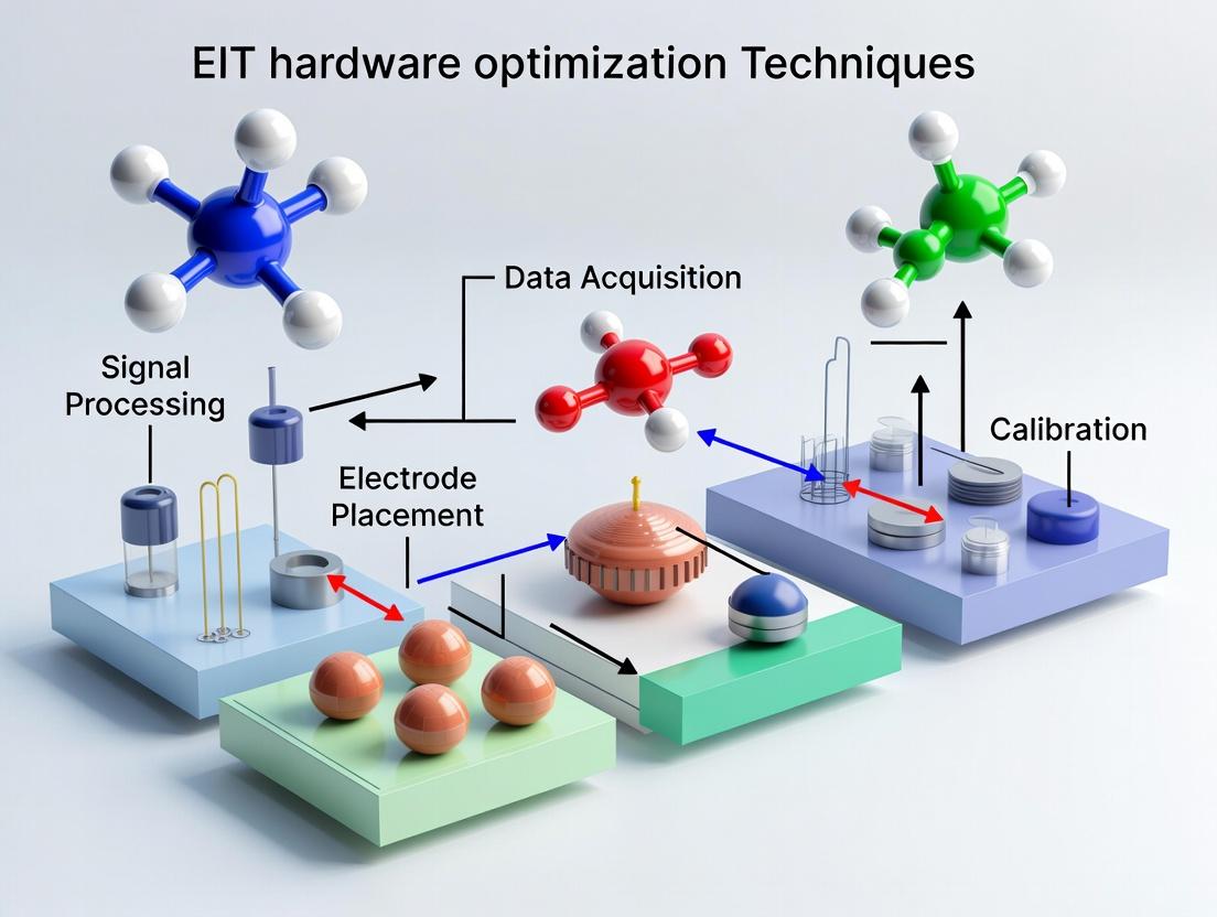
Advancing Biomedical Research: Cutting-Edge EIT Hardware Optimization Techniques for Precision Drug Development
This article provides a comprehensive guide to Electrical Impedance Tomography (EIT) hardware optimization, tailored for researchers and drug development professionals.
Advancing Biomedical Research: Cutting-Edge EIT Hardware Optimization Techniques for Precision Drug Development
Abstract
This article provides a comprehensive guide to Electrical Impedance Tomography (EIT) hardware optimization, tailored for researchers and drug development professionals. It explores the fundamental principles of EIT hardware, details modern methodological advancements and applications in pharmaceutical research (e.g., organ-on-chip, bioreactor monitoring), addresses common troubleshooting and optimization challenges, and validates performance through comparative analysis with other imaging modalities. The aim is to empower scientists with the knowledge to enhance data fidelity, temporal resolution, and system robustness for improved in vitro and preclinical studies.
Understanding EIT Hardware: Core Principles and System Components for Biomedical Applications
Technical Support Center
Troubleshooting Guides & FAQs
Q1: During in vivo EIT measurement of lung function, we observe severe signal drift and inconsistent impedance readings. What could be the cause and how can we resolve it?
A: This is frequently caused by unstable electrode-tissue contact impedance and motion artifact. The biophysical properties of living tissue (dynamic fluid content, perfusion, mechanical movement) directly challenge hardware stability.
- Protocol for Diagnosis & Resolution:
- Verify Electrode Gel & Skin Preparation: Use a high-conductivity, medical-grade hydrogel. Clean the skin with alcohol and, if protocol allows, lightly abrade the stratum corneum to reduce contact impedance.
- Implement Baseline Tracking: Program your EIT hardware to acquire a 30-second baseline before the experimental maneuver (e.g., ventilator cycle). Use this data to compute and subtract a moving average drift.
- Check Current Source Stability: A constant current source must maintain output across a wide range of load impedances (typical for tissue). Test by connecting known resistors (100Ω to 2kΩ) in place of electrodes and measure the applied current. Variation >1% indicates hardware limitation.
- Hardware Optimization Insight: This issue underscores the need for current sources with high output impedance (>1 MΩ at operating frequency) and active electrode guarding circuits to mitigate capacitive leakage paths, a direct design imperative from tissue's variable conductive/capacitive properties.
Q2: Our EIT system performs well on saline phantoms but shows poor spatial resolution and signal-to-noise ratio (SNR) in organotypic 3D cell culture samples. What hardware parameters should we investigate?
A: 3D tissue models present heterogeneous, frequency-dependent electrical properties distinct from homogeneous saline. Poor performance indicates hardware not optimized for the target biophysical environment.
- Protocol for Hardware Calibration & Assessment:
- Frequency Sweep Analysis: Characterize your sample. Use an impedance analyzer to measure the complex impedance of your 3D culture from 1 kHz to 1 MHz. Identify the beta-dispersion region where cellular membrane polarization effects are pronounced.
- Match Hardware Frequency: Configure your EIT system to operate at the frequency where the reactive (capacitive) component of your sample's impedance is significant (often 50-250 kHz for cell cultures). This maximizes contrast.
- Quantify SNR: Measure the RMS voltage (Vsignal) across electrodes during a known current injection. Then, short-circuit the input to the voltmeter (with current injection off) to measure RMS noise (Vnoise). Calculate SNR = 20*log10(Vsignal / Vnoise). Aim for >60 dB.
- Hardware Optimization Insight: This necessitates hardware with selectable, stable frequency generation and synchronous demodulation precisely tuned to the characteristic dispersion of the target tissue, optimizing for capacitance detection over simple conductance.
Q3: We encounter crosstalk between adjacent measurement channels in multi-frequency EIT, corrupting spectroscopic impedance data. How can this be minimized?
A: Crosstalk often arises from non-ideal multiplexer switching and shared ground return paths in the analog front-end, a critical hardware design flaw when measuring tissue's complex admittance.
- Protocol for Isolation Testing:
- Channel Isolation Test: Connect a sinusoidal current source (e.g., 100 µA, 100 kHz) to one measurement channel's input. Terminate all other channels with resistors equivalent to typical tissue impedance (e.g., 500Ω). Measure the voltage induced on the "quiet" adjacent channels. Crosstalk should be >80 dB below the driven signal.
- Improve Grounding Scheme: Implement a "star-point" ground for analog power supplies and reference grounds. Use separate return paths for current injection and voltage measurement circuits.
- Validate with Phantom: Create a two-compartment phantom with different conductivity solutions (e.g., 0.9% saline and 0.45% saline). Image with your multi-frequency protocol. Crosstalk manifests as spectral contamination where one region's impedance spectrum appears distorted near boundaries.
- Hardware Optimization Insight: This demands high channel-to-channel isolation (>100 dB) in multiplexers and fully differential, shielded signal paths. The design must account for the fact that tissue itself can become a coupling pathway at certain frequencies.
Table 1: Typical Electrical Properties of Biological Tissues at 10 kHz & 100 kHz
| Tissue Type | Conductivity (σ) at 10 kHz (S/m) | Relative Permittivity (ε_r) at 10 kHz | Conductivity (σ) at 100 kHz (S/m) | Relative Permittivity (ε_r) at 100 kHz | Key Biophysical Determinant |
|---|---|---|---|---|---|
| Lung (Inflated) | 0.05 - 0.12 | 1500 - 4000 | 0.08 - 0.18 | 800 - 1500 | Air volume fraction, perfusion |
| Liver | 0.03 - 0.07 | 2000 - 6000 | 0.06 - 0.12 | 1000 - 3000 | Cellular density, vascularity |
| Cardiac Muscle | 0.10 - 0.20 | 5000 - 15000 | 0.15 - 0.30 | 2000 - 6000 | Myocyte orientation, ion channel activity |
| 3D Cell Culture (High Density) | 0.15 - 0.30 | 10000 - 20000 | 0.25 - 0.40 | 4000 - 8000 | Extracellular matrix composition, cell-cell junctions |
| 0.9% Saline Phantom | ~1.4 | ~80 | ~1.4 | ~80 | Ionic concentration only |
Table 2: Target Hardware Specifications Informed by Tissue Properties
| Parameter | Target Specification | Rationale from Tissue Biophysics | ||
|---|---|---|---|---|
| Output Impedance of Current Source | >5 MΩ @ 50 kHz - 1 MHz | Maintains current constant despite large, variable tissue load impedance. | ||
| Common-Mode Rejection Ratio (CMRR) | >100 dB @ operating frequency | Rejects artifacts from shared body potential and external interference. | ||
| Input Impedance of Voltmeter | >1 GΩ | <10 pF | Minimizes signal loading on high-impedance electrode-tissue interfaces. | |
| Frequency Range | 1 kHz - 2 MHz (selectable) | To capture alpha, beta, and gamma dispersions of different tissue types. | ||
| Dynamic Range for Voltage Measurement | ±10 V, resolution < 1 µV | Accommodates large standing potentials & small impedance variation signals. |
Visualizations
Title: Troubleshooting EIT Hardware via Biophysical Workflow
Title: EIT System Block Diagram Informed by Tissue
The Scientist's Toolkit: Key Research Reagent Solutions
Table 3: Essential Materials for Validating EIT Hardware with Tissue Models
| Item | Function in Experiment | Relevance to Hardware Design |
|---|---|---|
| Agarose-Saline Phantoms (Varying [NaCl]) | Creates stable, homogeneous test objects with known conductivity. Baseline for isolating hardware performance from tissue complexity. | Validates basic hardware accuracy, linearity, and spatial resolution. |
| Layered Agar-Gelatin Phantoms | Mimics heterogeneous electrical properties (different conductivities/layers). Simulates organ boundaries. | Tests hardware ability to resolve contrast and handle abrupt impedance transitions. |
| Invitro 3D Cell Culture Spheroids/Organoids | Provides a biologically relevant, heterogeneous test bed with cell membranes and junctions. | Critical for testing hardware at frequencies targeting beta-dispersion and optimizing for capacitive sensing. |
| Standard Electrolyte Solution (e.g., 0.9% KCl) | Provides a stable, low-impedance calibration load. | Used for daily calibration of current source magnitude and voltage measurement gain. |
| Medical-Grade Hydrogel Electrode Paste | Ensures stable, low-impedance electrical interface to tissue or skin. | Minimizes variable contact impedance, allowing tissue properties to dominate the signal. |
| Impedance Analyzer (e.g., Keysight E4990A) | Gold-standard for measuring complex impedance of tissues and materials. | Provides "ground truth" data to benchmark the accuracy of the developed EIT hardware. |
Technical Support Center: Troubleshooting & FAQs
Q1: During multi-frequency EIT measurements, we observe significant crosstalk and inconsistent impedance readings across frequencies. What could be the cause and how can we mitigate this?
A: This is a common issue stemming from non-ideal behavior of the current source and electrode-skin interface. The primary causes are:
- Current Source Output Impedance: At higher frequencies, the output impedance of your Howland or mirror-based current source may drop, allowing signal leakage.
- Electrode Polarization Impedance (EPI): The EPI is highly frequency-dependent, causing voltage drops that vary with frequency and distort measurements.
Troubleshooting Protocol:
- Characterize Current Source: Use a precision resistor network (e.g., 1kΩ) in place of the electrode-tank setup. Measure the voltage across the resistor with a differential amplifier and oscilloscope across your frequency band (10 kHz - 1 MHz). Calculate output impedance (Zout = Vopen / I_short). A drop below 1 MΩ at higher frequencies confirms the issue.
- Bench Test Electrodes: In a saline phantom with controlled conductivity, perform a Electrochemical Impedance Spectroscopy (EIS) sweep on a single electrode pair to model the EPI.
Optimization Solution (Thesis Context): Implement a Howland current source with active guard drive to boost output impedance. Use Ag/AgCl electrodes with hydrogel to minimize EPI variance. For hardware optimization, consider a switchable parallel RC feedback in the current source to maintain high output impedance across the band.
Q2: We are experiencing poor Signal-to-Noise Ratio (SNR) in measured voltages, especially with adjacent drive patterns. What are the main noise sources and data acquisition strategies to improve this?
A: The dominant noise sources in EIT are:
- Front-End Electronic Noise: Voltage amplifier input-referred noise.
- Stochastic Biological Noise: Patient movement, blood flow.
- Powerline Interference (50/60 Hz).
- Quantization Noise from the ADC.
Experimental Protocol for Noise Floor Assessment:
- Short the differential amplifier inputs in your data acquisition (DAQ) system.
- Acquire data for 30 seconds at your standard sampling rate.
- Compute the Power Spectral Density (PSD) of the recorded signal. Identify peaks at 50/60 Hz and harmonics.
- Replace short with a phantom of known impedance. Acquire data and compute the standard deviation of the measured voltage over 100 frames. The ratio of signal (mean voltage) to noise (std. dev.) gives a practical SNR.
Optimization Solution (Thesis Context):
- Synchronous Demodulation: Use a dedicated IC (e.g., AD5933) or digital synchronous demodulation in FPGA/software to shift the signal band away from 1/f and powerline noise.
- Averaging: Average over N cycles. SNR improves by √N.
- Shielded Enclosure & Driven Guards: Essential for reducing capacitive pickup.
Table 1: Quantitative Noise Source Comparison & Mitigation Efficacy
| Noise Source | Typical Magnitude (Voltage Referred) | Effective Mitigation Strategy | Expected SNR Improvement |
|---|---|---|---|
| Front-End Amplifier | 1 - 10 µV RMS | Use low-noise instrumentation amps (e.g., INA828) | 20-40 dB |
| Powerline Interference | 100 µV - 1 mV | Synchronous Demodulation, Digital Notch Filters | 30-60 dB |
| Electrode Contact | Highly Variable (>1 mV) | Use abrasive skin prep, hydrogel | 10-30 dB |
| Quantization (16-bit ADC) | ~76 µV (for 5V range) | Oversampling & Averaging | 3 dB per doubling of samples |
Q3: Our voltage measurement circuit saturates when driven with a standard 1 mA current, despite using a high-gain instrumentation amplifier. What is the likely failure point?
A: Saturation is typically due to common-mode voltage overload. In a tetra-polar measurement, the driven electrodes establish a high common-mode voltage (often several volts) on the body/phantom, while the differential voltage of interest is in the millivolt range. The instrumentation amplifier has a limited Common-Mode Input Range specified in its datasheet.
Troubleshooting Guide:
- Measure Common-Mode Voltage: Use oscilloscope probes on each input of your differential amplifier (relative to system ground). You will likely see a large sine wave.
- Check Amp Datasheet: For a typical 5V single-supply IA, the input range may only extend to within 1.5V of the rails.
- Verify Bias Path: Ensure a proper DC bias path exists for the IA's input bias currents.
Solution: Implement a DC servo loop or AC coupling with very high-value resistors to bias the inputs correctly without attenuating the signal. The optimal hardware technique is to use a Driven Right Leg (DRL) circuit to actively suppress the common-mode voltage at its source.
The Scientist's Toolkit: Key Research Reagent Solutions
Table 2: Essential Materials for EIT Phantom Development & Electrode Characterization
| Item | Function in EIT Research | Example/Specification |
|---|---|---|
| Agarose or Phantoms | Creates stable, reproducible test medium with tunable conductivity. | 0.9-2% Agarose in saline with NaCl for conductivity, or inclusion objects. |
| Potassium Chloride (KCl) | Used for calibrating conductivity meters and making standardized saline solutions. | 0.1M KCl solution has a known conductivity of 12.88 mS/cm at 25°C. |
| Conductive Hydrogel | Reduces electrode-skin impedance and improves contact stability for in-vivo tests. | ECG/EEG grade hydrogel with chloride ions (e.g., SignaGel). |
| Ag/AgCl Electrode Pellets | Provides non-polarizable interface, minimizing polarization impedance and drift. | Sintered Ag/AgCl, 4-8 mm diameter for skin contact. |
| Isopropyl Alcohol & Abrasive Gel | Standard skin preparation to remove dead skin cells and oils, crucial for reducing contact impedance. | 70% IPA followed by light abrasion (NuPrep gel). |
| Precision Resistor Network | For validating current source output impedance and DAQ linearity. | 0.1% tolerance resistors, values from 100Ω to 2kΩ. |
Experimental & System Workflow Diagrams
Title: EIT Data Acquisition Sequential Workflow
Title: EIT Hardware Subsystem Interdependencies
Title: EIT System Troubleshooting Decision Tree
Technical Support & Troubleshooting Center
FAQ: Understanding and Optimizing Key EIT Hardware Metrics
Q1: During my EIT experiment, my reconstructed images appear grainy and unstable. Which metric is likely the problem, and how can I improve it? A1: This is a classic symptom of low Signal-to-Noise Ratio (SNR). A low SNR means your useful bioimpedance signal is being obscured by electronic noise, leading to poor image quality.
- Primary Fix (Hardware): Ensure all electrode connections are secure and use high-quality, gelled electrodes to reduce contact impedance. Use shielded cables and keep them away from power sources.
- Primary Fix (Protocol): Increase the amplitude of your injection current within safe physiological limits (typically < 1 mA RMS). Employ signal averaging over multiple measurement cycles.
- Diagnostic Test: Measure the standard deviation of the voltage signal over a short period with no active current injection. This estimates your noise floor.
Q2: My EIT system seems to miss fast physiological events. How are my hardware's Bandwidth settings involved? A2: Bandwidth determines the range of temporal frequencies your system can accurately measure. If the system bandwidth is too low, it will act as a low-pass filter, attenuating rapid impedance changes.
- Check: Verify the specification of your current source and voltage measurement circuitry. The system bandwidth is limited by the slowest component in the signal chain.
- Optimization: For dynamic processes like lung ventilation or blood flow, ensure your measurement frame rate is at least twice the highest frequency component of the physiological event (Nyquist criterion). Review and adjust any anti-aliasing filter settings in your analog front-end.
Q3: When switching from a high-conductivity to a low-conductivity phantom, my voltage readings saturate or become unresolvably small. What metric is failing? A3: This indicates insufficient Dynamic Range (DR). Your system cannot simultaneously handle the largest possible signal (from high conductivity) and resolve the smallest meaningful change (in low conductivity) without distortion.
- Troubleshooting: Check the input range of your analog-to-digital converter (ADC). For a given gain setting, a large signal may clip (saturate), while a small signal may be lost in quantization noise.
- Solution: Implement a programmable gain amplifier (PGA) in your front-end design. Automatically adjust the gain based on a preliminary signal scan to keep the measured voltage within the optimal range of the ADC.
Q4: How do SNR, Bandwidth, and Dynamic Range interact and constrain each other in EIT hardware design? A4: These metrics involve critical trade-offs central to hardware optimization:
- Bandwidth vs. SNR: Increasing measurement bandwidth typically admits more noise, reducing SNR. Narrowing bandwidth (averaging) improves SNR but reduces temporal resolution.
- Dynamic Range vs. Precision: A system with a very wide DR might use a lower-resolution ADC, reducing precision for small signal changes. Optimizing DR involves matching the gain to the expected signal.
- System Optimization Goal: The hardware must be tuned to provide sufficient DR and Bandwidth for the target application while maximizing SNR within those constraints.
Experimental Protocols & Methodologies
Protocol 1: Measuring System SNR for EIT Front-End Objective: Quantify the baseline noise performance of the voltage measurement channel. Procedure:
- Short-circuit the voltage measurement inputs of the EIT system.
- Acquire voltage data for 5 seconds at the intended operating sampling rate.
- Calculate the root-mean-square (RMS) value of this acquired data → This is
V_noise. - Connect a stable reference resistor (e.g., 100Ω) across a calibrated current source.
- Inject the standard operating current (e.g., 1 mA at 50 kHz) and measure the voltage.
- Calculate the RMS value of this signal → This is
V_signal. - Calculate SNR:
SNR (dB) = 20 * log10(V_signal / V_noise).
Protocol 2: Empirical Bandwidth Characterization Objective: Determine the effective -3dB bandwidth of the complete EIT signal chain. Procedure:
- Use a function generator to produce a sine wave, amplitude-modulated to your system's carrier frequency (e.g., 50 kHz).
- Inject this signal into the voltage measurement channel via a known resistive dummy load.
- Sweep the modulation frequency from 1 Hz to 10 kHz (or the system's max frame rate).
- Record the amplitude of the demodulated signal output by the system.
- Plot amplitude vs. modulation frequency. Identify the frequency at which the output power drops to half (-3dB) of its low-frequency value.
Protocol 3: Determining System Dynamic Range Objective: Find the range of input signals the system can measure without saturation or loss of resolution. Procedure:
- Define the Noise Floor (NF) using
V_noisefrom Protocol 1. - Gradually increase the amplitude of the test signal (from Protocol 2) until the system's output distorts (check Total Harmonic Distortion > 1%) or the ADC reaches its maximum code. This level is the Maximum Usable Signal (MUS).
- Calculate Dynamic Range:
DR (dB) = 20 * log10(MUS / NF).
Table 1: Typical Target Metrics for Bioimpedance Applications
| Application | Target SNR (dB) | Required Bandwidth | Needed Dynamic Range (dB) |
|---|---|---|---|
| Static Tissue Imaging | > 80 | Low (Single Freq.) | 60 - 70 |
| Thoracic EIT (Ventilation) | > 70 | Medium (1-50 Hz) | 70 - 80 |
| Cardiac EIT (Perfusion) | > 60 | High (50-200 Hz) | > 80 |
| Cell Culture Monitoring | > 90 | Very Low (DC-1 Hz) | 50 - 60 |
Table 2: Impact of Common Hardware Improvements on Key Metrics
| Hardware Modification | SNR Impact | Bandwidth Impact | Dynamic Range Impact | Primary Trade-off |
|---|---|---|---|---|
| Increased Injection Current | +++ | No change | Potential decrease (saturation) | Patient Safety, Linearity |
| Enhanced Shielding & Grounding | ++ | No change | No change | Cost, Complexity |
| Higher Resolution ADC | Slight + (at low signal) | Potential decrease | +++ | Cost, Data Rate |
| Programmable Gain Amplifier (PGA) | + (optimal gain) | No change | ++ | Complexity, Switching noise |
Visualizations
Title: Troubleshooting Flow for EIT Hardware Metrics
Title: EIT Hardware Signal Chain & Metric Locations
The Scientist's Toolkit: Essential Research Reagent Solutions
| Item | Function in EIT Hardware Research |
|---|---|
| Saline Phantoms (Varying Conductivity) | Stable, reproducible targets for system calibration and DR/SNR testing. |
| Pre-Gelled ECG Electrodes | Provide consistent, low-impedance skin contact for in vivo studies, reducing noise. |
| Programmable Resistor/Capacitor Networks | Mimic complex bioimpedance spectra for frequency response and bandwidth validation. |
| High-Precision Current Source IC | Key component for building injectors with high output impedance and stability. |
| Low-Noise Instrumentation Amplifier | Critical for the first gain stage in voltage measurement to maximize SNR. |
| Lock-in Amplifier (or Digital equivalent) | Enables precise demodulation of small signals at a specific frequency, enhancing SNR. |
| Calibrated Reference Resistors | Traceable standards for absolute impedance accuracy checks and gain calibration. |
| RF Shielding Enclosure (Faraday Cage) | Isolates system from ambient electromagnetic interference during noise floor tests. |
Technical Support Center: Troubleshooting & FAQs
This support center is designed to assist researchers conducting experiments within a thesis focused on EIT hardware optimization techniques. The guidance bridges fundamental principles of classic bench-top systems with the unique challenges of portable/wearable EIT hardware.
FAQ 1: Signal Integrity & Noise
- Q: My portable EIT system shows significantly higher baseline noise and inconsistent boundary voltage measurements compared to my bench-top reference system. How can I diagnose the source?
- A: This is a core challenge in hardware miniaturization. Follow this diagnostic protocol:
- Short-Circuit Test: Disconnect all electrodes and connect the measurement channels directly together via a low-impendance short. Acquire data. The residual signal is primarily internal electronic noise (amplifiers, ADC).
- Saline Bath Test: Use a homogeneous saline phantom (0.9% NaCl) with identical, evenly spaced electrodes. Compare the voltage measurements to a Finite Element Method (FEM) simulation of the same setup. Large deviations indicate poor electrode contact, channel mismatch, or stray capacitance.
- Protocol: For a 16-electrode system, use adjacent current injection and adjacent voltage measurement. Perform 10-frame averaging. Calculate the standard deviation of each voltage measurement across 100 frames on the homogeneous phantom. This matrix is your noise floor.
Quantitative Noise Comparison (Typical Values):
| Hardware Type | Voltage Measurement Noise (RMS) | Typical SNR (in phantom) | Current Source Frequency | Output Impedance |
|---|---|---|---|---|
| High-End Bench-top | 0.01 mV - 0.05 mV | 80 dB - 100 dB | 1 kHz - 1 MHz | > 1 MΩ |
| Portable System | 0.05 mV - 0.2 mV | 60 dB - 80 dB | 10 kHz - 250 kHz | 100 kΩ - 1 MΩ |
| Wearable System | 0.1 mV - 1 mV | 50 dB - 70 dB | 10 kHz - 100 kHz | 10 kΩ - 100 kΩ |
FAQ 2: Electrode-Skin Interface for Wearables
- Q: In wearable thoracic monitoring, I observe gradual signal drift and motion artifacts. What optimization strategies can I implement at the hardware level?
- A: The electrode-skin interface is a major source of instability. Implement a multi-factor experimental protocol:
- Electrode Characterization: Measure the interface impedance spectrum (e.g., 1 kHz to 100 kHz) for each electrode type (gel Ag/AgCl, dry, textile) in situ on the subject before EIT data collection.
- Drift Test: Have the subject remain still. Record EIT data for 10 minutes. Plot the average boundary voltage for a single measurement channel over time. The slope indicates drift.
- Motion Artifact Test: Have the subject perform controlled breathing, then a torso twist. Compare the voltage waveform's temporal standard deviation during quiet segments vs. motion segments.
Experimental Protocol: Electrode-Skin Interface Stability Assessment Objective: Quantify the impact of different electrode types on signal stability for wearable EIT. Materials: See "The Scientist's Toolkit" below. Procedure:
- Prepare the skin site per manufacturer guidelines for each electrode type.
- Attach electrodes in a 16-electrode belt configuration around the thorax.
- Connect electrodes to an EIT system capable of measuring impedance (frequency range: 1 kHz-100 kHz).
- Phase 1 (Static): With subject seated and breathing normally, record EIT data at 1 frame/sec for 5 minutes.
- Phase 2 (Dynamic): Instruct subject to perform torso rotations (5 cycles) and deep coughs (3 times). Record data.
- Data Analysis: For each electrode type, calculate: a) Baseline drift (% change from start to end of Phase 1), b) Motion Artifact Index (MAI = std. dev. of voltages during motion / std. dev. during quiet breathing in Phase 2).
The Scientist's Toolkit: Key Research Reagent Solutions
| Item | Function in EIT Hardware Research |
|---|---|
| Calibrated Test Load (e.g., 500Ω ±0.1%) | Provides a known, stable impedance to verify current source accuracy and voltage measurement precision of the EIT front-end. |
| Multi-Frequency Saline Phantom | Homogeneous phantom with known conductivity profile across frequencies. Essential for testing system frequency response and validating reconstruction algorithms. |
| IEC-Torso Simulator Gel | Standardized conductive gel with stable, tissue-mimicking impedance properties. Used for reproducible electrode-skin interface testing. |
| Programmable Impedance Array | A board with digitally switchable precision resistors. Allows for rapid, programmable creation of known, complex impedance distributions to test image reconstruction speed and accuracy. |
| Shielded Electrode Cables (Twisted Pair) | Minimizes capacitive coupling and electromagnetic interference (EMI), crucial for high-fidelity signal acquisition in unshielded portable environments. |
| Skin Impedance Spectrometer | Dedicated device to measure electrode-skin impedance magnitude and phase across a frequency sweep. Critical for characterizing interface stability. |
Diagram: EIT Hardware Optimization Workflow
Diagram: Key EIT Hardware Subsystem Relationships
Modern EIT Hardware Architectures and Their Application in Drug Development Workflows
Technical Support Center
Troubleshooting Guides & FAQs
Q1: High contact impedance is causing excessive noise in our EIT measurements with a flexible electrode array. What are the primary causes and solutions? A: High contact impedance often stems from poor skin preparation, dried electrolyte gel, or insufficient pressure from the flexible array. First, clean the skin site with an alcohol wipe and abrade gently with conductive paste. For chronic dry-out, use a hydrogel with higher humectant content (e.g., 20% glycerol). Ensure the flexible substrate applies a uniform pressure of 5-15 kPa. If the issue persists, check for micro-cracks in the electrode traces using a microscope.
Q2: Our microfabricated Pt-black electrodes show a rapid increase in impedance over repeated sterilization cycles. How can this be mitigated? A: This indicates degradation of the porous Pt-black layer. Autoclaving (steam sterilization) is not recommended. Instead, use low-temperature hydrogen peroxide plasma (e.g., Sterrad cycle) or ethylene oxide gas. For liquid sterilization, immerse in 70% ethanol for no longer than 30 minutes. As a preventive measure, consider applying a thin, conformal coating of Parylene-C (≈2 µm) post-fabrication to stabilize the nanostructure.
Q3: During long-term bioimpedance monitoring, our flexible array develops inconsistent channel drift. What is the protocol to diagnose the issue? A: Follow this systematic protocol: 1. Bench Test: Measure the impedance of each electrode in a standardized 0.9% NaCl solution using an impedance analyzer at 1 kHz, 10 kHz, and 100 kHz. Compare to baseline values. 2. Inspect Interconnects: Use a multimeter in continuity mode to check for intermittent connections between the electrode pad and the connector, especially after repeated flexing. 3. Validate Circuit: Isolate the array from the EIT system and test with a known resistive phantom to confirm the issue is with the array, not the instrumentation. 4. Check for Delamination: Use optical microscopy to examine the electrode-skin interface for uneven adhesion or sweat accumulation under the array.
Q4: What is the recommended protocol for characterizing the performance of a new microfabricated electrode array for thoracic EIT? A: Title: Protocol for Microfabricated Electrode Array Characterization Objective: To quantitatively assess key performance metrics of a new microfabricated EIT electrode array. Materials: See "Research Reagent Solutions" table. Methodology: 1. Electrochemical Impedance Spectroscopy (EIS): Immerse array in phosphate-buffered saline (PBS). Apply a 10 mV RMS sinusoidal signal across a frequency range of 1 Hz to 1 MHz. Record magnitude and phase. 2. Stability Test: Apply a constant voltage (200 mV) in PBS and record current over 12 hours. Calculate drift rate (%/hour). 3. Flex Durability: Mount array on a motorized flexion jig. Cycle through a 30° bend for 10,000 cycles. Repeat EIS every 1,000 cycles. 4. Contact Impedance Mapping: On a standardized skin simulant gel, measure the contact impedance of all electrodes at 50 kHz. Calculate the mean and standard deviation.
Q5: How do we manage varying contact impedances across channels to prevent reconstruction artifacts? A: Implement active or passive compensation. Passive: Incorporate individual, tunable shunt capacitors in parallel with each measurement channel to help balance phase. Active (Recommended): Use a driven-right-leg (DRL) circuit or individual electrode shielding with guard drives to reduce common-mode voltage and effective impedance. All modern EIT data acquisition systems should include real-time impedance monitoring and software-based compensation algorithms (e.g., based on a parallel RC model) to correct measurements before image reconstruction.
Table 1: Comparative Performance of Common Electrode Coatings for Microfabricated Sensors
| Coating Material | Typical Impedance at 1 kHz (in PBS) | Stability (Drift over 12 hrs) | Recommended Sterilization Method | Key Advantage |
|---|---|---|---|---|
| Platinum Black | 100 - 500 Ω | < 2% | H2O2 Plasma | High surface area, low noise |
| Iridium Oxide | 1 - 10 kΩ | < 5% | Cold Ethanol | High charge injection capacity |
| PEDOT:PSS | 10 - 50 kΩ | < 10% (if hydrated) | UV Light (≤30 min) | Excellent flexibility, mixed ionic/electronic conduction |
| Gold (Plain) | 50 - 200 kΩ | < 1% | Autoclave | Chemically inert, stable |
Table 2: Troubleshooting Matrix for Contact Impedance Issues
| Symptom | Most Likely Cause | Immediate Action | Long-term Solution |
|---|---|---|---|
| Sudden >100% impedance spike on one channel | Broken trace or dry gel pocket | Replace electrode/gel; inspect for physical damage | Redesign flex circuit strain relief; switch gel formula |
| Gradual impedance rise on all channels | Gel drying | Rehydrate with saline mist or reapply gel | Use encapsulated hydrogel pods; reduce experiment duration |
| High impedance & phase shift at high freq (>100 kHz) | Poor electrode-electrolyte interface | Ensure full wetting of porous coating | Re-electrodeposit Pt-black; add surfactant to electrolyte |
| Unstable, fluctuating readings | Motion artifact or poor adhesion | Secure array with medical adhesive film | Use stretchable, adhesive substrate (e.g., silicone-based) |
Experimental Protocol Detail
Protocol: Electrochemical Deposition of Platinum Black for Impedance Reduction Purpose: To create a high-surface-area, low-impedance coating on microfabricated platinum electrodes. Reagents: 1% Chloroplatinic acid (H2PtCl6) solution, 0.01% Lead acetate (Pb(CH3COO)2) solution, Concentrated HCl, DI water. Equipment: Potentiostat/Galvanostat, Three-electrode cell (Pt working, Pt counter, Ag/AgCl reference), Magnetic stirrer. Steps: 1. Clean the substrate electrodes in piranha solution (3:1 H2SO4:H2O2) for 1 minute. CAUTION: Highly exothermic. 2. Rinse thoroughly with DI water. 3. Prepare plating bath: Mix 100 mL of 1% H2PtCl6 with 0.3 mL of 0.01% lead acetate and 2 drops of HCl. 4. Place electrode in bath with constant stirring. Connect to potentiostat. 5. Apply a constant current density of -10 mA/cm² for 2-5 minutes (until dark black coating forms). 6. Rinse and store in 0.9% saline. Characterize via EIS.
Diagrams
Title: Diagnostic Workflow for High Contact Impedance
Title: Validation Workflow for New Flexible EIT Electrode Array
The Scientist's Toolkit: Research Reagent Solutions
Table 3: Essential Materials for Advanced Electrode Development & Testing
| Item & Example Product | Function in Research | Key Specification for EIT |
|---|---|---|
| Conductive Hydrogel (e.g., Parker Labs Ten20) | Provides stable, low-impedance interface between skin and electrode. | Adhesive strength ≥ 0.5 N/cm, Impedance at 50 kHz: < 2 kΩ·cm² |
| Skin Simulant Gel (e.g., AGAR based with NaCl) | Phantoms for standardized in-vitro impedance testing. | Conductivity: 0.2 - 1.0 S/m (mimicking tissue), Stable hydration. |
| Parylene-C Deposition System (e.g., Specialty Coating Systems) | Applies conformal, pinhole-free insulating/biocompatible coating. | Thickness control: 0.1 - 10 µm, High vapor-phase penetration. |
| Electroplating Solution (e.g., Sigma-Aldrich Pt Black Plating Kit) | Deposits nanostructured Pt-black to lower electrode impedance. | Contains lead acetate catalyst for uniform, adherent black deposit. |
| Flexible Substrate (e.g., Polyimide film, ~25µm) | Base material for microfabricated flexible arrays. | High flex endurance (>100k cycles), Stable dielectric properties. |
| Stretchable Conductor (e.g., Ag/AgCl flake in silicone) | Creates stretchable interconnects for highly conformable arrays. | Resistance change < 20% at 30% strain, Biostable. |
| Impedance Analyzer (e.g., Zurich Instruments MFIA) | Gold-standard for electrode/interface electrochemical characterization. | Frequency range: 1 mHz to 5 MHz, Low current measurement capability. |
Troubleshooting Guides & FAQs
Q1: My measured signal-to-noise ratio (SNR) is significantly lower than expected when using a high-precision current source with a lock-in amplifier for EIT measurements. What are the primary culprits?
A: This common issue in EIT hardware optimization typically stems from three areas:
- Ground Loops & Stray Impedances: Improper grounding creates extraneous current paths, injecting mains-frequency noise (50/60 Hz and harmonics). This noise is often within the lock-in's detection bandwidth.
- Current Source Instability: The output impedance of the current source may be insufficient at higher frequencies, or its internal noise (e.g., voltage noise,
1/fnoise) may be corrupting the excitation signal. - Lock-in Amplifier Configuration Errors: Incorrect filter settings (time constant, roll-off), improper reference phase adjustment, or input overload can drastically reduce SNR.
Protocol for Diagnosis:
- Step 1: Disconnect the device under test (DUT, e.g., electrochemical cell). Terminate the current source output into a precision reference resistor (e.g., 1 kΩ ±0.01%). Measure the voltage across this resistor directly with the lock-in.
- Step 2: If SNR improves, the issue is with the DUT interface or stray impedances. If SNR remains poor, the issue is in the source/measurement chain.
- Step 3: Systematically check grounding: use a single-point ground, ensure all shields are properly connected, and employ differential inputs on the lock-in.
- Step 4: Verify lock-in settings: optimize the time constant (increase for lower noise, but slower response), ensure the reference phase is adjusted for null quadrature (Q) component, and confirm input voltages are within the linear range.
Q2: I observe a persistent drift in the in-phase (X) output of my lock-in amplifier over time, complicating long-term EIT monitoring. How can this be mitigated?
A: Drift in the DC output of a lock-in is often due to temperature-induced changes in component values.
Protocol for Mitigation:
- Environmental Stabilization: Place the current source and front-end preamplifiers in a temperature-stabilized enclosure. Allow a 1-2 hour warm-up period for all instruments before critical measurements.
- AC Coupling & Modulation: Use the current source to apply an AC excitation current. Ensure all signal paths are AC-coupled (use coupling capacitors) to block thermo-electric DC offsets. The lock-in will then measure only at the modulation frequency.
- Differential Measurement: For the voltage measurement, use a high-impedance differential preamplifier before the lock-in to reject common-mode drift.
- Regular Nulling: Implement a periodic auto-zeroing routine in your software, where the excitation is momentarily disconnected, and the lock-in's offset is measured and subtracted.
Q3: When scaling my current source to higher frequencies (>100 kHz) for broadband EIT, the output distorts and the magnitude drops. What steps should I take?
A: This indicates bandwidth limitations and impedance matching problems.
Protocol for Bandwidth Optimization:
- Step 1: Characterize the open-loop gain and output impedance of your current source circuit versus frequency using a network analyzer or a scope with signal generator.
- Step 2: Implement feedback loop compensation to ensure stability at the target frequencies. This may require adjusting feedback capacitor values.
- Step 3: Minimize parasitic capacitance at the output node. Use short, shielded cables and consider a "Howland" or "modified Howland" current pump topology with high-speed operational amplifiers.
- Step 4: Match the cable impedance. For very high frequencies, use 50Ω coaxial cables and ensure the output stage can drive this load, or use a dedicated RF current amplifier.
Q4: How do I accurately calibrate the combined gain of my current source and lock-in amplifier measurement chain for quantitative EIT analysis?
A: Absolute calibration is critical for extracting accurate conductivity values.
Experimental Calibration Protocol:
- Prepare Calibration Standards: Use a set of high-precision, non-inductive resistors (e.g., 100Ω, 1kΩ, 10kΩ) with known tolerance (±0.05% or better).
- Measurement Sequence: Replace the DUT with each calibration resistor
R_cal. Apply your standard AC excitation currentI_ex. Measure the resulting voltage amplitudeV_meswith the lock-in. - Calculate System Gain: For each resistor, the theoretical voltage is
V_calc = I_ex * R_cal. The system gain factorGisV_mes / V_calc. AverageGover all resistors. - Apply to DUT: For an unknown DUT impedance
Z_dut, the measured lock-in voltageV_dutrelates as|Z_dut| = (V_dut / I_ex) / G.
Table: Typical Performance Metrics for EIT Hardware Components
| Component | Key Parameter | Typical Target Specification for Low-Noise EIT | Common Issue if Out of Spec |
|---|---|---|---|
| Precision Current Source | Output Impedance | >1 MΩ at 10 kHz, >100 kΩ at 100 kHz | Signal attenuation with DUT load |
| Output Noise Density | < 100 pA/√Hz at 1 kHz | Degrades overall system SNR | |
| Bandwidth (-3 dB) | >10x your maximum excitation frequency | Distortion and phase shift at high freq. | |
| Lock-in Amplifier | Input Voltage Noise | < 5 nV/√Hz | Limits minimum detectable signal |
| Harmonic Rejection | >80 dB at 2f, 3f | Susceptibility to non-linear DUT signals | |
| Time Constant Range | 100 µs to 100 ks | Limits noise filtering and measurement speed | |
| Front-End Preamplifier | Common-Mode Rejection Ratio (CMRR) | >100 dB at excitation frequency | Pick-up of ground loop noise |
| Input Impedance | >10^9 Ω in parallel with <10 pF | Loads the DUT, causing signal drop |
The Scientist's Toolkit: Key Research Reagent Solutions & Materials
| Item | Function in EIT Hardware Optimization |
|---|---|
| Precision Metal-Film Resistor Set (e.g., 0.01% tolerance) | Provides stable, known impedance values for system calibration and current source feedback networks. |
| Low-Noise Operational Amplifiers (e.g., OPAx211, ADA4522) | Core component for building current sources and preamplifiers; chosen for low 1/f noise, voltage noise, and high gain-bandwidth. |
| Low-ESR Capacitors (Polypropylene, NP0/C0G Ceramic) | Used in feedback loops and filter stages; stable capacitance vs. temperature/voltage is crucial for reproducible frequency response. |
| Shielded Twisted-Pair or Coaxial Cables | Minimizes electromagnetic interference (EMI) pick-up on sensitive analog voltage measurement lines. |
| Electrochemical Test Cell (3-Electrode) | Standardized DUT containing working, counter, and reference electrodes for controlled drug interaction studies. |
| Phosphate Buffered Saline (PBS) Solution | Standard electrolyte for simulating physiological conditions in in-vitro drug development experiments. |
| Faraday Cage / Electromagnetic Enclosure | Metallic enclosure that blocks external RFI/EMI, essential for measuring very low-amplitude signals. |
Multifrequency and Broadband EIT (MFEIT/BEIT) Hardware for Spectral Bioimpedance Analysis
Technical Support Center: Troubleshooting & FAQs
Frequently Asked Questions (FAQs)
Q1: What are the most common sources of measurement noise in MFEIT/BEIT systems, and how can they be mitigated? A: Primary noise sources include stray capacitance, electromagnetic interference (EMI), and electrode-skin contact impedance instability. Mitigation involves:
- Using driven-shield cables to minimize stray capacitance.
- Enclosing the system in a Faraday cage and using twisted-pair cables for EMI.
- Employing high-quality, pre-gelled Ag/AgCl electrodes and ensuring proper skin preparation (cleansing, light abrasion) to stabilize contact impedance.
Q2: Our system shows inconsistent impedance readings at higher frequencies (>1 MHz). What could be the cause? A: This is typically due to signal integrity issues. Key checks:
- Cable Length & Matching: Ensure coaxial cables are of equal length and properly impedance-matched (usually 50 Ω) to prevent standing waves.
- Amplifier Slew Rate: Verify that your current source and voltage amplifier have sufficient slew rate and bandwidth for the target frequencies.
- PCB Layout: Inspect for parasitic capacitances/inductances in the front-end PCB layout; use guard traces and proper grounding planes.
Q3: How do we validate the accuracy of our custom-built MFEIT system? A: Follow a standardized protocol using passive phantoms with known, stable electrical properties:
- Resistive Phantoms: Use precision resistor networks to validate amplitude accuracy across frequencies.
- RC Phantoms: Utilize resistor-capacitor networks mimicking tissue dispersions (e.g., Cole-Cole models) to validate phase accuracy.
- Compare system output against reference measurements from a commercial impedance analyzer (e.g., Keysight E4990A).
Q4: What is the impact of electrode number and arrangement on spectral image reconstruction? A: Increased electrode count improves spatial resolution but adds hardware complexity and data throughput demands. For spectral analysis, a consistent, symmetric arrangement (e.g., 16-32 electrodes in a circular array) is crucial to avoid spatial aliasing artifacts that corrupt frequency-dependent parameter extraction.
Q5: How can we synchronize multiple frequency sources in a broadband excitation system? A: Use a single, stable master clock (e.g., a low-jitter crystal oscillator) to derive all digital waveform generation (via Direct Digital Synthesis chips or FPGA). This ensures phase coherence between different frequency components, which is essential for accurate complex impedance calculation.
Troubleshooting Guides
Issue: Poor Signal-to-Noise Ratio (SNR) in Reconstructed Images
- Step 1: Measure the raw voltage data across a known calibration load. If SNR is poor here, the issue is hardware-related.
- Step 2 (Hardware Check):
- Increase excitation current within safe, regulatory limits (typically 1-10 mA pk-pk).
- Check all solder joints and connectors for integrity.
- Verify that the analog front-end (AFE) amplification stages are free from oscillation (check with an oscilloscope).
- Step 3 (Software/Protocol Check): If raw data is good, the issue is in reconstruction. Increase the number of signal averages (frame averaging) in your acquisition protocol. Ensure the reconstruction algorithm uses an appropriate regularization parameter weighted by noise estimates.
Issue: Ghosting or Smearing Artifacts in Spectral Images
- Step 1: This often indicates system timing or phase drift. Perform a repeated measurement on a stable phantom. Plot the phase of a fixed channel over time.
- Step 2: If phase drifts, recalibrate the system. Implement a periodic "active calibration" protocol in your experiment where the system injects signals into internal calibration loads to correct for gain/phase drift over time and temperature.
- Step 3: Ensure all digital filters (anti-aliasing, reconstruction) have linear phase characteristics to avoid introducing frequency-dependent spatial distortions.
Issue: Inconsistent Results Between Successive Scans on the Same Subject
- Step 1: Electrode Contact: This is the most likely cause. Reapply all electrodes. Consider using electrode contact impedance monitoring channels; discard data from any electrode where contact impedance varies by >5% during the scan.
- Step 2: Physiological Motion: Implement gating or triggering synchronized with the subject's respiration or cardiac cycle if measuring the thorax.
- Step 3: System Warm-up: Allow the electronic system (especially precision voltage references and oscillators) to warm up for a stable period (e.g., 30 minutes) before commencing experiments.
Experimental Protocols & Data
Protocol 1: System Performance Characterization
Objective: Quantify the basic electrical performance of an MFEIT/BEIT system.
Methodology:
- Connect the system outputs to a high-precision, wideband load (e.g., a 100Ω ±0.1% resistor).
- Program the system to sweep through its operational frequency range (e.g., 10 kHz to 2 MHz, 20 steps per decade).
- Measure the output current with a calibrated current probe and the voltage across the load with a differential voltage probe, both connected to a calibrated digital oscilloscope.
- Calculate amplitude impedance (|Z|) and phase (∠Z) at each frequency.
- Repeat with a known RC phantom (e.g., 100Ω resistor in parallel with a 100pF capacitor).
Quantitative Performance Metrics Table:
| Metric | Target Specification | Measurement Method | Typical Value for Optimized System |
|---|---|---|---|
| Output Impedance | >1 MΩ ∥ <5 pF | Measure voltage drop with variable load | >1.5 MΩ at 100 kHz |
| Total Harmonic Distortion (THD) | < -80 dB at 1 mA | Spectrum analysis of output current | < -85 dB @ 500 kHz |
| Common Mode Rejection Ratio (CMRR) | > 80 dB | Apply common-mode signal, measure output | > 90 dB @ 100 kHz |
| Noise Floor (Voltage Referred) | < 10 nV/√Hz | Short input, measure spectral density | ~5 nV/√Hz @ 10 kHz |
| Phase Stability | < 0.1° drift over 1 hr | Repeated measurement on stable RC load | < 0.05° drift |
Protocol 2: Spectral Bioimpedance Phantom Experiment
Objective: Acquire multifrequency EIT data for a phantom with spectrally varying regions to test image reconstruction algorithms.
Methodology:
- Phantom Preparation: Create a saline background (0.9% NaCl, ~150 mS/m). Introduce inclusions filled with solutions mimicking different tissue types (e.g., agar with varying NaCl/Cellulose concentrations to create α- and β-dispersions).
- Setup: Arrange a 16-electrode ring array around the phantom. Connect to the MFEIT system.
- Data Acquisition: Use a simultaneous multi-frequency excitation waveform (e.g., a sum of 10, 50, 100, 500 kHz sinusoids). Acquire voltage data for all independent current injection patterns.
- Reconstruction: Use a finite element model (FEM) of the phantom. Reconstruct separate conductivity images at each frequency using a regularized Gauss-Newton solver.
- Analysis: Extract conductivity spectra from regions of interest (ROI) corresponding to inclusions and fit to a Cole-Cole model to extract parameters (R∞, R0, α, τ).
Visualizations
MFBEIT Hardware Signal Pathway
Spectral EIT Experimental Workflow
The Scientist's Toolkit: Key Research Reagents & Materials
| Item | Function in MFEIT/BEIT Research | Specification Notes |
|---|---|---|
| Ag/AgCl Electrodes (Gelled) | Provide stable, low-impedance, and non-polarizable contact with tissue/phanto m. | Pre-gelled, adhesive, disposable. Contact impedance < 1 kΩ at 10 kHz. |
| Phantom Materials (NaCl, Agar, Cellulose) | Create physical models with known, tunable electrical properties for system validation. | NaCl sets conductivity. Agar creates solid matrix. Cellulose mimics β-dispersion. |
| Precision Resistor/Capacitor Kits | Build calibration networks and simple RC phantoms for basic system testing. | Tolerance < 0.1%, low temperature coefficient. Stable up to several MHz. |
| Conductive Electrode Gel | Used with dry electrodes or to improve contact in phantom studies. | High conductivity, non-corrosive, stable pH. |
| Faraday Cage | Shields the sensitive measurement system from external electromagnetic interference. | Mesh or solid enclosure, properly grounded. |
| Calibrated Impedance Analyzer | Gold-standard instrument for validating the accuracy of the EIT system's measurements. | E.g., Keysight E4990A; used to characterize phantom properties. |
| High-Fidelity Data Acquisition Card | Digitizes analog voltages from the AFE with high resolution and sampling rate. | 24-bit ADC, simultaneous sampling, >2 MS/s aggregate rate. |
| FPGA Development Board | Implements real-time digital signal processing (demodulation, filtering) and control logic. | Sufficient I/O, DSP blocks, and memory for multi-channel systems. |
Integrating EIT with Organ-on-Chip Platforms and Bioreactors for Real-Time Monitoring
Technical Support Center: Troubleshooting & FAQs
Frequently Asked Questions (FAQ)
Q1: Our EIT measurements show unstable contact impedance, causing significant noise in the reconstructed images. What are the primary causes and solutions? A: Unstable electrode contact is a common issue in EIT-OoC integration. Causes include: (1) Electrode delamination or fouling from cell culture media, (2) Inconsistent gel or media bridge between the 3D tissue and the planar electrode array, (3) Electrode corrosion (e.g., Ag/AgCl in long-term culture). Solutions within an optimization thesis framework include:
- Protocol: Implement a daily pre-measurement calibration routine involving a 1 kHz impedance sweep across all electrode pairs to identify and exclude faulty channels.
- Hardware Optimization: Apply a thin, stable coating of conductive polymer (e.g., PEDOT:PSS) or nanoporous gold on electrodes to enhance biocompatibility and stability.
- Algorithm Adjustment: Use a time-difference imaging protocol that references a baseline measurement taken immediately after a media change when conditions are most stable.
Q2: How do we differentiate the impedance signal from cell barrier function changes versus cell mass (growth/death) in a monolayer model? A: This requires a multi-frequency EIT (MF-EIT) approach and careful protocol design.
- Protocol: Conduct a frequency sweep from 1 kHz to 1 MHz. Monitor the phase angle (θ) and the real (Z') and imaginary (Z'') components of impedance.
- Interpretation: At low frequencies (e.g., 1-10 kHz), current flows extracellularly, sensitive to barrier integrity (e.g., Trans-Epithelial Electrical Resistance, TEER). At high frequencies (e.g., 100 kHz-1 MHz), current penetrates cell membranes, sensitive to intracellular volume and viability.
- Analysis: A decrease in low-frequency |Z| with stable high-frequency |Z| suggests barrier disruption. A concurrent drop across all frequencies suggests cell detachment or death.
Q3: What is the optimal electrode configuration and number for a perfusion bioreactor containing a 3D spheroid? A: This is a core hardware optimization question. The trade-off is between spatial resolution and system complexity.
Table 1: Electrode Configuration Trade-offs for 3D Bioreactors
| Configuration | Electrode Count (Typical) | Advantage | Disadvantage | Best For |
|---|---|---|---|---|
| Planar (2D) | 8-16 | Simple integration into chip lid/base. Easy to fabricate. | Poor sensitivity to depth/vertical changes. | Monolayers, thin tissue slices. |
| Circumferential | 16-32 | Excellent uniform sensitivity field around a 3D construct. | Requires custom bioreactor chamber. Complex wiring. | Spheroids, organoids in cylindrical chambers. |
| Opposing Paddle | 4-8 | Very simple, can be dipped into media. | Very low spatial resolution, highly inhomogeneous sensitivity. | Bulk conductivity monitoring only. |
Q4: We observe signal drift over 72-hour cultures. Is this biological or an artifact? A: Likely both. Systematic drift must be characterized and minimized.
- Artifact Sources & Mitigation: (1) Evaporation: Use a humidity-controlled incubator or perfusion system with an oil overlay. (2) Electrode Polarization: Use non-polarizable electrodes (Ag/AgCl) or low-amplitude current injection (<1 mA). (3) Temperature Fluctuation: Implement an in-line temperature probe and correct conductivity values using a known temperature coefficient (≈2%/°C for saline).
- Biological Validation Protocol: Correlate EIT drift with a daily endpoint assay (e.g., lactate production, glucose consumption, or supernatant biomarkers). A correlated drift confirms biological origin.
Troubleshooting Guide: Common Error Codes & Issues
Issue: EIT System returns "Voltage Saturation" or "Overrange" error during measurement.
- Check 1: Electrode Contact: Ensure all electrodes are immersed in conductive medium (culture media). A disconnected electrode causes infinite impedance.
- Check 2: Current Injection Amplitude: Reduce the injected current amplitude by 50%. Start with 100 µA - 500 µA for cell culture media.
- Check 3: Shorts: Inspect for salt bridges or metal debris causing short circuits between adjacent electrodes.
Issue: Reconstructed images are persistently blurry with poor feature distinction.
- Check 1: Forward Model Mismatch: The computational model of your bioreactor's geometry must match the physical setup. Verify electrode positions in the model are accurate to within 1 mm.
- Check 2: Regularization Strength: The regularization parameter (λ) is too high. Systematically reduce λ in your inverse solver (e.g., Gauss-Newton) until features appear, but before noise explodes.
- Check 3: Signal-to-Noise Ratio (SNR): Increase the number of measurement frame averages. Use 10-50 averages for slow biological processes.
Issue: Perfusion flow causes unacceptable noise spikes in the impedance data.
- Solution 1: Gating Protocol: Synchronize data acquisition with a paused flow system. Acquire EIT data during brief (e.g., 30 sec), static intervals triggered by a programmable pump.
- Solution 2: Signal Processing: Apply a low-pass digital filter (e.g., Butterworth, 0.1 Hz cutoff) to the time-series data from each electrode pair to remove high-frequency flow turbulence noise.
- Hardware Optimization: Design flow channels that are symmetric relative to the electrode array to minimize periodic flow artifacts.
Experimental Protocol: Validating EIT for Monolayer Barrier Integrity
Title: Protocol for Correlating MF-EIT with TEER in a Gut-on-Chip Model. Objective: To establish a quantitative relationship between traditional TEER and MF-EIT parameters for real-time, non-invasive monitoring. Duration: 5-7 days.
Chip Preparation:
- Seed Caco-2 cells onto a porous polyester membrane in an Organ-on-Chip device pre-fitted with 8 planar gold electrodes (4 top, 4 bottom).
- Culture under constant perfusion (100 µL/h) for 7-10 days until confluent.
Baseline Measurement (Day 0):
- MF-EIT Scan: Using a bioimpedance analyzer, perform a sweep from 500 Hz to 1 MHz. Record |Z| and phase at 1 kHz (Z1k) and 100 kHz (Z100k). Calculate the normalized index: Barrier Index (BI) = Z1k / Z100k.
- Gold Standard TEER: Disconnect from EIT, measure TEER using a handheld epithelial voltohmmeter. Apply formula: TEER (Ω·cm²) = (Resistancesample - Resistanceblank) × Membrane Area.
Intervention & Monitoring (Days 1-2):
- Introduce 5 mM EDTA (chelating agent) to the perfusion medium to disrupt tight junctions.
- Real-time EIT: Acquire single-frequency (1 kHz) EIT data every 15 minutes.
- Endpoint Validation: Every 4 hours, pause flow, perform a full MF-EIT scan and a TEER measurement.
Data Analysis:
- Plot TEER vs. EIT-derived Barrier Index (BI) over time.
- Perform linear regression to derive a conversion function: TEER (estimated) = m × BI + c.
- Use this function to calibrate subsequent real-time, EIT-only experiments.
Title: Workflow for Validating EIT Against TEER
The Scientist's Toolkit: Key Research Reagent Solutions
Table 2: Essential Materials for EIT-Integrated Organ-on-Chip Experiments
| Item | Function & Rationale | Example/Specification |
|---|---|---|
| Conductive Electrode Coating | Improves electrode-electrolyte interface, reduces polarization noise, enhances stability in long-term culture. | PEDOT:PSS, Nanoporous Gold, Platinum Black. |
| Reference Electrodes | Provides stable potential for voltage measurements in perfusion systems. | Agar-salt bridge (3M KCl) with Ag/AgCl wires, or miniaturized integrated Ag/AgCl. |
| Calibration Solutions | For system validation and conductivity calibration. | Phosphate Buffered Saline (PBS) at known conductivities (e.g., 0.1 S/m, 1.5 S/m). |
| Barrier Modulating Agents | Positive & negative controls for barrier function experiments. | Disruptor: EDTA (5-10 mM), TNF-α (10-100 ng/mL). Enhancer: Dexamethasone (1 µM). |
| Viability/Cytotoxicity Assay | Endpoint validation of EIT-derived cell mass signals. | ATP-based luminescence (e.g., CellTiter-Glo 3D) for spheroids; Calcein-AM/EthD-1 live/dead imaging. |
| Perfusion-Compatible Tubing | Chemically inert, non-gas permeable, prevents bubble formation. | Platinum-cured silicone tubing or fluoropolymer (PFA, FEP) with low gas permeability. |
| Impedance Matching Gel/Medium | For stable contact between 3D constructs and electrodes. | Serum-free medium with 0.2-0.5% agarose or Matrigel to reduce drift. |
| Data Acquisition & Inverse Solver Software | Hardware control, image reconstruction, and time-series analysis. | Custom MATLAB/Python with EIDORS toolkit, or vendor-specific software (e.g., Swisstom APT). |
Title: Signaling Pathway from Disruption to EIT Signal
Solving Common EIT Hardware Challenges: A Practical Guide to Noise Reduction and Calibration
Technical Support & Troubleshooting Center
Welcome to the EIT Hardware Optimization Technical Support Center. This resource, developed under the thesis "Advanced Noise Mitigation in High-Precision Electrical Impedance Tomography Hardware," provides targeted guidance for researchers encountering noise-related issues in sensitive bioimpedance measurements, particularly in drug development applications.
Troubleshooting Guides & FAQs
Q1: My EIT system shows erratic impedance readings at high frequencies (>500 kHz). The issue worsens when I move my hand near the electrode cables. What is the likely cause and how can I fix it? A: This is a classic symptom of stray capacitance coupling, often from unshielded cables or improper guarding.
- Diagnosis: Measure the baseline noise with inputs shorted. Then, wave a hand near cables. A significant spike confirms stray capacitive pickup.
- Mitigation Protocol:
- Replace standard cables with double-shielded, coaxial cables. Connect the outer shield to chassis ground and the inner shield to guard driver output if available.
- Implement active guarding: Drive the cable shield at the same potential as the signal conductor to eliminate the potential gradient.
- Keep cables short and fixed. Use cable ties to secure them in place, minimizing movement-induced capacitance changes.
- Insert a Faraday cage around the measurement setup (e.g., the tissue culture plate or microfluidic device).
Q2: We observe periodic spikes or a baseline "hum" in our time-series EIT data, coinciding with the building's HVAC cycle or other lab equipment. Is this EMI and how do we isolate it? A: Yes, this indicates electromagnetic interference (EMI) from power lines or switched-mode power supplies.
- Diagnosis: Use a spectrum analyzer on the EIT output. Look for peaks at 50/60 Hz (mains) and their harmonics.
- Mitigation Protocol:
- Power Conditioning: Use a linear power supply for your EIT front-end instead of a noisy switching supply. Employ a mains isolation transformer with an electrostatic shield.
- Differential Signaling: Ensure your analog front-end uses a high Common-Mode Rejection Ratio (CMRR >100 dB at 50Hz) instrumentation amplifier.
- Twisted Pair Wiring: For any non-coaxial signal lines (e.g., to reference electrodes), use tightly twisted pairs to cancel magnetically induced interference.
- Spatial Separation: Physically move the EIT system and its electrodes away from suspected noise sources (motors, computers, UPS units).
Q3: During long-term perfusion experiments, our reconstructed conductivity images drift slowly over several hours, compromising dose-response analysis. What should we check? A: This points strongly to thermal drift affecting electronic component stability and/or the sample itself.
- Diagnosis: Log the ambient temperature near the electronics and the sample chamber. Correlate temperature changes with baseline impedance drift in a control saline solution.
- Mitigation Protocol:
- Component Selection: Use amplifiers and precision resistors with low temperature coefficients (e.g., <10 ppm/°C).
- Thermal Management: Install temperature-stabilizing enclosures for critical analog circuits (e.g., using Peltier elements with PID control).
- Experimental Control: Enclose the entire sample chamber in a thermally insulated box. Use a perfusion bath with an in-line heater/cooler regulated by a feedback-controlled thermostat.
- Software Compensation: Implement a periodic auto-zero/calibration routine that injects a known reference impedance and corrects gain/drift in software.
Q4: We see inconsistent results between replicates. Noise seems random. How can we systematically identify the dominant noise source? A: Follow a structured diagnostic workflow to isolate the contribution of each noise type.
Diagram Title: Systematic Diagnostic Workflow for EIT Noise Source Identification
Quantitative Noise Source Comparison
The following table summarizes typical characteristics and magnitudes of key noise sources, as quantified in our thesis research.
| Noise Source | Typical Frequency Range | Magnitude (in a 1V, 100kHz system) | Primary Impact on EIT Data |
|---|---|---|---|
| Stray Capacitance | High (>100 kHz) | Can induce >10 mV offset/unpredictable coupling | Image blurring, phase errors at high frequency. |
| Mains EMI (50/60 Hz) | Very Low (50/60 Hz & harmonics) | 1-100 mV pickup without shielding | Baseline stripes in time-series, reconstruction artifacts. |
| Broadband EMI | Wideband (kHz to MHz) | Variable, based on environment | Random spikes, increased noise floor. |
| Thermal Drift | Very Low (<0.1 Hz) | 50-500 µV/°C in op-amps; % change in sample | Slow conductivity drift, false temporal trends. |
| Intrinsic (Johnson) Noise | Broadband (White) | ~1 µV/√Hz for 1kΩ source @ 25°C | Fundamental limit to resolution and SNR. |
Experimental Protocol: Guarded, Shielded Cable Performance Test
Objective: Quantify the reduction in stray capacitive noise achieved by implementing active guarding and double shielding.
- Setup: Configure a voltage-driven, 2-electrode impedance measurement circuit at 1 MHz.
- Control: Use a 1-meter unshielded twisted pair (UTP) cable to connect a 1 kΩ test resistor.
- Intervention 1: Replace UTP with a 1-meter single-shielded coaxial cable. Connect shield to ground.
- Intervention 2: Use a double-shielded coaxial cable. Connect outer shield to ground, inner shield to a guard driver (unity-gain buffer from signal source).
- Measurement: For each configuration, measure the peak-to-peak noise voltage (Vpp) and RMS noise (Vrms) at the receiver over a 30-second interval. Introduce a standard capacitive disturbance by moving a grounded metal plate near the cable.
- Analysis: Calculate the noise reduction factor relative to the control for each configuration.
The Scientist's Toolkit: Key Reagents & Materials for Low-Noise EIT Experiments
| Item | Function in Noise Mitigation |
|---|---|
| Phosphate-Buffered Saline (PBS), 0.1X | Standardized, low-conductivity calibration solution for baseline system characterization. |
| Agarose Phantoms (0.5-2%) | Stable, homogeneous test phantoms with known conductivity for long-term drift tests. |
| Conductive Silver Epoxy | Creates low-impedance, stable connections between electrodes and cables, minimizing contact noise. |
| Electrically Conductive Shielding Paint | Used to create ad-hoc Faraday cages on custom sample holders or enclosures. |
| Temperature Calibration Thermistor | High-accuracy (±0.01°C) sensor for real-time thermal drift correlation and compensation. |
| SMA/BNC-terminated Coaxial Cables | Provide consistent, shielded connections; prefer SMA for frequencies >1 MHz. |
| Toroidal Ferrite Cores (Mix 31/43) | Snap-on cores to suppress common-mode high-frequency noise on cables (EMI chokes). |
| Isothermal Chamber (DIY or Commercial) | An insulated box with passive/active temperature stabilization for the sample stage. |
Technical Support Center: Troubleshooting & FAQs
Q1: My bioimpedance measurements show high variability between repeated electrode placements. What is the primary cause and how can I mitigate it?
A: The primary cause is inconsistent electrode-skin contact impedance, often due to dead skin cells (stratum corneum), poor adhesion, or uneven pressure. Mitigation strategies include:
- Skin Preparation: Clean the site with 70% isopropyl alcohol and gently abrade the skin with fine-grade sandpaper or Nuprep gel to reduce the stratum corneum's high resistance.
- Electrode Gel: Use a high-conductivity, wet gel electrode or hydrogel with chloride ions (e.g., Ag/AgCl) to ensure a stable ionic interface.
- Pressure Control: Employ electrode holders or straps to apply consistent, gentle pressure. Avoid overtightening, which can cause ischemia and drift.
Q2: We observe significant low-frequency drift and DC offset in our EIT data during long-term monitoring. What contact-related issues could be responsible?
A: This is typically caused by electrochemical changes at the electrode-solution interface.
- Cause: Polarization of non-polarizable electrodes (e.g., bare metals) or drying of the electrode gel/solution alters the half-cell potential.
- Solution: Use properly chlorided Ag/AgCl electrodes, which are nearly non-polarizable. Ensure hydrogel pads are sealed (e.g., with medical tape) to prevent drying. For solution measurements, maintain a stable electrolyte concentration and temperature.
Q3: How can I quantitatively assess the quality of my electrode contact before starting an EIT experiment?
A: Perform a single-frequency impedance sweep at a representative frequency (e.g., 10 kHz).
- Protocol:
- Connect electrodes to an impedance analyzer.
- Measure the magnitude and phase of each electrode pair.
- Calculate the contact impedance. Consistent, moderately low values (e.g., < 2 kΩ at 10 kHz for skin) with low standard deviation across electrodes indicate good contact.
- Acceptance Criteria: Reject electrodes with impedance values > 3 standard deviations from the mean or phase angles indicative of purely capacitive coupling.
Q4: In cell culture or solution EIT, how do I prevent electrode polarization artifacts at higher frequencies?
A: Electrode polarization impedance dominates at lower frequencies. To minimize its impact:
- Electrode Material: Use gold or platinum-black coated electrodes to increase the effective surface area and reduce polarization impedance.
- Frequency Selection: For EIT, operate in a frequency range where the measured impedance is dominated by the solution/tissue, not the electrode interface. This is identified via electrochemical impedance spectroscopy (EIS).
- Protocol: Perform an EIS scan from 1 Hz to 1 MHz on your electrode system in the relevant solution. The "flattened" region in the impedance magnitude plot indicates the suitable frequency range for EIT measurements.
Table 1: Common Contact Artifacts and Mitigation Strategies
| Artifact Symptom | Likely Cause | Recommended Solution |
|---|---|---|
| High Noise & Variance | High, variable contact impedance | Skin abrasion, conductive gel, uniform pressure |
| Low-Frequency Drift | Drying gel, electrode polarization | Use Ag/AgCl electrodes, seal hydrogel, maintain hydration |
| Unstable Baseline | Electrochemical changes, poor adhesion | Ensure full chloridation of Ag/AgCl, use adhesive overlays |
| Capacitive Phase Shift | Insulating layer (dead skin, biofilm) | Clean/abrade skin, sterilize & clean solution electrodes |
| Nonlinear Response | Excessive current density | Use larger electrodes or lower injection current |
Table 2: Electrode Contact Impedance Benchmarks (Typical Values)
| Electrode Type | Application | Target Impedance (at 10 kHz) | Key Metric |
|---|---|---|---|
| Ag/AgCl Hydrogel | Chest Skin | < 2 kΩ | Consistency (< 10% variance across array) |
| Gold-plated | Saline Solution | < 500 Ω | Polarization Impedance (should be minimal) |
| Platinum Black | Cell Culture Medium | < 1 kΩ | Stability over time (drift < 5%/hour) |
| Stainless Steel | Long-term Wearable | < 3 kΩ | Motion Artifact Resilience |
Experimental Protocol: Standardized Electrode-Skin Contact Assessment Title: Quantifying Electrode-Skin Interface Stability for EIT. Objective: To establish a reproducible protocol for assessing and preparing electrode-skin contact to minimize baseline artifacts in thoracic EIT. Materials: See Scientist's Toolkit below. Procedure:
- Site Marking: Mark electrode positions on the skin using a template.
- Skin Preparation: Wipe each site with 70% isopropyl alcohol. Gently abrade with single-use, fine-grit (240-400) sandpaper until the skin is slightly pink.
- Impedance Check: Apply a single electrode to one site. Using a two-electrode setup with a distal reference, measure impedance magnitude and phase at 1 kHz, 10 kHz, and 100 kHz.
- Baseline Recording: Apply all electrodes. Record 5 minutes of baseline EIT data at 1 frame/sec.
- Analysis: Calculate the mean and standard deviation of initial contact impedance across all electrodes. In the baseline data, compute the standard deviation of each pixel's impedance over time. A stable contact yields a low temporal standard deviation map.
The Scientist's Toolkit: Key Research Reagent Solutions
| Item | Function in Mitigating Contact Artifacts |
|---|---|
| Nuprep Skin Prep Gel | Abrasive, conductive gel to remove dead stratum corneum and lower initial contact impedance. |
| SignaGel Electrode Gel | High-conductivity, chloride-rich wet gel for Ag/AgCl electrodes to maintain stable ionic interface. |
| Redux Creme | Post-experiment skin cream to soothe abraded skin and maintain participant comfort in studies. |
| KCl Solution (0.9% - 3M) | Standard electrolyte for conditioning and testing Ag/AgCl electrodes; provides stable reference potential. |
| Platinum Black Plating Solution | Used to electroplate electrodes, increasing surface area and drastically reducing polarization impedance. |
| Hydrogel Adhesive Overlays | Transparent dressings to secure electrodes, prevent gel drying, and minimize motion-induced contact changes. |
Troubleshooting Contact Artifacts Workflow
Artifact Causation Pathway
FAQs Continued
Q5: What is the optimal method for chloriding silver electrodes for stable solution measurements?
A: Use electrochemical chloridation.
- Protocol:
- Clean silver wire/electrode with isopropanol and rinse.
- Prepare a 0.1M HCl or 0.9% NaCl solution.
- Connect the Ag electrode as the anode and a platinum or carbon cathode to a constant current source.
- Apply a current density of 0.5 mA/cm² for 30-60 seconds. The electrode will turn a uniform dull gray/purple (AgCl layer).
- Rinse and store in a KCl solution to maintain the layer.
Q6: How does electrode contact instability specifically compromise drug development research using EIT?
A: In drug development, EIT may monitor tissue perfusion or edema. Contact artifacts can:
- Mimic or Mask Pharmacodynamic Responses: Drift can be mistaken for a slow physiological change.
- Reduce Statistical Power: Increased noise requires larger sample sizes to detect drug effects.
- Invalidate Longitudinal Studies: Unstable baselines prevent comparison across time points. This directly impacts the reliability of efficacy and safety assessments, underscoring the need for the hardware optimization techniques central to this thesis.
Technical Support Center
Troubleshooting Guides & FAQs
Q1: During phantom-based validation, we observe inconsistent impedance readings across repeated measurements with the same phantom. What are the primary causes and solutions?
A: Inconsistent readings typically stem from electrode contact instability or environmental drift. First, ensure all electrode connections are secure and the contact gel is uniformly applied and not dehydrated. Second, verify laboratory temperature and humidity are stable; fluctuations >2°C or >10% RH can cause significant baseline drift. Third, perform a system self-test and baseline reset before each validation run. The recommended protocol is: 1) Power cycle the EIT system, 2) Execute internal self-calibration (refer to sys_cal command), 3) Measure system offset with open/short calibration loads, 4) Proceed with phantom measurement. If inconsistency persists (>5% variation), check for phantom electrolyte degradation or air bubbles.
Q2: How do we quantitatively distinguish between true system drift and random measurement noise in long-term monitoring experiments?
A: Implement a dual-reference protocol. Use a stable, sealed reference phantom measured at the start and end of each experimental session. Analyze the data using Allan deviation. True system drift manifests as a rising trend in the Allan deviation plot at longer averaging times, while white noise decreases. Calculate the drift coefficient (ΔZ/ΔT) from the reference phantom data. If the coefficient exceeds 0.1% per hour for your system's typical frequency, schedule a full recalibration.
Q3: What is the detailed protocol for performing a full system calibration and drift compensation sequence?
A: Follow this workflow:
- Pre-conditioning: Power on the system and allow a 30-minute warm-up.
- Open/Short/Load Calibration: Connect calibration standards to all channels. Execute the 3-point calibration procedure in the software to characterize the system's transfer function.
- Reference Phantom Measurement: Image a known validation phantom (e.g., cylindrical phantom with off-center conductive target). Record mean amplitude and phase for the target region.
- Drift Compensation During Experiment: For sessions >1 hour, inject a brief reference signal (e.g., a known resistor across channel 1-2) every 30 minutes. Use the change in this reading to apply a linear correction factor to all experimental data.
- Post-Experiment Validation: Re-measure the reference phantom. If results deviate >2% from step 3, flag the experimental data for potential compensation or exclusion.
Q4: Our reconstructed images show gradual geometric distortion over weeks, even with regular phantom checks. What component failure might this indicate?
A: Progressive geometric distortion often points to analog front-end component aging, specifically in the voltage-controlled current source (VCCS) or the multiplexer switches. This can cause channel-dependent gain/phase errors. To diagnose, run a channel symmetry test: measure a perfectly centered, homogeneous phantom. The impedance magnitude for symmetrical electrode pairs (e.g., 1-9, 2-10) should match within 1.5%. A table of deviations will identify failing channels. Replace the analog board or contact technical support if deviations exceed 3%.
Key Experimental Protocols
Protocol 1: Monthly Comprehensive System Validation
- Objective: Quantify overall system performance and detect gradual drift.
- Materials: Homogeneous saline phantom, target phantom (with known inclusion), calibration loads.
- Procedure:
- Perform 3-point electrical calibration.
- Measure homogeneous phantom. Calculate signal-to-noise ratio (SNR) and reciprocity error.
- Image target phantom. Calculate image metrics: Position Error (PE), Radius Error (RE), and Contrast-to-Noise Ratio (CNR).
- Compare metrics to baseline values from system acceptance. A >10% degradation in CNR or >20% increase in PE triggers investigative maintenance.
Protocol 2: Real-Time Drift Compensation for Longitudinal Studies
- Objective: Correct for intra-session drift without interrupting subject/experiment.
- Methodology:
- Dedicate two electrodes to a stable, embedded reference impedance within the sensor or subject setup.
- Acquire a brief measurement of this reference impedance at a fixed interval (e.g., every 10 frames).
- Model the drift as a linear function of time using the reference data.
- Apply the inverse drift function to all measurement data during reconstruction.
Table 1: Acceptable Performance Metrics for EIT System Validation
| Metric | Calculation | Acceptable Threshold | Corrective Action if Failed |
|---|---|---|---|
| Reciprocity Error | |V_ab->cd - V_cd->ab| / |V_ab->cd| |
< 0.5% | Check electrode contacts & current source symmetry. |
| Signal-to-Noise Ratio (SNR) | Mean(Signal) / StdDev(Noise) |
> 80 dB | Check grounding, shield cables, replace batteries. |
| Position Error (PE) | |Actual Pos - Reconstructed Pos| |
< 10% of diameter | Re-run full system calibration (Open/Short/Load). |
| Amplitude Drift (1hr) | (Z_final - Z_initial) / Z_initial |
< 0.5% | Improve temperature control; check component heating. |
Table 2: Drift Compensation Algorithm Comparison
| Method | Principle | Advantages | Limitations | Best For |
|---|---|---|---|---|
| Linear Interpolation | Corrects data based on linear drift of reference measurements. | Simple, low computational cost. | Assumes linear drift; ineffective for sudden shifts. | Short-term (<2hr) stable experiments. |
| Kalman Filtering | Uses a state-space model to estimate and correct true impedance. | Robust to noise; can handle non-linear trends. | Complex to implement; requires tuning. | Long-term, high-noise monitoring. |
| Reference Electrode | Uses measurement from a stable, dedicated channel as a divisor. | Real-time, continuous correction. | Requires a perfectly stable reference. | Systems with built-in reference impedance. |
Diagrams
EIT System Validation Workflow
Diagnostic Logic for EIT System Issues
The Scientist's Toolkit: Research Reagent Solutions
| Item | Function & Importance in Calibration/Validation |
|---|---|
| Geometric Validation Phantoms | Agar or PVC cylinders with precisely positioned conductive/inclusion targets. Provide ground truth for assessing image reconstruction accuracy and spatial resolution. |
| Stable Electrolyte Solution | 0.9% NaCl with 1% agar or surfactant. Creates a homogeneous, stable medium for baseline SNR and reciprocity measurements. Consistency is critical. |
| Calibration Load Set | Precision resistors (e.g., 100Ω, 1kΩ) and capacitors for Open/Short/Load calibration. Characterizes system's front-end transfer function. |
| Reference Impedance Module | A small, temperature-stable resistor-capacitor network. Serves as an embedded drift monitor for real-time compensation during long experiments. |
| High-Purity Contact Gel | Electrode-skin interface gel with consistent ionic conductivity. Minimizes variable contact impedance, a major source of noise and drift. |
| Environmental Logger | Precise thermometer/hygrometer. Monitors lab conditions to correlate environmental fluctuations with measured system drift. |
Troubleshooting Guides and FAQs
Q1: During high-speed EIT data acquisition, we observe a significant drop in SNR and image artifacts. What are the primary hardware checks?
A: This typically indicates a trade-off where speed optimization has compromised accuracy. Follow this protocol:
- Check Current Source Stability: At high multiplexing rates, the current source may not reach steady-state. Verify with an oscilloscope across a known calibration resistor.
- Verify Amplifier Settling Time: The instrumentation amplifier requires sufficient time to settle after each channel switch. If the sampling rate is too high, the reading is taken before the signal stabilizes.
- Assess Power Supply Noise: High-speed switching can introduce coupled noise. Use a spectrum analyzer to check for increased noise in the 50/60 Hz line frequency and its harmonics.
Experimental Protocol for Diagnosis:
- Objective: Quantify the relationship between data acquisition speed and measurement accuracy.
- Method: Use a stable, known phantom (e.g., a saline tank with fixed, off-center conductive target). Acquire data at sequentially higher frame rates (e.g., 10, 50, 100 fps). For each rate, compute the standard deviation of voltage measurements on a stable channel over 1000 frames (noise proxy) and the positional error of the reconstructed target.
- Expected Outcome: A table quantifying the "accuracy cost" of increased speed, guiding an optimal configuration.
Q2: Our system uses a high-resolution, focused electrode array. How can we reconfigure for broader organ coverage without a full hardware rebuild?
A: This is a classic resolution vs. coverage trade-off. Two primary strategies can be implemented:
- Strategic Electrode Multiplexing: Reconfigure the switching matrix to group adjacent electrodes, effectively creating larger "virtual electrodes." This increases coverage at the expense of spatial resolution. The multiplexing pattern must be symmetrically designed to maintain model accuracy.
- Multi-Frequency Protocol Optimization: Use a lower base frequency for broader, deeper tissue coverage (due to its better penetration) to establish a baseline conductivity map. Then, intermittently interleave higher-frequency measurements on the central focused array for detailed resolution of the region of interest.
Experimental Protocol for Coverage Optimization:
- Objective: Maximize field-of-view (FOV) coverage while maintaining acceptable resolution for a large organ (e.g., lung).
- Method: Implement a two-stage scanning protocol on a large, heterogeneous phantom.
- Stage 1 (Broad Coverage): Use every 4th electrode (or grouped electrodes) to perform a full scan cycle. Reconstruct a low-resolution image.
- Stage 2 (Focused Zoom): For regions of interest identified in Stage 1, activate all electrodes in that sub-array for a high-resolution scan.
- Expected Outcome: A hybrid imaging workflow that balances global coverage with local detail.
Data Presentation
Table 1: Impact of Acquisition Speed on Measurement Fidelity
| Frame Rate (fps) | Voltage Noise (µV RMS) | Target Position Error (mm) | Recommended Use Case |
|---|---|---|---|
| 10 | 12.5 | 0.8 | Baseline calibration, static imaging |
| 50 | 18.7 | 1.5 | Standard dynamic monitoring |
| 100 | 41.2 | 3.9 | Fast transient capture (trade-off accepted) |
| 200 | 105.0 | 8.7 | Not recommended for quantitative imaging |
Table 2: Electrode Configuration Trade-offs
| Configuration | Electrodes Active | Effective Coverage | Spatial Resolution | Primary Application |
|---|---|---|---|---|
| Focused Array | 32 (dense cluster) | 20% of FOV | High (sub-cm) | Localized tumor monitoring |
| Broad Array | 16 (evenly spaced) | 80% of FOV | Low (2-3 cm) | Whole-organ perfusion scan |
| Multiplexed Virtual | 16 physical -> 8 virtual | 65% of FOV | Medium (1-2 cm) | Compromise for thoracic imaging |
Visualizations
High-Speed EIT Hardware Troubleshooting Workflow
Resolution vs. Coverage Decision Logic in EIT
The Scientist's Toolkit: Key Research Reagent Solutions
| Item | Function in EIT Hardware Optimization Research |
|---|---|
| Calibrated Saline Phantoms | Stable, known-conductivity targets for quantifying system accuracy and spatial resolution under different configurations. |
| Programmable Multiplexer Board | Allows dynamic reconfiguration of electrode patterns to test coverage vs. resolution strategies without physical changes. |
| Precision Current Source (1mA, 50kHz-1MHz) | Generates the injection signal; stability and bandwidth are critical for testing speed-accuracy limits. |
| Lock-in Amplifier (Reference) | Used to validate voltage measurements from custom EIT front-ends, providing a gold standard for accuracy. |
| Conductive Gel & Electrode Arrays (Ag/AgCl) | Standardized interface materials; consistent electrode-skin contact impedance is vital for reproducible results. |
| Network Analyzer | Characterizes the full frequency response of the analog front-end, identifying bandwidth bottlenecks affecting speed. |
Benchmarking EIT Hardware Performance: Validation Strategies and Comparative Analysis with Other Modalities
Troubleshooting Guides & FAQs
Q1: During a conductivity change experiment, our system shows a high signal-to-noise ratio (SNR) but poor quantitative accuracy compared to a reference standard. What could be the cause? A: This often indicates a calibration or systematic error rather than random noise. First, verify that your standardized phantom's electrical properties are certified and traceable. Second, ensure your hardware's current source output and voltage measurement gain are calibrated using a precision resistive network. Follow the Protocol for Basic System Calibration (P-BSC-01) below.
- Protocol P-BSC-01: Basic System Calibration for Quantitative Accuracy
- Materials: Precision reference resistors (e.g., 100Ω, 500Ω), calibrated digital multimeter (DMM), data acquisition (DAQ) interface.
- Current Source Calibration: Disconnect electrodes. Connect the current source output to the DMM in series with a reference resistor (e.g., 100Ω). Command a known current (e.g., 1mA RMS). Measure the voltage drop across the resistor with the DMM. Calculate actual current: Iactual = VDMM / Rreference. Adjust the source gain in software until Iactual matches the commanded value. Repeat for all current amplitude settings.
- Voltage Measurement Calibration: Apply a known, stable voltage (from a calibrated source) directly to the voltage measurement channel inputs. Record the system's measured value. Calculate a per-channel gain correction factor. Apply this factor to all subsequent experimental voltage measurements.
- Validation: Test the calibrated system on a multi-compartment phantom with known conductivity contrasts. Accuracy should improve to within ±5% of known values.
Q2: We observe significant drift in boundary voltage measurements over a 1-hour dynamic imaging experiment. How can we isolate the cause? A: Drift can originate from the hardware front-end or environmental factors. Perform a systematic isolation test.
| Potential Cause | Diagnostic Test | Expected Outcome if Cause is Isolated |
|---|---|---|
| Temperature Drift in Electronics | Enclose system in temperature-stable environment. Run measurement on a fixed, stable passive resistor network for 60 mins. | Drift persists on the passive network. |
| Electrode-Polarization Impedance Drift | Replace electrodes with direct, soldered connections to a stable resistor phantom. Run the same long-term test. | Drift is eliminated, pointing to electrode/electrolyte interface. |
| Power Supply Instability | Monitor system's internal voltage rails with an oscilloscope during operation. | Correlation between rail noise/fluctuation and measurement drift. |
- Protocol for Drift Isolation (P-DI-01):
- Construct a simple, sealed 8-resistor network mimicking a 16-electrode ring.
- First, measure with standard electrodes and agar phantom for 60 mins (Baseline).
- Second, replace the agar/saline with the sealed resistor network, keeping electrodes. Measure for 60 mins.
- Third, remove electrodes and solder wires directly to the resistor network terminals. Measure for 60 mins.
- Compare the drift magnitude (e.g., % change from baseline voltage) across the three tests to isolate the problem layer.
Q3: What are the key metrics and standardized phantoms we should use for a comprehensive hardware performance paper? A: A complete evaluation requires multiple phantoms and metrics. The table below summarizes the core set.
| Evaluation Category | Standardized Phantom Type | Key Quantitative Metrics | Target Value (Benchmark) |
|---|---|---|---|
| Basic System Performance | Uniform Saline Tank (Geometrically Simple) | Signal-to-Noise Ratio (SNR), Total Harmonic Distortion (THD) | SNR > 80 dB, THD < -60 dB |
| Spatial Resolution | Contrast Inclusion Phantom (e.g., rods in tank) | Contrast-to-Noise Ratio (CNR), Point Spread Function (PSF) Width | CNR > 5 for 10% contrast inclusion |
| Quantitative Accuracy | Multi-Compartment Phantom (Certified conductivities) | Image Error (IE), Relative Error (RE) | IE < 10%, RE < 5% for known contrasts |
| Temporal Performance | Dynamic Mechanical Actuator (Moving target) | Frame Rate, Temporal SNR | Consistent with sampling theorem |
- Protocol for Spatial Resolution Assessment (P-SRA-01):
- Use a phantom with insulating and conducting rods of varying diameters (e.g., 5mm, 10mm, 15mm).
- Collect data for all rod positions and types.
- Reconstruct images using a standard algorithm (e.g., Gauss-Newton).
- Calculate CNR for each rod: CNR = \|μroi - μbackground\| / σ_background, where μ is mean conductivity, σ is standard deviation.
- Plot CNR vs. rod diameter to determine the minimum resolvable feature size.
The Scientist's Toolkit: Essential Research Reagents & Materials
| Item | Function in EIT Hardware Evaluation |
|---|---|
| Certified Saline Solution (0.9% NaCl, 20°C) | Provides a uniform, predictable medium for baseline measurements and calibration. |
| Agar or Polyvinyl Alcohol (PVA) Gel | Creates stable, shape-retaining phantoms with tunable conductivity via NaCl/KCl doping. |
| Precision Reference Resistors (0.1% tolerance) | Enables direct calibration of current sources and voltage measurement chains. |
| Conductive Rubber or Ag/AgCl Electrode Arrays | Standardized interface for reliable current injection and voltage sensing. |
| Geometrically Precise Tank (e.g., PMMA Cylinder) | Ensures reproducible electrode positioning and finite element model (FEM) matching. |
| Data Acquisition (DAQ) System with Synchronized Sampling | Critical for multi-channel voltage measurement with precise timing and low phase drift. |
| Calibrated Conductivity Meter | Validates the bulk conductivity of phantom materials against a traceable standard. |
EIT Hardware Troubleshooting & Optimization Workflow
Core Components of EIT Hardware Performance Evaluation
Technical Support Center
Troubleshooting Guides & FAQs
Q1: My EIT system shows poor signal-to-noise ratio (SNR) in murine cardiac imaging. What are the primary hardware optimization steps?
A: Poor SNR in small-animal cardiac EIT is often due to electrode contact impedance and motion artifact.
- Check Electrodes: Use needle electrodes (29-33 gauge) with conductive gel. Ensure consistent insertion depth. Measure contact impedance per channel; target < 5 kΩ at 50 kHz.
- Synchronize with Physiology: Hardware triggering is mandatory. Use an ECG module to gate data acquisition to the cardiac cycle's diastolic phase (stable period). Average over 10-20 gated cycles.
- Shield & Ground: Enclose the animal cradle in a Faraday cage. Use a driven-right-leg circuit or common ground point to reduce common-mode noise.
Q2: During longitudinal tumor therapy monitoring with EIT and ultrasound, the conductivity changes diverge after Day 10. Which modality is more reliable?
A: This divergence is expected and relates to the biophysical parameter measured.
- EIT measures bulk tissue electrical conductivity, sensitive to ionic content (e.g., necrosis, edema) and cell membrane density.
- Ultrasound measures mechanical impedance, sensitive to tissue density and stiffness.
- Protocol: Perform coregistered imaging and sacrifice cohorts at divergence points for histology (H&E for necrosis, Masson's Trichrome for fibrosis). Correlation tables from recent studies show:
| Post-Treatment Day | EIT Conductivity Trend | Ultrasound Echogenicity Trend | Likely Histological Correlation |
|---|---|---|---|
| 1-5 | ↑ 15-25% | ↓ Slight | Edema, vasodilation |
| 5-10 | ↓ Towards baseline | ↑ 10-15% | Early necrosis, immune infiltration |
| 10+ | ↓↓ >30% from baseline | ↑↑ or highly heterogeneous | Late necrosis/cyst formation vs. Fibrosis/Scarring |
EIT better tracks necrotic fluidization, while ultrasound better tracks fibrotic solidification. The "gold standard" depends on the therapy's intended mechanism.
Q3: How do I coregister EIT and OCT data for skin lesion imaging, given their different coordinate systems?
A: Coregistration requires a multimodal phantom and fiducial markers.
- Fabricate a Phantom: Create an agar gel with 0.9% NaCl and 1% gelatin. Embed 3-4 stainless steel pins (200 µm diameter) at known coordinates as fiducials.
- Imaging Protocol:
- Mount the phantom/animal on a translational stage.
- OCT Scan: Acquire a 3D volume. The pins appear as hyper-reflective shadows with distinct attenuation tails.
- EIT Scan: Use a planar 16-electrode array. The pins appear as localized minima in surface voltage measurements.
- Registration: Use a point-based affine transformation. In your software (e.g., MATLAB), extract pin centroids from both datasets. Compute the transformation matrix that minimizes the mean square error between the two point sets. Apply this matrix to the EIT conductivity map to overlay it on the OCT structural map.
Q4: For dynamic contrast-enhanced (DCE) imaging, can I use the same iodinated agent for EIT and CT?
A: No. Iodinated agents are radio-opaque but not highly conductive.
- CT/Iodinated US: Use Iohexol or Iobitridol.
- EIT DCE: You must use an ionic contrast agent like Hypertonic Saline (5-10% NaCl) or Ionic Iodinated Contrast (e.g., Diatrizoate). The key is a high ion concentration transient to alter local conductivity.
- Injection Protocol: Bolus injection of 50-100 µL of 7% NaCl via tail vein catheter. Frame rate for EIT must be high (>10 fps). The first-pass kinetics will differ from CT due to the different sensing mechanism.
The Scientist's Toolkit: Key Research Reagent Solutions
| Item | Function in Preclinical Multimodal Imaging |
|---|---|
| Conductive Agar Gel (0.5% NaCl, 2% Agar) | Standardized coupling medium for EIT electrode arrays; ensures stable contact impedance. |
| Isoflurane/O₂ Anesthesia System | Maintains stable physiology during longitudinal studies; vital for gated cardiac/respiratory imaging. |
| ECG/Respiratory Gating Module | Hardware trigger source for EIT, ultrasound, and OCT to acquire data at specific physiological phases. |
| Multimodal Imaging Phantom | Custom agar/silica sphere phantom with known electrical & scattering properties for system validation and coregistration. |
| Ionic Contrast Agent (e.g., 7% NaCl) | Bolus for DCE-EIT to map perfusion via conductivity change. |
| Heparinized Saline (10 IU/mL) | Flush for catheter lines during contrast agent injection to prevent clotting. |
| Disposable 30G Needle Electrodes | For percutaneous EIT in rodents; minimal tissue damage, stable impedance. |
| Ultrasound Gel (Heated) | Acoustic coupling medium; must be non-conductive and wiped clean before EIT to prevent current shunting. |
| Fiducial Markers (TiO₂/ Carbon Fiber) | Used for spatial coregistration of EIT with OCT/Photoacoustic systems. |
Experimental Protocol: Concurrent EIT/US Imaging of Tumor Response
Title: Longitudinal Monitoring of Chemotherapy Response in a Murine Xenograft Model.
Objective: To correlate EIT-derived conductivity and US-derived shear wave velocity with histological endpoints.
Materials: SCID mice, MDA-MB-231 cell line, Doxorubicin, EIT system (100 kHz, 16-electrode ring array), High-frequency US with elastography module.
Methodology:
- Day -7: Implant 1x10⁶ cells subcutaneously in flank.
- Day 0: Randomize into Treatment (n=8) and Control (n=8) groups. Acquire baseline EIT and US images under 2% isoflurane.
- EIT Protocol: Anesthetize, place mouse in prone position inside electrode array. Inject 10 µA RMS current at 100 kHz. Acquire 10-frame average. Reconstruct using Gauss-Newton solver with Tikhonov regularization.
- US Protocol: Apply warmed gel. Acquire B-mode, Power Doppler, and Shear Wave Elastography (SWE) maps of the tumor.
- Treatment: Administer Doxorubicin (5 mg/kg i.p.) to Treatment group.
- Longitudinal Imaging: Repeat multimodal imaging on Days 2, 5, 9, and 14 post-treatment.
- Terminal Endpoint: On Day 14, perform final imaging, then euthanize. Excise tumor, section, and stain with H&E and Trichrome.
- Analysis: Coregister EIT and US regions of interest (ROI). Calculate mean conductivity and mean shear wave velocity per ROI per time point. Perform correlation analysis with histology scores (e.g., % necrosis, % fibrosis).
| Parameter | Electrical Impedance Tomography (EIT) | High-Frequency Ultrasound (HFUS) | Optical Coherence Tomography (OCT) | Optical Imaging (Bioluminescence/Fluorescence) |
|---|---|---|---|---|
| Primary Contrast | Tissue Electrical Conductivity (σ) & Permittivity (ε) | Acoustic Impedance (Z), Shear Stiffness | Backscattered Light (Microstructural) | Photon Emission (Luciferase) or Fluorescence |
| Typical Resolution | 5-15% of field diameter (e.g., 1-3 mm in mouse) | 30-100 µm axial, 100-200 µm lateral | 1-15 µm axial, 3-30 µm lateral | 1-3 mm (surface), >5 mm (deep) |
| Penetration Depth | Full body (small animal) | 2-4 cm (≥20 MHz) | 1-3 mm (in scattering tissue) | Several cm (BLI), <1 cm (Fluo., tissue dependent) |
| Key Hardware Components | Current source, electrode array, voltage amplifier, multiplexer | Ultrasound transducer, pulser, beamformer, RF amplifier | Superluminescent diode, interferometer, spectrometer | CCD/CMOS camera, lenses, filter sets, light-tight box |
| Quantitative Output | Absolute/Δ Conductivity (S/m) | B-mode Intensity, SWE Velocity (m/s), Blood Flow (ml/min) | Refractive Index, Scattering Coefficient | Radiance (p/s/cm²/sr) or Efficiency (% ID/g) |
| Primary Preclinical Use | Lung/ cardiac function, tumor ablation monitoring, brain edema | Tumor growth, blood flow, fibrosis, cardiac function | Skin/eye, intravascular, brain cortex (optical window) | Cell tracking, gene expression, drug bioavailability |
Visualization: Multimodal Imaging Decision Workflow
Diagram Title: Preclinical Imaging Modality Selection Workflow
Visualization: EIT Hardware Optimization Pathway
Diagram Title: EIT Hardware Optimization Thesis Framework
Technical Support Center
Troubleshooting Guides
Issue Category: Signal Quality and Noise
Q1: Why is my EIT measurement showing inconsistent or unusually high impedance readings?
- A: Inconsistent or high impedance can be caused by several factors:
- Electrode Contact: Ensure all electrodes have consistent, stable contact with the 3D culture medium. Air bubbles or debris at the electrode-tissue interface are a common cause.
- Culture Medium Conductivity: Verify the conductivity and temperature of your cell culture medium. Uncontrolled evaporation or temperature fluctuations can alter baseline conductivity. Use a conductivity meter before each experiment.
- Hardware Connection: Check all cable connections and shielding for integrity. Loose connections can introduce significant noise.
- A: Inconsistent or high impedance can be caused by several factors:
Q2: What causes high levels of noise in the reconstructed EIT images?
- A: High noise often stems from electrical interference or system drift.
- Environmental Noise: Operate the system inside a Faraday cage if possible. Ensure all peripheral equipment (pumps, heaters) is properly grounded and located away from the measurement setup.
- System Calibration: Perform a full system calibration (open, short, and known load) before each experimental session. Drift in the current source or voltage amplifiers can degrade signal-to-noise ratio.
- Electrode Degradation: Inspect electrodes for corrosion or plating degradation, which increases contact impedance and noise.
- A: High noise often stems from electrical interference or system drift.
Issue Category: Data Interpretation & Biological Relevance
Q3: How do I distinguish a true drug-induced cellular response from an artifact caused by medium exchange or drug addition?
- A: This requires careful control experiments.
- Control Protocol: Always run a parallel control experiment where an equal volume of vehicle (e.g., DMSO, PBS) is added to an identical 3D culture. The EIT response from the control should be subtracted from the drug-treated response.
- Timing: Initiate EIT monitoring at least 30 minutes before drug addition to establish a stable baseline. Post-addition, monitor for at least 60-90 minutes to capture transient and sustained phases.
- Signal Frequency: Analyze responses across multiple EIT frequencies. True cellular impedance changes (from membrane integrity or cell-cell adhesion) often show characteristic frequency dispersion, while bulk medium artifacts do not.
- A: This requires careful control experiments.
Q4: The impedance change in my 3D spheroid is less pronounced than in 2D monolayer studies. Is this expected?
- A: Yes, this is a common observation and relates to the measurement geometry.
- Current Path: In a 3D spheroid surrounded by conductive medium, a significant portion of the injected current bypasses the cellular aggregate. The sensitivity of the measurement to intracellular changes is thus lower.
- Optimization Strategy (Thesis Context): This highlights the need for hardware and reconstruction algorithm optimization for 3D cultures, a core focus of our thesis research. Using higher density electrode arrays and differential imaging protocols (post-drug vs. pre-drug) can improve sensitivity.
- A: Yes, this is a common observation and relates to the measurement geometry.
Frequently Asked Questions (FAQs)
Q: What is the optimal electrode configuration for a standard 96-well plate format 3D culture?
- A: For a single well, a 4-electrode (tetrapolar) setup is minimum. For higher resolution, implement a 16-electrode ring array around the well perimeter. Ensure electrodes are made of biocompatible gold or platinum to avoid ion toxicity.
Q: How often should I calibrate my EIT system for longitudinal studies?
- A: Perform a full electronic calibration at the start of each day. Additionally, perform a "biological calibration" using a standardized phantom (e.g., agarose gel with known conductivity) at the beginning and end of each experimental run to check for drift.
Q: Can I use EIT to monitor specific ion channel activity?
- A: EIT measures bulk impedance changes and is not ion-specific. However, by using pharmacological modulators (agonists/antagonists) of specific channels and observing correlated impedance shifts, you can infer activity. Combine EIT with fluorescent dyes for validation.
Q: What is the recommended sampling rate for monitoring fast cellular responses?
- A: A frame rate of 1-10 Hz is typically sufficient for most drug-induced responses (apoptosis, barrier function changes). For monitoring faster events like contractions, rates >50 Hz may be needed, requiring hardware with high-speed data acquisition capabilities.
Table 1: Typical Impedance Changes for Common Drug-Induced Responses in 3D Cultures
| Drug/Condition | Target Pathway | Expected ΔZ (Magnitude) | Time Scale | Primary Frequency Dependency |
|---|---|---|---|---|
| Histamine | Endothelial Barrier Disruption | -5% to -15% | 2-10 minutes | High (>100 kHz) |
| Cytochalasin D | Actin Disruption / Cell Detachment | -10% to -25% | 30-90 minutes | Mid (10-100 kHz) |
| Staurosporine | Apoptosis | +8% to +20% | 2-6 hours | Low (<10 kHz) |
| Triton X-100 | Complete Lysis (Control) | -40% to -60% | 1-5 minutes | All frequencies |
| Vehicle (DMSO) | Handling Control | -1% to +2% (noise) | Immediate | None |
Table 2: Comparison of EIT Hardware Configurations for 3D Culture Monitoring
| Parameter | Basic 2-Channel System | Optimized 16-Channel System (Thesis Focus) | Clinical Bioimpedance System |
|---|---|---|---|
| Max Frame Rate | 1 fps | 100 fps | 0.5 fps |
| Noise Floor | 1 mΩ | 0.1 mΩ | 5 mΩ |
| Electrode Channels | 4 | 16-64 | 8 |
| Suitability for 3D | Low (Low Sensitivity) | High (Optimized Geometry) | Medium |
| Cost | Low | Medium-High | Very High |
Experimental Protocols
Protocol 1: Baseline Validation of EIT System with 3D Culture Phantoms
- Phantom Preparation: Create agarose phantoms (1-2% w/v) with known concentrations of NaCl to mimic the conductivity of standard cell culture medium (~1.5 S/m). Mold into spheroid shapes.
- System Setup: Place phantom in measurement chamber with integrated electrode array. Connect to EIT hardware inside a grounded enclosure.
- Calibration: Perform open, short, and load calibration using precision resistors.
- Data Acquisition: Acquire EIT data at frequencies from 100 Hz to 1 MHz. Use a constant current amplitude (e.g., 100 µA RMS).
- Validation: Reconstructed phantom conductivity should be within 5% of the value measured by a commercial conductivity meter.
Protocol 2: Monitoring Drug Response in HepG2 Spheroids
- 3D Culture: Seed HepG2 cells in ultra-low attachment U-bottom 96-well plates (2000 cells/well). Culture for 96 hours to form compact spheroids (~500 µm diameter).
- EIT Chamber Transfer: Carefully transfer one spheroid, with 200 µL of its medium, to a custom EIT measurement chamber pre-fitted with a circumferential 16-electrode array.
- Baseline Acquisition: Mount the chamber on the stage (37°C). Acquire EIT data at 10 kHz and 100 kHz every 30 seconds for 30 minutes to establish a stable baseline.
- Drug Intervention: Gently add 2 µL of 100x drug stock (e.g., 1 mM Staurosporine in DMSO) to the well. For control, add 2 µL of 0.1% DMSO.
- Longitudinal Monitoring: Continue EIT measurement for 6-24 hours, maintaining temperature.
- Endpoint Assay: Correlate impedance changes with endpoint viability assay (e.g., CellTiter-Glo 3D).
Visualizations
Title: Signaling Pathway Linking Drug-Induced Apoptosis to EIT Readout
Title: Experimental Workflow for Drug Monitoring with EIT in 3D Cultures
The Scientist's Toolkit: Research Reagent Solutions
Table 3: Essential Materials for EIT Monitoring of 3D Drug Responses
| Item | Function | Example Product/ Specification |
|---|---|---|
| Ultra-Low Attachment (ULA) Plates | Promotes the formation of single, uniform 3D spheroids without cell adhesion to the plate bottom. | Corning Spheroid Microplates |
| Custom EIT Chamber with Electrode Array | Holds 3D culture and provides stable, multi-frequency electrical contact. Must be sterilizable. | Custom-made, 16-electrode gold-plated array, chamber volume ~300 µL. |
| Biocompatible Electrolyte | Provides stable, physiological conductivity for measurements. Must support cell health. | Phenol-red free cell culture medium, with 10mM HEPES for pH stability. |
| Conductivity Standard Phantom | Validates EIT system performance and reconstruction accuracy prior to biological experiments. | 1% Agarose gel with 0.9% NaCl (~1.5 S/m at 25°C). |
| Validated Pharmacological Agents | Positive and negative controls for cellular impedance responses. | Positive Control: Triton X-100 (lysis). Apoptosis Inducer: Staurosporine. Barrier Disruptor: Histamine. |
| Endpoint Viability Assay (3D Optimized) | Correlates final EIT readings with quantitative cell viability. | CellTiter-Glo 3D Cell Viability Assay (ATP quantification). |
| Faraday Enclosure | Shields the sensitive EIT measurement from ambient electromagnetic interference. | Custom or commercial grounded metal mesh cage. |
Technical Support Center
Frequently Asked Questions (FAQs)
Q1: During a concurrent EIT-CT experiment, my EIT reconstructed image shows severe artifacts when the subject moves slightly. What could be the cause and solution? A1: This is typically an electrode boundary misalignment issue. EIT reconstruction relies on a precise finite element model (FEM) of the domain and electrode positions. Even sub-millimeter movement post-CT scan degrades accuracy.
- Troubleshooting Protocol:
- Verify: Use the CT scan to create the initial FEM mesh. Ensure the physical electrode positions on the subject are marked and tracked via a stereoscopic camera system during EIT data acquisition.
- Correct: Implement a boundary shape correction algorithm. Use the CT-derived boundary as a prior and apply a modified Newton-Raphson reconstruction that iteratively updates both conductivity and electrode positions.
- Prevent: Use flexible electrode belts with integrated fiducial markers visible in both EIT and CT modalities for automatic co-registration.
Q2: When fusing EIT electrical conductivity data with Optical Coherence Tomography (OCT) scans, the spatial resolutions are vastly different. How do I meaningfully register these datasets? A2: This requires a multi-scale registration framework. EIT provides global functional data at low resolution (~5-10% of domain diameter), while OCT provides high-resolution local structural data.
- Troubleshooting Protocol:
- Upsample EIT Data: Use a Gaussian Process or diffusion-based interpolation constrained by the EIT FEM mesh to generate a continuous conductivity field.
- Define Landmarks: Identify common structural features (e.g., tissue layer boundaries in a phantom, vessel locations) that can be inferred from EIT conductivity gradients and directly seen in OCT.
- Apply Transform: Compute a non-rigid transformation (e.g., B-spline) using these landmarks. Validate by checking the alignment of known inclusions in a multi-modal phantom.
Q3: My EIT system introduces high-frequency noise into my simultaneous EEG measurements, corrupting neural signals. How can I isolate or mitigate this interference? A3: This is electromagnetic interference (EMI) from the EIT current injection circuitry.
- Troubleshooting Protocol:
- Shielding & Grounding: Enclose the EIT current source in a grounded Faraday cage. Use shielded, twisted-pair wires for all EIT electrodes. Ensure a single, common star-point ground for both EIT and EEG systems.
- Temporal Separation: If real-time fusion is not critical, implement time-division multiplexing. Interleave EIT current injection and EEG acquisition in alternating millisecond blocks, synchronizing via a hardware trigger.
- Post-Processing: Apply a adaptive filter (e.g., LMS filter) using the injected EIT current pattern as a reference signal to subtract the interference from the EEG stream.
Q4: In EIT-Ultrasound fusion for tumor monitoring, how do I calibrate the EIT conductivity values to correspond to specific tissue types identified by ultrasound? A4: This requires a pixel-wise calibration based on a supervised learning model.
- Troubleshooting Protocol:
- Create a Phantom: Develop a multi-compartment phantom with materials of known, stable conductivities (e.g., agar gels with varying saline concentrations) that also have distinct echogenicities for ultrasound.
- Acquire Paired Data: Collect synchronized EIT and US images of the phantom. Segment the US image to create a ground-truth mask of different regions.
- Train a Model: For each region in the US mask, extract the corresponding EIT conductivity values. Build a lookup table or a simple regression model (Conductivity = f(US echogenicity, EIT frequency)).
- Apply In Vivo: Use the trained model to color-code EIT conductivity maps with tissue labels informed by the concurrent US scan.
Table 1: Common Multi-Modal EIT Pairings and Key Integration Parameters
| Primary Modality | Complementary EIT Role | Key Fusion Challenge | Typical Co-Registration Method | Achievable Spatial Resolution (EIT) |
|---|---|---|---|---|
| Computed Tomography (CT) | Adds functional conductivity contrast to structural images. | Electrode boundary alignment; Motion artifacts. | Fiducial markers; Surface scan-based mesh warping. | 5-10% of field diameter. |
| Magnetic Resonance Imaging (MRI) | Provides concurrent electrical property mapping (MREIT). | EMI from EIT system disturbing MRI; Long acquisition time. | Hardware synchronization; Current injection during specific MRI sequences. | 2-5% (higher with MREIT). |
| Ultrasound (US) | Adds functional data to cheap, real-time structural scans. | Differing spatial resolutions; Soft tissue contrast in US. | Landmark-based affine transformation; Shared transducer/electrode array. | 7-15% of field diameter. |
| Electroencephalography (EEG) | Adds depth-resolved conductivity/activity to scalp potentials. | EIT current injection corrupting EEG signals. | Time-division multiplexing; Adaptive filtering. | 10-20% of field diameter. |
Table 2: Quantitative Impact of Hardware Optimization on Fusion Accuracy
| Optimization Technique | EIT Hardware Parameter Improved | Measured Improvement in Fusion Metric | Experimental Setup (Phantom) |
|---|---|---|---|
| Active Electrode Shielding | Signal-to-Noise Ratio (SNR) | SNR increased by 15-20 dB, leading to a ~30% reduction in co-registration error. | Saline tank with insulating targets; EIT-US fusion. |
| Wide-Band Current Source | Frequency Range (1 kHz - 1 MHz) | Enabled discrimination of 2 additional tissue types in fused EIT-MRI images based on multi-frequency spectroscopy. | Multi-layer agar phantom with varied ionic concentrations. |
| High-Precision Digital Sync | Inter-Modal Timing Jitter | Reduced temporal misalignment to <1µs, improving dynamic fusion correlation coefficient (R²) from 0.75 to 0.94. | Dynamic perfusion phantom with pulsatile flow. |
| 32 to 128 Channel Expansion | Number of Independent Measurements | Increased spatial resolution by 2.1x, allowing EIT to match US-defined boundary with <3% volumetric error. | Phantom with complex, ultrasound-visible inclusions. |
Experimental Protocols
Protocol 1: Validating Co-Registration Accuracy in EIT-CT Fusion Objective: To quantify the accuracy of electrode boundary alignment using fiducial markers. Materials: Multi-modal phantom, 16-electrode EIT system, micro-CT scanner, radio-opaque fiducial markers. Methodology:
- Attach fiducial markers to the EIT electrode positions on the phantom.
- Acquire a high-resolution CT scan of the phantom. Reconstruct a 3D model.
- Extract the marker positions from CT to create the "ground truth" FEM mesh.
- Perform an EIT scan. Reconstruct image using (a) the CT-derived mesh and (b) a default circular mesh.
- Introduce a known conductivity target. Compare the reconstructed target position, shape, and conductivity contrast between methods (a) and (b) against the known CT geometry. Validation Metric: Calculate the Target Position Error (TPE) as the Euclidean distance between the reconstructed and CT-verified target centroids.
Protocol 2: Assessing Functional-Structural Fusion Benefit for Drug Delivery Monitoring Objective: To demonstrate the added value of EIT-determined conductivity in monitoring tissue hydration during ultrasound-mediated drug delivery. Materials: Ex vivo tissue model, US transducer, EIT system, osmotic agent. Methodology:
- Acquire baseline co-registered EIT and US images.
- Apply focused ultrasound to a region to increase local perfusion.
- Introduce an osmotic agent as a drug surrogate.
- Acquire time-lapsed fused EIT-US data. US tracks structural changes (e.g., edema); EIT tracks conductivity changes from ionic shift.
- Correlate the temporal evolution of the EIT conductivity map with the US-measured region expansion. Validation Metric: Compute the Temporal Correlation Coefficient between the growth of the US-hyperechoic region and the area of significant EIT conductivity change.
Diagrams
EIT Multi-Modal Data Fusion Workflow
Hardware Optimization Feedback Loop
The Scientist's Toolkit: Research Reagent & Essential Materials
Table 3: Key Materials for Multi-Modal EIT Phantom Development & Validation
| Item | Function in Research | Example Specification/Brand |
|---|---|---|
| Agarose Powder | Base for creating stable, tunable conductivity phantoms. Allows embedding of structures. | Low-electroendosmosis (EEO) agarose, e.g., Sigma-Aldrich A9539. |
| Potassium Chloride (KCl) | Ionic component to precisely set phantom conductivity to mimic biological tissues (0.1 - 2 S/m). | Analytical grade, used to make stock saline solutions. |
| Graphite Powder/Stainless Steel Inclusions | Simulate tumors or regions of high conductivity contrast in phantom. | -325 mesh graphite powder; 1-3mm steel beads. |
| Radio-Opaque Fiducial Markers (for CT/MRI) | Enable precise spatial co-registration between EIT electrodes and medical images. | Iodine-based beads (CT) or Gadolinium-filled capsules (MRI). |
| Ultrasound Scattering Agent | Provides echogenicity in phantoms for US fusion experiments. | Silicon dioxide powder (SiO2) or hollow glass microspheres. |
| Flexible Carbon Electrode Array | Conformal electrode belt for in-vivo studies, improves contact and reduces motion artifact. | Custom-made belts with embedded snap connectors. |
| Multi-Frequency EIT System | Hardware capable of spectroscopic EIT (sEIT) to extract more tissue parameters for fusion. | Systems like Swisstom Pioneer or KHU Mark 2.5. |
| Synchronization Hub | Critical hardware for sub-millisecond temporal alignment of data streams from different devices. | National Instruments DAQ with digital trigger I/O. |
Conclusion
Optimizing EIT hardware is pivotal for unlocking its full potential as a robust, non-invasive, and real-time monitoring tool in biomedical research and drug development. From foundational design principles to advanced application-specific architectures, each optimization step enhances data quality and reliability. Effective troubleshooting ensures system robustness, while rigorous validation establishes credibility against established modalities. The future of EIT lies in further miniaturization, higher-density sensor arrays, seamless integration with microfluidic and organ-on-chip systems, and AI-driven hardware control. These advancements promise to provide unprecedented insights into dynamic physiological and pharmacological processes, accelerating therapeutic discovery and translation to clinical impact.