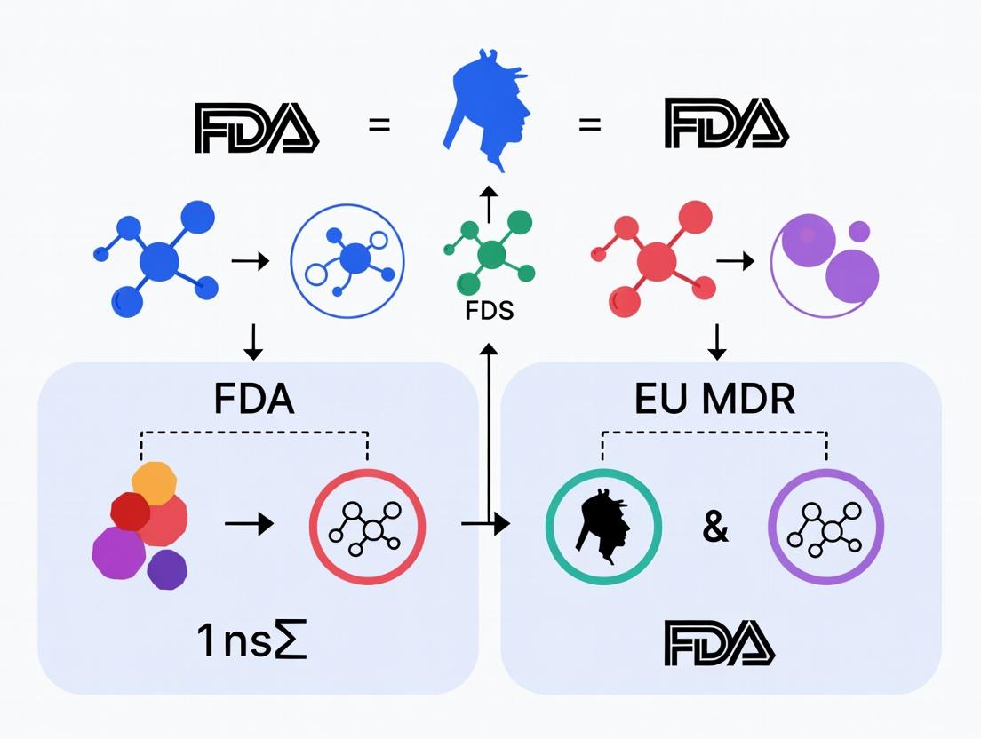
Biocompatibility Standards Decoded: Navigating the Critical Differences Between FDA ISO 10993 and EU MDR Requirements for Medical Implants
This article provides a detailed comparative analysis of biocompatibility requirements for medical implants under the US FDA (guided by ISO 10993-1:2018) and the European Union's Medical Device Regulation (MDR 2017/745).
Biocompatibility Standards Decoded: Navigating the Critical Differences Between FDA ISO 10993 and EU MDR Requirements for Medical Implants
Abstract
This article provides a detailed comparative analysis of biocompatibility requirements for medical implants under the US FDA (guided by ISO 10993-1:2018) and the European Union's Medical Device Regulation (MDR 2017/745). Tailored for researchers, scientists, and drug development professionals, it explores foundational regulatory philosophies, practical testing methodologies, common compliance challenges, and strategic validation approaches. The analysis clarifies key distinctions in risk classification, data requirements, and the evolving emphasis on chemical characterization and biological evaluation plans, offering a roadmap for global market entry and regulatory success.
Regulatory Philosophies Unveiled: Core Principles of FDA and EU MDR Biocompatibility Frameworks
The evaluation of long-term implant biocompatibility sits at the intersection of material science, biology, and regulatory strategy. A critical component of regulatory submissions—both to the US FDA under ISO 10993-1 and to the EU under the more proactive, life-cycle focused Medical Device Regulation (MDR)—is the comparative performance data of the novel material against established alternatives. This guide compares key testing paradigms through experimental data.
Comparison Guide:In VitroCytotoxicity Assays
While both FDA and EU MDR require biocompatibility testing, the EU MDR emphasizes a more rigorous scientific justification and continuous post-market surveillance. The following in vitro assays form the initial screening bedrock.
Table 1: Comparative Performance of Common Cytotoxicity Assays for Polymer Implants
| Assay Type | Test Material (Sample) | Reference Control (Negative) | Positive Control | Key Metric & Result | Advantage for Regulatory Submission |
|---|---|---|---|---|---|
| Elution (Extract) Test | Ultra-High Molecular Weight Polyethylene (UHMWPE) extract | Polyethylene (HDPE) extract | Latex extract | Cell Viability (MTT assay): 98.2% ± 3.1% vs. Control (100%) | Excellent for screening leachables; aligns with FDA's use of extracts. |
| Direct Contact Test | Polydimethylsiloxane (PDMS) disc | Medical-grade silicone disc | Copper disc | Zone of Inhibition: 0 mm (no cytotoxicity) | EU MDR values direct physiological simulation. Demonstrates device-form effect. |
| Indirect Contact (Agar Diffusion) | Polyetheretherketone (PEEK) particle layer | Polypropylene layer | Zinc diethyldithiocarbamate layer | Reactivity Grade: 0 (None) per ISO 10993-5 | Historically accepted; useful for dense, non-porous materials. |
Experimental Protocol: MTT Elution Assay for ISO 10993-5 Compliance
Methodology:
- Extract Preparation: The test material (e.g., UHMWPE) is sterilized and extracted in cell culture medium (e.g., RPMI 1640 with serum) at a surface area-to-volume ratio of 3 cm²/mL for 24±2 hours at 37°C.
- Cell Culture: L929 mouse fibroblast cells are seeded in a 96-well plate at a density of 1 x 10⁴ cells/well and incubated for 24 hours to form a near-confluent monolayer.
- Exposure: The culture medium is replaced with 100 µL of the material extract. Negative (HDPE extract) and positive (latex or phenol solution) controls are run in parallel.
- Incubation: Cells are incubated with the extract for 48 hours at 37°C in a 5% CO₂ atmosphere.
- Viability Measurement: 10 µL of MTT (3-(4,5-dimethylthiazol-2-yl)-2,5-diphenyltetrazolium bromide) solution (5 mg/mL) is added to each well. After 4 hours, the medium is removed, and 100 µL of dimethyl sulfoxide (DMSO) is added to solubilize the formazan crystals.
- Data Analysis: The optical density (OD) is measured at 570 nm using a plate reader. Cell viability is calculated as: (ODTest / ODNegative Control) x 100%. A viability > 70% is typically considered non-cytotoxic.
Comparison Guide:In VivoSensitization & Irritation
Moving beyond in vitro screens, in vivo tests provide critical systemic interaction data, increasingly scrutinized under EU MDR's emphasis on animal welfare (3Rs principle).
Table 2: Comparison of Sensitization Assay Performance
| Assay Name | Test Material | Adjuvant/Procedure | Key Endpoint & Result (vs. Control) | Regulatory Preference & Rationale |
|---|---|---|---|---|
| Guinea Pig Maximization Test (GPMT) | Polyurethane film extract (in saline & paraffin oil) | Freund's Complete Adjuvant | Mean Challenge Score: 0.4 (Grade: Weak) | Traditional FDA benchmark. Potent, but less favored by EU MDR due to animal welfare. |
| Local Lymph Node Assay (LLNA) | Methacrylate monomers from bone cement | No adjuvant required | Stimulation Index (SI): 2.1 (EC₃ = 12% vol/vol) | Favored by both FDA (alternative) and EU MDR. Quantitative, reduces animal suffering (3Rs). |
| Murine Sensitization Test (MST) | Nickel ions (as positive control benchmark) | -- | SI Threshold: ≥ 2.7 for positive classification | Emerging in vitro alternative. Gaining traction for EU MDR submissions seeking state-of-the-art methods. |
Experimental Protocol: Local Lymph Node Assay (LLNA)
Methodology:
- Dosing: Female CBA/J mice (n=4/group) receive 25 µL of the test material extract (at three concentrations) or controls (vehicle, positive sensitizer) applied to the dorsum of both ears daily for three consecutive days.
- Pulsing: On day 6, all mice receive an intravenous injection of ³H-thymidine or bromodeoxyuridine (BrdU).
- Sacrifice & Analysis: Five hours post-injection, the draining auricular lymph nodes are excised and pooled for each mouse.
- Measurement (³H-thymidine): A single-cell suspension is prepared. DNA incorporation of ³H-thymidine is measured by beta-scintillation counting, expressed as disintegrations per minute (DPM).
- Data Calculation: The Stimulation Index (SI) is calculated for each dose group as: Mean DPMTest / Mean DPMVehicle Control. An SI ≥ 3 is historically considered a positive sensitization response, though EC₃ (concentration needed to elicit SI=3) is a more precise metric.
Visualization: Biocompatibility Assessment Workflow
Title: Biocompatibility Testing Workflow for FDA & EU MDR
The Scientist's Toolkit: Key Research Reagent Solutions
Table 3: Essential Materials for Implant Biocompatibility Testing
| Item | Function & Application in Biocompatibility Research |
|---|---|
| L929 Mouse Fibroblast Cell Line | Standardized cell model for cytotoxicity assays (ISO 10993-5). Provides reproducible baseline reactivity data. |
| MTT/XTT Cell Viability Assay Kits | Colorimetric assays to quantify mitochondrial activity and cell health after exposure to material extracts. |
| Freund's Complete Adjuvant (FCA) | Immunopotentiator used in the classical Guinea Pig Maximization Test to enhance sensitization response. |
| BrdU (Bromodeoxyuridine) ELISA Kit | Alternative to radioactive ³H-thymidine for measuring cell proliferation in the LLNA, aligning with 3Rs. |
| Medical-Grade Silicone (e.g., PDMS) | Common negative control material for irritation and implantation studies due to its well-established safety profile. |
| Dimethyl Sulfoxide (DMSO) | Solvent for preparing extracts of materials with low polarity and for solubilizing formazan crystals in MTT assay. |
| ISO 10993-12 Standardized Solvents | Saline, vegetable oil, and other vehicles specified for creating biologically relevant material extracts. |
| Polyethylene (HDPE) Particles | Standard reference and control material for particle-induced inflammation studies in long-term implantation models. |
Within the broader thesis on FDA versus EU MDR requirements for implant biocompatibility, the FDA's 2020 guidance, "Use of International Standard ISO 10993-1, 'Biological evaluation of medical devices Part 1: Evaluation and testing within a risk management process'," represents a pivotal framework. This guide compares the testing strategies and outcomes driven by this risk-based approach against traditional, prescriptive testing paradigms, providing experimental data to illustrate the shift.
Comparison of Testing Strategies and Outcomes
The FDA's risk-based approach, aligning with ISO 10993-1:2018, emphasizes a chemically and biologically informed assessment over a standard checklist. The table below compares key aspects of this approach against a traditional, prescriptive testing model.
Table 1: Comparison of Prescriptive vs. Risk-Based Biocompatibility Strategies
| Aspect | Traditional Prescriptive Approach | FDA/ISO 10993-1 Risk-Based Approach |
|---|---|---|
| Philosophy | Checklist-based; apply standard test battery. | Risk management-driven; testing justifies safety. |
| Initial Step | Immediate in vivo testing. | Thorough chemical characterization (ISO 10993-18). |
| Test Selection | Fixed based on contact duration and type. | Justified by material chemistry, medical device nature, and biological risks. |
| In Vivo Reliance | High; default for endpoints like irritation. | Reduced; in vitro and chemical data replace animal use where possible. |
| Key Guidance | FDA Blue Book Memo G95-1 (superseded). | FDA Guidance (2020) & ISO 10993-1:2018. |
| EU MDR Alignment | Lower; conflicts with Annex I GSPRs requiring risk reduction. | High; aligns with MDR's risk management requirements (Annex I). |
Table 2: Experimental Data: In Vitro vs. In Vivo Irritation Testing for a Polymer Implant
| Test Method | Protocol Summary | Key Endpoint | Result for Example Material | Time to Result | Regulatory Acceptance |
|---|---|---|---|---|---|
| In Vivo (Draize) | Intracutaneous injection of extracts in rabbits. | Mean scores for erythema/eschar & edema at 24, 48, 72h. | Mean Score: 0.4 (Non-irritant) | 3 days + animal acclimation | Fully accepted under FDA & MDR with justification. |
| In Vitro (Reconstructed Human Epidermis - RHE) | Apply extract to 3D epidermis model (EpDerm). | Cell viability via MTT reduction. | % Viability: 98% (Non-irritant) | 1-3 days | Accepted per FDA guidance with proper validation; aligns with MDR's desire for alternatives. |
Detailed Experimental Protocols
Protocol 1: Chemical Characterization per ISO 10993-18 for Risk Assessment
- Objective: Identify and quantify extractable/leachable chemicals from device materials.
- Materials: Finished device, appropriate extraction solvents (e.g., saline, ethanol/water), analytical equipment (GC-MS, LC-MS, ICP-MS).
- Procedure:
- Extraction: Use exhaustive or simulated-use extraction. Mill or cut device to increase surface area. Extract at defined temperature and duration (e.g., 70°C for 24h).
- Analysis: Analyze extracts via:
- GC-MS: For volatile and semi-volatile organics.
- LC-MS: For non-volatile organics (e.g., additives, degradants).
- ICP-MS: For elemental impurities.
- Risk Assessment: Quantify all identified substances. Compare to established safety thresholds (e.g., Analytical Evaluation Threshold (AET), Threshold of Toxicological Concern (TTC), ICH Q3D elements). Justify any required biological testing based on gaps.
Protocol 2: In Vitro Cytotoxicity (ISO 10993-5) – Elution Method
- Objective: Assess the cytotoxic potential of device extracts.
- Materials: L-929 or BALB/3T3 fibroblast cells, complete growth medium, device extracts, multi-well plates, MTT reagent, spectrophotometer.
- Procedure:
- Prepare extracts per ISO 10993-12.
- Seed cells in a 96-well plate and incubate for 24h to form a near-confluent monolayer.
- Replace culture medium with device extracts (100 µL/well). Include negative (HDPE) and positive (latex) controls.
- Incubate for 24-48 hours at 37°C, 5% CO₂.
- Add MTT reagent and incubate to allow viable cells to form formazan crystals.
- Solubilize crystals and measure absorbance at 570 nm.
- Calculate cell viability relative to the negative control. A reduction of >30% is considered a positive cytotoxic result.
Visualizing the Risk-Based Assessment Workflow
Title: FDA/ISO 10993 Risk-Based Biological Evaluation Workflow
The Scientist's Toolkit: Key Research Reagent Solutions
Table 3: Essential Materials for Chemical & Biological Evaluation
| Item | Function in Biocompatibility Assessment |
|---|---|
| Certified Reference Standards | For accurate quantification of extractables (e.g., BPA, DEHP, antioxidants) via GC/LC-MS. Critical for toxicological risk assessment. |
| Reconstructed Human Epidermis (RHE) Models | In vitro 3D tissue models (e.g., EpDerm, EpiDerm) for assessing skin irritation/corrosion, replacing in vivo rabbit tests. |
| Validated Cell Lines (L-929, BALB/3T3) | Standardized mammalian fibroblasts for cytotoxicity testing (ISO 10993-5). |
| Pyrogen Testing Reagents | Limulus Amebocyte Lysate (LAL) for bacterial endotoxin testing, replacing rabbit pyrogen test. |
| Simulated Body Fluids | Extraction media (e.g., saline, with/without ethanol) that mimic physiological conditions for leachable studies. |
| Positive Control Materials | Standardized materials (e.g., latex, zinc diethyldithiocarbamate) to validate the responsiveness of biological test systems. |
The global regulatory landscape for medical devices, particularly implants, is bifurcating. While the U.S. FDA primarily relies on a risk-based biocompatibility framework guided by ISO 10993, the EU MDR mandates a more comprehensive, systematic, and lifecycle-oriented approach through its Annex I General Safety and Performance Requirements (GSPRs). This comparison guide analyzes the paradigm shift from the Medical Devices Directive (MDD) to the Medical Device Regulation (MDR), focusing on implications for implant biocompatibility research.
Core Comparison: MDD Essential Requirements vs. MDR GSPRs for Biocompatibility
The transition represents a shift from general principles to explicit, detailed, and verified requirements.
Table 1: Key Changes Impacting Biocompatibility Research
| Aspect | MDD (Directive 93/42/EEC) | EU MDR (2017/745) Annex I GSPRs | Impact on Research & Testing |
|---|---|---|---|
| Legal Form | Directive (transposed into national law) | Regulation (directly applicable) | Harmonized, non-negotiable requirements across EU. |
| Structure | 13 Essential Requirements (ERs) | 23 Chapters with ~100 detailed GSPRs | More granular, specific demands for proof of safety. |
| Biocompatibility Focus | Implicit in ERs 1, 2, 3, 5, 7. Relied heavily on harmonized standards (e.g., ISO 10993). | Explicit in GSPR 10.2, 10.4, 10.5, 17, 18. Requires a defined biological evaluation plan per ISO 10993-1. | Plan must be established a priori. Evaluation is continuous across the lifecycle. |
| Proof Requirement | Presumption of conformity via standards. | Heightened Scrutiny: Requires "sufficient clinical evidence" and justification for all material choices. Reliance on standards alone is insufficient. | Increased need for chemical characterization (ISO 10993-18), toxicological risk assessment (ISO 10993-17), and often clinical data. |
| Risk Management Link | Loosely connected (EN ISO 14971). | Fully integrated (GSPR 3, 8). Biological evaluation must be an integral part of the risk management process. | Biocompatibility is not a checklist but a risk-based, iterative process documented in the Risk Management File. |
| Material Documentation | General requirements for material safety. | Specific requirements (GSPR 10.4, 18.4) for material and substance identification, including CMR/Endocrine disruptors >0.1% w/w. | Mandatory supply chain disclosure. Requires analytical chemistry (e.g., GC-MS, ICP-MS) to identify leachables. |
Experimental Data Comparison: The Case of Orthopedic Implant Biocompatibility
A comparative study simulating the evidence generation for a titanium alloy spinal implant under MDD vs. MDR frameworks illustrates the heightened data requirements.
Table 2: Simulated Testing Scope & Data Requirements Comparison
| Test Area (ISO 10993 series) | Typical MDD-Compliant Submission (Presumption of Conformity) | MDR-Compliant Submission (GSPR 10.2 & 10.4) | Supporting Experimental Data/Justification |
|---|---|---|---|
| Chemical Characterization | Limited extractables study using 1-2 solvents. | Full material composition & exhaustive extractables/leachables study. | Data: ICP-MS identified [Ni] = 0.08% w/w (<0.1% threshold). LC-QTOF-MS detected 2 novel leachable processing aids (≤ 5 ppm). Justification: Toxicological risk assessment required for novel leachables. |
| Cytotoxicity | 72-hour elution test with mouse fibroblasts (L929). | Same base test, plus direct contact test with human osteoblast cells for relevance. | Data: MDD Test: >90% viability (pass). MDR Addendum: Osteoblast metabolic activity showed 15% decrease at 24h, normalizing by 72h. Justified as non-adverse transient effect. |
| Sensitization | ISO 10993-10 Guinea Pig Maximization Test. | Consider additive in-vitro assay (e.g., h-CLAT) per GSPR 10.5 (reduce animal use). | Data: GPMT: Negative. h-CLAT (in-vitro): Positive for one leachable. Conclusion: Conflicting data triggered expanded chemical analysis and a justification based on exposure dose being below threshold. |
| Implantation | 4-week rabbit muscle implantation study. | 12-week osseointegration study in relevant bone model (sheep), plus histomorphometry. | Data: New bone-to-implant contact (BIC) at 12 weeks: 45% ± 8%. Required to verify GSPR 17.1 (intended performance) and long-term biological safety. |
| Clinical Evidence | Possibly literature-based equivalence. | Required as part of "sufficient clinical evidence" under Heightened Scrutiny. | Data: Prospective clinical follow-up (24 months) showing 96% implant survivorship. Paired with explant analysis (SEM/EDX) confirming no abnormal corrosion. |
Detailed Experimental Protocols
Protocol 1: Exhaustive Extraction for Chemical Characterization (Per ISO 10993-18)
- Objective: Identify and quantify all leachable substances.
- Materials: Implant material (powdered), Mili-Q water, 50% ethanol/water, hexane, Soxhlet extractor, GC-MS, LC-HRMS, ICP-MS.
- Method:
- Sample Prep: Powder material to increase surface area. Accurately weigh triplicate samples.
- Exhaustive Extraction: Use Soxhlet extraction with polar (50% ethanol) and non-polar (hexane) solvents for 72 hours each.
- Analysis:
- Volatiles: Headspace GC-MS.
- Semi-Volatiles: GC-MS of extract concentrates.
- Non-Volatiles: Liquid Chromatography-High Resolution Mass Spectrometry (LC-HRMS).
- Elements: Inductively Coupled Plasma Mass Spectrometry (ICP-MS).
- Data Processing: Identify unknowns via spectral libraries. Quantify against calibrated standards.
Protocol 2: Enhanced Cytotoxicity & Cell Function Assay (Per ISO 10993-5 & -12)
- Objective: Assess cytotoxicity and specific cell-type response.
- Materials: L929 mouse fibroblast cell line, primary human osteoblasts (hOB), extraction media (as above), cell culture reagents, MTT/XTT assay kit, fluorescent live/dead stain.
- Method:
- Eluate Preparation: Extract device per ISO 10993-12 at 37°C for 72h at 3 cm²/mL surface area-to-volume ratio.
- Co-culture: Plate L929 and hOB cells in separate 96-well plates. Expose to 100%, 50%, 25% eluate concentrations (n=6).
- Viability Assessment:
- At 24h and 72h, perform MTT assay (absorbance at 570nm) for metabolic activity.
- At 72h, perform live/dead staining (Calcein-AM/EthD-1) for membrane integrity.
- Analysis: Calculate % viability relative to negative control. Statistically compare groups (ANOVA).
Protocol 3: Histomorphometric Analysis of Osseointegration (Per ISO 10993-6)
- Objective: Quantify bone ingrowth and interface response.
- Materials: Sheep tibia model with implanted device, undecalcified histology setup, hard tissue microtome, toluidine blue stain, light microscope with morphometry software.
- Method:
- Explant & Processing: Retrieve implant-bone block at study endpoint. Fix in formalin, dehydrate in ethanol, embed in PMMA resin.
- Sectioning: Cut ~50 μm longitudinal sections across the implant using a diamond saw. Polish and stain with toluidine blue.
- Morphometry: Under light microscope, capture images of bone-implant interface. Using software, measure:
- Bone-to-Implant Contact (%): (Length of bone in direct contact with implant / Total implant perimeter) x 100.
- Bone Area Fraction (%): Area of bone within first 500 μm from implant interface / Total area of region of interest.
Diagrams
Title: MDD vs MDR Biocompatibility Evaluation Workflow
Title: MDR Chemical Characterization & Risk Assessment Pathway
The Scientist's Toolkit: Key Research Reagent Solutions
Table 3: Essential Materials for MDR-Compliant Biocompatibility Research
| Item / Reagent | Function in MDR Context | Example Product/Catalog |
|---|---|---|
| Reference Materials | Critical for quantifying leachables in chemical characterization (ISO 10993-18). | USP <661> plastic additives standards; NIST traceable elemental standards. |
| In-Vitro Sensitization Assay Kits | To fulfill GSPR 10.5 (reduce animal testing) for sensitization assessment. | h-CLAT assay kit (e.g., MatTek Epiderm SIT). |
| Primary Human Cell Lines | Provide biologically relevant data beyond standard fibroblast lines, strengthening evidence. | Human osteoblasts (hOB), mesenchymal stem cells (hMSC) from reputable banks (e.g., Lonza, ATCC). |
| Histology Embedding Resin (e.g., PMMA) | For undecalcified sectioning of bone-implant interfaces for histomorphometry per ISO 10993-6. | Technovit 7200 VLC resin (Kulzer). |
| Automated Morphometry Software | To generate quantitative, reproducible data on bone ingrowth (BIC, BA%) for performance claims (GSPR 17.1). | BioQuant Osteo, Olympus cellSens. |
| LC-HRMS & ICP-MS Systems | Essential analytical platforms for exhaustive chemical characterization as required by GSPR 10.4 and 18.4. | Thermo Fisher Orbitrap LC-MS; Agilent 7900 ICP-MS. |
The global regulatory landscape for implantable medical devices dictates stringent biocompatibility evaluations. The triggers for a full assessment, however, differ significantly between the U.S. Food and Drug Administration (FDA) and the European Union Medical Device Regulation (EU MDR). This guide compares these regulatory triggers and the associated experimental expectations.
Regulatory Trigger Framework: FDA vs. EU MDR
The core divergence lies in the foundational approach: FDA reliance on a risk-based, matrix-driven standard (ISO 10993-1) versus the EU MDR’s integration of biocompatibility within a broader safety and risk management process.
Table 1: Key Regulatory Trigger Comparison
| Trigger Factor | FDA Approach (via ISO 10993-1) | EU MDR Approach |
|---|---|---|
| Primary Driver | Material classification & bodily contact (nature, duration, frequency). | Integration into General Safety & Performance Requirements (Annex I), requiring a risk management process per ISO 14971. |
| Assessment Start Point | Largely prescriptive based on contact matrix. A new material or change in contact duration triggers re-evaluation. | Justification required for any material of human or animal origin, or that is intentionally resorbable. Implicitly required for all patient-contact components. |
| "Full Assessment" Threshold | Required for: Permanent implants (>30 days), blood contact devices, and novel materials without established safety profiles. | Required when risks from chemical constituents cannot be adequately controlled by design or manufacturing, and thus require characterization and biological evaluation. |
| Acceptance of Existing Data | Possible via a "master file" or literature for well-established materials (e.g., USP Class VI polymers). Stricter for novel leachables. | Requires demonstration of "sufficiently low" risk. Historical data alone is often insufficient; new testing per state-of-the-art is frequently mandated. |
| Toxicological Risk Assessment | Follows ISO 10993-17; required to set allowable limits for leachables. | Mandated per ISO 10993-17 and integrated into the overall risk management file. More explicit requirement for cumulative exposure assessment from multiple material sources. |
Experimental Data & Protocol Comparison
Both frameworks ultimately require similar experimental endpoints (cytotoxicity, sensitization, irritation, systemic toxicity, genotoxicity, implantation) but the trigger and justification for testing differs.
Table 2: Representative Experimental Data Requirements for a Permanent Polymer Implant
| Test (ISO 10993 series) | Typical FDA-Triggered Data Package | Typical EU MDR-Triggered Data Package | Supporting Experimental Protocol Summary |
|---|---|---|---|
| Cytotoxicity (ISO 10993-5) | Required. Direct contact or extract elution assay on mammalian fibroblast cells (e.g., L929). | Required. Same base test, but may require testing on multiple extractants (polar, non-polar). | Protocol: Per ISO 10993-5. Prepare device extract in serum-supplemented media (37°C, 24h). Apply to L929 monolayer. Assess cell viability after 24-48h via MTT assay or microscopic evaluation of morphological changes. >70% viability is typically acceptable. |
| Sensitization (ISO 10993-10) | Required. Maximization Test (GPMT) or Local Lymph Node Assay (LLNA). | Required. Prefers LLNA or equivalent in vitro methods (e.g., h-CLAT) aligned with 3Rs principle. | Protocol (LLNA): Mice (CBA/J strain) receive topical application of device extract or controls on ears for three consecutive days. Proliferation is measured via radioactive thymidine incorporation in auricular lymph nodes. A Stimulation Index ≥3 indicates potential sensitization. |
| Genotoxicity (ISO 10993-3) | Required. Battery of in vitro tests: Ames test + Mouse Lymphoma or Chromosomal Aberration assay. | Required. Identical base battery. May require additional in vivo follow-up if in vitro results are positive or for materials with known mutagenic precursors. | Protocol (Ames Test): Per OECD 471. Device extracts are incubated with Salmonella typhimurium strains (TA98, TA100, etc.) with/without metabolic activation (S9 mix). Revertant colony count is compared to control. A dose-responsive increase indicates mutagenicity. |
| Implantation (ISO 10993-6) | Required for permanent implants. 12-26 week study in rodents or rabbits. | Required. Similar duration. Greater emphasis on correlating findings with chemical characterization (ISO 10993-18) – linking leachables to biological response. | Protocol: Per ISO 10993-6. Implant material or miniature device is surgically placed in subcutaneous or muscle tissue of rats. Explant at endpoint (e.g., 26 weeks) for histopathology. Tissue response is scored for inflammation, fibrosis, necrosis, and capsule thickness. |
Logical Workflow for Biocompatibility Assessment Trigger
The following diagram illustrates the divergent decision pathways under FDA and EU MDR frameworks.
Diagram 1: Regulatory decision pathways for biocompatibility assessment.
The Scientist's Toolkit: Key Research Reagent Solutions
Table 3: Essential Materials for Core Biocompatibility Experiments
| Item | Function in Experimental Protocol |
|---|---|
| L929 Mouse Fibroblast Cell Line | Standardized cell model for in vitro cytotoxicity testing (ISO 10993-5). |
| MTT Reagent (3-(4,5-Dimethylthiazol-2-yl)-2,5-diphenyltetrazolium bromide) | Yellow tetrazole reduced to purple formazan by living cell mitochondria; used to quantify cytotoxicity. |
| Salmonella typhimurium TA98 & TA100 Strains | Genetically modified bacterial strains used in the Ames test to detect frameshift and base-pair mutagens. |
| Rat Serum (with S9 Metabolic Activation Fraction) | Provides mammalian liver enzymes for metabolic activation of pro-mutagens in genotoxicity assays. |
| Polyurethane Film, USP Class VI Verified | Common negative control material for cytotoxicity and irritation tests, providing a benchmark. |
| Zinc Diethyldithiocarbamate (ZDEC) | Standard positive control material for sensitization testing (e.g., in LLNA). |
| High-Density Polyethylene (HDPE) with BHT | Standard positive control material for in vitro cytotoxicity testing. |
| PBS & MEM Elution Media | Polar and non-polar extraction vehicles for preparing device extracts to simulate clinical exposure. |
This guide compares product performance testing within the regulatory frameworks of the U.S. Food and Drug Administration (FDA) and the European Union Medical Device Regulation (EU MDR 2017/745). Both require biocompatibility assessment based on ISO 10993-1, "Biological evaluation of medical devices," but differ in interpretation, categorization, and specific endpoints. This article objectively compares experimental data and methodologies relevant to these requirements.
Regulatory Comparison: FDA vs. EU MDR Device Categorization and Contact Durations
While both systems utilize the matrix from ISO 10993-1, differences emerge in application and risk classification.
Table 1: Comparison of Key Categorization and Endpoint Requirements
| Aspect | FDA (Using ISO 10993-1 & Guidance) | EU MDR (Using ISO 10993-1 & Annex I GSPRs) |
|---|---|---|
| Governing Principle | Safety-based; "least burdensome approach" | Risk-based; fulfillment of General Safety and Performance Requirements (GSPRs) |
| Device Categorization | Based on nature of body contact (surface, external communicating, implant) and contact duration. | Similar categorization, but intrinsically linked to device class (I, IIa, IIb, III) under MDR Article 51. |
| Contact Duration Definitions | Transient: ≤24 h; Short-term: 24 h to 30 d; Long-term: >30 d. | Limited: ≤24 h (Transient), >24 h to ≤30 d (Short-term); Prolonged: >30 d to ≤10 y; Permanent: >10 y. |
| Critical Differentiation | Primarily focuses on the three duration categories from ISO 10993-1. | Explicitly considers "Permanent" (>10 years) as a distinct category, emphasizing long-term risk management. |
| Endpoint Selection Driver | Contact category and duration, material review, and clinical use. | Device class, contact/duration, and the need to prove compliance with all relevant GSPRs (e.g., 10.4.1 on leakage, 10.4.2 on degradation). |
Biological Endpoints and Performance Comparison
The following experimental data is illustrative for a long-term (>30 days) implantable cardiovascular device (e.g., a stent or pacemaker component).
Table 2: Comparison of Key Biological Endpoint Test Results for a Novel Polymer vs. a Marketed Control
| Endpoint System (ISO 10993-20) | Test Method (ISO Standard) | Marketed Control (Mean Result) | Novel Test Polymer (Mean Result) | Key Performance Insight |
|---|---|---|---|---|
| Cytotoxicity | ISO 10993-5 (Extract) | Grade 1 (Non-cytotoxic) | Grade 0 (Non-cytotoxic) | Both materials meet the non-cytotoxic requirement. Novel polymer shows marginally better cell viability. |
| Sensitization | ISO 10993-10 (GPMT) | 0% Sensitization Rate | 0% Sensitization Rate | Equivalent performance; no potential for skin sensitization. |
| Irritation/Intracutaneous Reactivity | ISO 10993-10 (Extract) | Irritation Index: 0.2 | Irritation Index: 0.1 | Both well below threshold (≤1.0). Novel polymer shows minimal reactivity. |
| Systemic Toxicity | ISO 10993-11 (Acute, Extract) | No adverse systemic effects | No adverse systemic effects | Equivalent performance in acute systemic toxicity. |
| Subchronic Toxicity | ISO 10993-11 (90-Day Implant, Rodent) | No test article-related mortality. Mild local inflammation at 30d, resolving by 90d. | No test article-related mortality. Minimal inflammation at all timepoints. | Novel polymer demonstrates improved local tissue compatibility over a 90-day period. |
| Genotoxicity In vitro | ISO 10993-3 (Ames, MLA) | Negative in all assays | Negative in all assays | Equivalent performance; no mutagenic potential detected. |
| Implantation | ISO 10993-6 (Muscle/Bone, 12w) | Mean Histopath Score: 3.2 (Moderate reaction) | Mean Histopath Score: 2.1 (Mild reaction) | Novel polymer elicits a significantly milder chronic inflammatory response. |
Detailed Experimental Protocols
Protocol 1: Subchronic Toxicity (90-Day Implant Study)
Objective: Evaluate local and systemic effects following prolonged implantation. Methodology (ISO 10993-11):
- Test Article Preparation: Sterilize implant samples (e.g., 2mm x 5mm rods) of both novel and control polymers.
- Animal Model: Use rodents (e.g., rats, n=10/group). Anesthetize animals.
- Implantation: Surgically implant one test or control article into a paravertebral muscle pouch per animal. Include a sham surgery control group.
- Clinical Observations: Monitor daily for signs of systemic toxicity (weight, behavior, food consumption).
- Termination: Euthanize animals at 90 days. Perform gross necropsy.
- Tissue Analysis: Excise implant site with surrounding tissue. Process for histopathology (H&E staining).
- Scoring: Use a standardized semi-quantitative scoring system (e.g., ISO 10993-6: 0-4 for inflammation, fibrosis, necrosis).
- Statistical Analysis: Compare group means using appropriate tests (e.g., ANOVA).
Protocol 2: In Vivo Implantation Test for Local Effects
Objective: Assess the local tissue response after implantation. Methodology (ISO 10993-6):
- Study Design: Utilize a rodent or rabbit model. Implant materials in muscle or subcutaneous tissue.
- Time Points: Include multiple explant intervals (e.g., 1, 4, 12 weeks) to assess reaction progression.
- Control Articles: Implant negative control (e.g., high-density polyethylene) and positive control (e.g., organotinstabilized PVC) materials in separate sites/animals.
- Histopathological Preparation: Fix explanted tissue, section, and stain with H&E and special stains for connective tissue (e.g., Masson's Trichrome).
- Evaluation: A blinded pathologist evaluates slides for inflammation (polymorphonuclear cells, lymphocytes, plasma cells, macrophages, giant cells), fibrosis, necrosis, and fatty infiltration.
- Response Classification: The overall biological response is classified as non-irritant, slight, moderate, or severe irritant based on comparative scores against controls.
Visualization of Key Concepts
Title: FDA vs EU MDR Biocompatibility Assessment Pathways
Title: ISO 10993-1: From Device Contact to Endpoint Selection
The Scientist's Toolkit: Research Reagent Solutions for Biocompatibility Testing
Table 3: Essential Materials and Reagents for Featured Experiments
| Item | Function/Brief Explanation |
|---|---|
| L929 Mouse Fibroblast Cell Line | Standardized cell model per ISO 10993-5 for evaluating cytotoxicity via extract or direct contact tests. |
| Minimum Essential Medium (MEM) Eluent | Serum-free medium used for preparing device extracts for cytotoxicity and other in vitro tests to avoid interference. |
| Guinea Pigs (Dunkin-Hartley strain) | Preferred in vivo model for Magnusson-Kligman Guinea Pig Maximization Test (GPMT) for sensitization potential. |
| High-Density Polyethylene (HDPE) Rods | Standardized negative control material for implantation studies (ISO 10993-6). |
| Organotin-Stabilized Polyvinyl Chloride (PVC) | Standardized positive control material for implantation studies to provoke a recognizable tissue response. |
| Histopathology Stains (H&E, Masson's Trichrome) | Hematoxylin and Eosin for general morphology; Trichrome for collagen/fibrosis assessment in implantation sites. |
| Saline and Cottonseed Oil | Standard polar and non-polar vehicles for preparing device extracts for systemic toxicity tests. |
| S9 Metabolic Activation Mix | Liver homogenate fraction used in in vitro genotoxicity assays (Ames, MLA) to simulate mammalian metabolic processes. |
| Standard Reference Materials (e.g., USP PE) | Certified materials used for system suitability and calibration of test models. |
From Theory to Testing: Implementing Biological Evaluation Plans for FDA and EU MDR Compliance
Within the thesis that the FDA's biocompatibility framework (guided by ISO 10993 and specific guidance documents) and the EU's Medical Device Regulation (MDR 2017/745) necessitate distinct strategic emphases in implant research, this guide compares methodologies for key biological endpoint evaluations. A compliant BEP must satisfy both, often through a single, robust testing program designed to meet the more stringent of the two requirements.
Comparison of Cytotoxicity Assessment: Elution vs. Direct Contact Methods
Cytotoxicity testing, a mandatory first step, is approached similarly, but the EU MDR often demands more comprehensive justification for test selection.
Table 1: Cytotoxicity Test Method Comparison
| Parameter | FDA / ISO 10993-5 Preferable Method | Alternative / Supplementary Method (Common in EU MDR Dossiers) | Key Experimental Data Insight |
|---|---|---|---|
| Primary Test | Elution (Extract) Test using L929 mouse fibroblast cells. | Direct Contact Test using NHDF-Neo human dermal fibroblasts. | Elution test LC50 > 80% viability; Direct contact may show <1 mm zone of inhibition. |
| Exposure Time | 24-hour extract exposure to cells for 24-48 hrs. | Device component placed directly on cell monolayer for 24 hrs. | Direct contact provides more severe mechanical & chemical stress. |
| Endpoint Readout | Quantitative (MTT/XTT assay): Cell viability %. | Qualitative & Quantitative: Microscopy for lysis & MTT assay. | Data must show > 70% viability (ISO) vs. ≥ 2 out of 3 cultures unaffected (USP). |
| MDR-Specific Nuance | Accepted but may require rationale for extractant choice. | Often favored for solid implants; better simulates clinical use. | Supports "state-of-the-art" requirement under MDR Annex I. |
Experimental Protocol for Enhanced Direct Contact Test (Per MDR Expectations):
- Cell Culture: Seed Normal Human Dermal Fibroblasts (NHDF-Neo) in a 12-well plate at 2.5 x 10^4 cells/cm². Cultivate in DMEM+10% FBS until 80% confluent.
- Sample Preparation: Sterilize three representative implant samples (e.g., 5mm x 5mm) per ISO 10993-12. Use one negative control (HDPE) and one positive control (Latex).
- Direct Contact: Carefully place one test sample directly onto the cell monolayer in the center of each well. Add minimal medium to prevent drying.
- Incubation: Incubate plates at 37°C, 5% CO₂ for 24 ± 2 hours.
- Viability Assessment: Remove samples. Perform MTT assay: Add 0.5 mg/mL MTT reagent, incubate 2 hrs, solubilize formazan crystals with DMSO, measure absorbance at 570 nm.
- Microscopic Evaluation: Prior to MTT, observe under phase-contrast microscope. Grade reactivity: 0 (none), 1 (mild), 2 (moderate), 3 (severe), 4 (extreme) per ISO 10993-5.
Comparison of Sensitization Assessment: ISO Guinea Pig vs. In Vitro Alternatives
The shift toward alternative methods is critical. While the FDA's ISO 10993-10 recognizes the GPMT, the EU MDR strongly encourages non-animal methods per Annex I (Requirements 10, 11).
Table 2: Sensitization Test Strategy Comparison
| Parameter | Traditional Animal Method (Accepted by FDA & MDR) | OECD-Validated In Vitro Alternative (Key for MDR Compliance) | Supporting Data Correlation |
|---|---|---|---|
| Standard Test | Guinea Pig Maximization Test (GPMT) per ISO 10993-10. | Direct Peptide Reactivity Assay (DPRA) (OECD 442C). | DPRA predicts GPMT outcome with ~85% accuracy for many chemicals. |
| Key Metric | Incidence of erythema in test vs. control animals. | % Depletion of model peptides (Cysteine, Lysine). | Cysteine depletion > 6.38% often correlates with sensitizer potential. |
| Regulatory Stance | FDA accepts; requires strong justification if not used. | MDR mandates first consideration; DPRA is part of a defined approach (OECD 497). | Data from DPRA + h-CLAT + KeratinoSens can form a WoE assessment. |
| Strategic Use in BEP | May be needed for novel materials or complex leachables. | Essential for demonstrating adherence to "alternative first" MDR principle. | In vitro data required to justify not using alternatives under MDR. |
Experimental Protocol for Direct Peptide Reactivity Assay (DPRA):
- Peptide Preparation: Prepare 0.667 mM solutions of model peptides in phosphate buffer: Cysteine (Cys) and Lysine (Lys).
- Test Article Preparation: Dissolve or suspend test material at 100 mM in solvent (e.g., Acetonitrile:Water 1:1). Prepare serial dilutions.
- Reaction: Mix 25 µL of peptide solution with 25 µL of test article solution (final conc. typically 5 mM). Include vehicle and positive controls (e.g., Hexy Cinnamic Aldehyde).
- Incubation: Incubate plates at 25°C for 24 hours in the dark.
- Analysis: Quantify remaining peptide via High-Performance Liquid Chromatography (HPLC) with UV detection (220 nm for Cys, 210 nm for Lys).
- Calculation: Calculate % peptide depletion:
[(Mean control peak area - Mean test peak area) / Mean control peak area] x 100. Classify per OECD TG 442C prediction model.
Pathway: Integrated Biological Evaluation Plan Workflow
Diagram 1: BEP Development Logic Flow
The Scientist's Toolkit: Key Research Reagent Solutions
| Reagent / Material | Function in Biocompatibility Testing |
|---|---|
| L929 Mouse Fibroblast Cell Line | Standardized model for cytotoxicity elution tests per ISO 10993-5 and USP. |
| Normal Human Dermal Fibroblasts (NHDF-Neo) | More clinically relevant human cell model for direct contact tests, valued in MDR dossiers. |
| MTT (3-(4,5-Dimethylthiazol-2-yl)-2,5-diphenyltetrazolium bromide) | Tetrazolium salt reduced by mitochondrial enzymes to formazan, quantifying viable cells. |
| DPRA Peptides (Cysteine & Lysine) | Synthetic model peptides used to predict protein-binding potential of chemicals (sensitization). |
| Reconstructed Human Epidermis (RhE) Models | 3D tissue models (e.g., EpiDerm) used for in vitro skin irritation/corrosion testing, reducing animal use. |
| h-CLAT (human Cell Line Activation Test) THP-1 Cells | Monocytic cell line used to assess the in vitro potential to induce skin sensitization via CD86/CD54 expression. |
| LLNA Reference Controls | Hexy Cinnamic Aldehyde (strong sensitizer) and Salicylic Acid (non-sensitizer) for assay validation. |
| Simulated Body Fluids (e.g., SBF) | Ionic solution mimicking human blood plasma for in vitro bioactivity or degradation studies of implants. |
This guide compares the performance of analytical techniques and study designs for extractable and leachable (E&L) assessment of implants, framed within the context of diverging FDA and EU MDR requirements for biocompatibility.
Comparison of Analytical Technique Performance for E&L Studies
Table 1: Sensitivity and Applicability of Key Analytical Techniques
| Analytical Technique | Typical Detection Limit (ppb) | Ideal Application for Implants | Key Limitation |
|---|---|---|---|
| Headspace GC-MS | 1 - 50 | Volatile organic compounds (e.g., residual solvents) | Non-volatile compounds not detected |
| Pyrolysis GC-MS | 10 - 100 | Polymer backbone characterization, non-volatile additives | Complex data interpretation, semi-quantitative |
| LC-UV/MS (QTOF) | 0.1 - 10 (MS) / 50 - 100 (UV) | Semi-volatile and non-volatile organics (e.g., antioxidants, plasticizers) | Requires method development; UV lacks specificity |
| ICP-MS/OES | 0.001 - 1 (ICP-MS) / 1 - 100 (OES) | Elemental impurities, catalyst residues | Does not provide speciation information |
| FTIR / ATR-FTIR | ~1000 (1%) | Functional group identification, polymer surface characterization | Low sensitivity, qualitative for unknowns |
Table 2: Comparison of Simulated Extraction Study Designs
| Study Design Parameter | Exhaustive Extraction (ISO 10993-12/18) | Accelerated/Simulated-Use Extraction | Controlled Exhaustive Extraction |
|---|---|---|---|
| Primary Goal | Identify all potential extractables | Mimic clinical leachables profile | Bridge exhaustive data to clinical conditions |
| Solvents | Polar, non-polar, (e.g., water, hexane, ethanol) | Simulating solvents (e.g., PBS, simulated body fluid) | Exhaustive solvents, followed by simulated-use |
| Time/Temperature | Elevated (e.g., 50-70°C for 72h) | Physiological (37°C) for prolonged periods (e.g., 30-90 days) | Exhaustive conditions first, then simulated-use |
| Regulatory Alignment | EU MDR (Emphasis on worst-case) | FDA (Emphasis on clinically relevant) | Hybrid approach for both jurisdictions |
| Data Output | Complete extractables profile; worst-case AET | Predictive leachables profile; risk-based assessment | Correlation between extractables and leachables |
Experimental Protocols for Key E&L Studies
Protocol 1: Exhaustive Extraction for Polymer-Based Implant
- Sample Preparation: Cut implant material into pieces with high surface-area-to-volume ratio (<5mm dimension). Weigh precisely.
- Extraction Solvents: Use water (polar), 2-propanol (polar-protic), and n-hexane (non-polar) at a ratio of 3 cm²/mL or 0.2 g/mL.
- Extraction Conditions: Place in sealed vial, incubate at 70°C ± 2°C for 72 hours. Include blank controls for each solvent.
- Analysis: Analyze cooled extracts by:
- LC-QTOF-MS (ESI+ and ESI-) with a C18 column for semi/non-volatiles.
- Headspace GC-MS (with static or dynamic concentration) for volatiles.
- ICP-MS for elemental analysis.
- Analytical Evaluation Threshold (AET): Calculate based on a permitted exposure limit (e.g., 1.5 µg/day SCT) and sample surface area/weight.
Protocol 2: Simulated-Use Leachable Study for a Cardiovascular Implant
- Simulating Medium: Use phosphate-buffered saline (PBS, pH 7.4) with 4% ethanol to simulate blood solubility.
- Extraction Conditions: Incubate intact, sterilized implant in medium at 37°C for 30 days. Use a volume to simulate clinical dose (e.g., implant surface area to blood volume ratio).
- Time Points: Sample aliquots at 24h, 72h, 7 days, and 30 days.
- Analysis: Analyze time-point samples using highly sensitive LC-MS/MS (MRM mode) and GC-MS for targeted compounds identified in exhaustive studies. Perform non-targeted screening with LC-QTOF.
- Control: Use medium-only controls incubated under identical conditions.
Visualizations
Title: FDA vs EU MDR Regulatory Pathways for Implant E&L Studies
Title: Chemical Characterization Workflow from Extraction to Report
The Scientist's Toolkit: Key Research Reagent Solutions
Table 3: Essential Materials for E&L Studies of Implants
| Item | Function in E&L Studies | Key Consideration |
|---|---|---|
| Simulated Body Fluids (PBS, SBF) | Simulating medium for clinically relevant leachables studies. | Must match pH, ionic strength; may require additives like surfactants or ethanol. |
| High-Purity Extraction Solvents (Water, Hexane, IPA) | Exhaustive extraction to obtain worst-case extractables profile. | Must be MS/ICP-MS grade to avoid interference from solvent impurities. |
| Certified Reference Standards | Identification and quantification of targeted leachables (e.g., DEHP, BHT, Irganox). | Critical for developing sensitive and accurate LC-MS/MS or GC-MS methods. |
| Internal Standards (Deuterated/Surrogates) | Compensates for matrix effects and analytical variability during quantification. | Should be added at the beginning of extraction to track recovery. |
| Soxhlet Extraction Apparatus | Traditional method for performing controlled exhaustive extractions. | Preferred for some regulators for exhaustive extraction of polymers. |
| Inert Sample Vials/Containers (Glass, PTFE-lined caps) | Holds extraction mixtures without introducing interference. | Must be pre-cleaned and screened to avoid contaminating the sample. |
| Proprietary Polymer Databases (for GC-MS, FTIR) | Spectral libraries for rapid identification of polymer additives and breakdown products. | Reduces time for unknown identification; requires regular updating. |
This guide compares the application of ISO 10993-17:2023 for deriving allowable limits of leachable substances within the contrasting regulatory frameworks of the U.S. FDA and the European Union Medical Device Regulation (EU MDR 2017/745). The focus is on the practical experimental approaches for generating the necessary toxicological risk assessment (TRA) data.
Regulatory Context Comparison: FDA vs. EU MDR
The foundational principles of biocompatibility assessment are aligned via ISO 10993 standards. However, the regulatory implementation and emphasis differ.
Table 1: Key Regulatory Contexts for ISO 10993-17 Application
| Aspect | U.S. FDA (CDRH) | EU MDR (Notified Body) |
|---|---|---|
| Primary Guidance | ISO 10993-1 (Biocompatibility Evaluation), FDA's "Use of International Standard ISO 10993-1". | EN ISO 10993-1, mandated by MDR Annex I (General Safety and Performance Requirements). |
| Risk Management Standard | ISO 14971 is recognized and expected. | ISO 14971 is harmonized (EN ISO 14971), making its application legally obligatory. |
| Toxicological Risk Assessment (TRA) | Expected as part of the biological evaluation. ISO 10993-17 is a recognized consensus standard. | Explicitly required by MDR Annex I, Chapter II (6.2). ISO 10993-17 is a critical tool for demonstrating conformity. |
| Threshold Approach | Accepts the Threshold of Toxicological Concern (TTC) and Permitted Daily Exposure (PDE) concepts from ISO 10993-17. | Similarly accepts TTC/PDE but may demand more stringent justification for certain high-risk device categories (e.g., long-term implants). |
| Data Acceptability | Prefers data from GLP-compliant laboratories. Published literature and in silico data (Q)SAR may be used with justification. | Requires data per MEDDEV 2.7/1 rev 4, which outlines a detailed hierarchy of evidence. (Q)SAR and read-across require robust scientific justification. |
Comparative Analysis of Methodologies for Allowable Limit Derivation
ISO 10993-17 outlines two primary methods: the TTC-based screening method and the more substance-specific PDE method. The choice impacts experimental design.
Table 2: Comparison of TTC vs. PDE Methodologies
| Feature | TTC-Based Screening Method | PDE (Permitted Daily Exposure) Method |
|---|---|---|
| Definition | A generic, conservative exposure threshold below which no significant risk is expected for any unstudied chemical. | A substance-specific dose derived from key toxicological studies, unlikely to cause adverse effects over a lifetime. |
| Applicability | Ideal for unidentified or unknown leachables, or known substances with no adequate toxicity data. | Used for identified leachables with sufficient hazard data (e.g., from repeated-dose, reproductive, carcinogenicity studies). |
| Default Value | 1.5 µg/day (for systemic exposure for devices with contact >24h ≤30 days). Other thresholds exist for different durations and routes. | No default; calculated per substance using No-Observed-Adverse-Effect-Level (NOAEL) or benchmark dose, and application of adjustment factors. |
| Data Requirement | Minimal. Requires only an estimate of total exposure to all leachables. | Extensive. Requires a robust point of departure (POD) from relevant studies and justification for all adjustment factors (e.g., species, duration, database). |
| Regulatory Perception | Accepted as a screening tool. Exceeding the TTC triggers a need for identification and a more specific TRA (PDE). | The gold standard. Provides a defendable, tailored limit but is resource-intensive. |
Experimental Protocols for TRA Data Generation
To move from TTC to a PDE, key toxicological data must be generated or sourced.
Protocol 1: Extractables & Leachables (E&L) Analysis for Identification & Quantification
- Objective: Identify and quantify chemical entities released from a medical device/material.
- Methodology: (1) Simulated Extraction: Use exaggerated conditions (e.g., solvents like ethanol/water mixture, elevated temperature) to exhaustively extract substances for identification (Extractables). (2) Clinical-Use Extraction: Use solvents and conditions mimicking clinical use (e.g., saline at 37°C) to quantify substances likely to reach the patient (Leachables).
- Analytical Techniques: Gas Chromatography-Mass Spectrometry (GC-MS), Liquid Chromatography-Mass Spectrometry (LC-MS), Inductively Coupled Plasma Mass Spectrometry (ICP-MS) for metals.
- Data Output: A list of leachables with daily exposure estimates (µg/day).
Protocol 2:In Silico(Q)SAR Analysis for Hazard Screening
- Objective: Predict the genotoxicity and systemic toxicity endpoints for identified leachables lacking experimental data.
- Methodology: Use two complementary (Q)SAR software tools (one rule-based, one statistical) following the ICH M7 guideline principles. Databases are queried for structural alerts for mutagenicity (e.g., in Ames test) and other toxicity endpoints.
- Acceptance: Positive predictions for mutagenicity typically require the compound to be controlled to a more stringent limit (TTC for mutagenic compounds: 1.5 µg/day total from all sources). Non-mutagenic alerts may trigger the need for targeted in vitro testing.
Protocol 3:In VitroCytotoxicity Assay (ISO 10993-5)
- Objective: Assess the basal biological reactivity of leachables or extracts.
- Methodology:
- Prepare device extracts per ISO 10993-12.
- Expose mammalian cell lines (e.g., L-929 mouse fibroblasts) to serial dilutions of the extract.
- Assess cell viability after a defined period (e.g., 24-72h) using an endpoint like MTT/XTT (metabolic activity) or Neutral Red Uptake (membrane integrity).
- Calculate the percentage of viability relative to controls. An extract causing a reduction of >30% is typically considered cytotoxic.
Workflow and Pathway Diagrams
TRA Decision Workflow per ISO 10993-17
PDE Derivation from a NOAEL
The Scientist's Toolkit: Key Research Reagent Solutions
Table 3: Essential Materials for TRA Experimental Work
| Item | Function in TRA | Example/Note |
|---|---|---|
| Simulated Body Fluids | Extraction solvents for leachables testing per ISO 10993-12. | Saline, phosphate buffered saline (PBS), ethanol/water mixtures. |
| Cell Lines for Cytotoxicity | Biological indicators for in vitro reactivity (ISO 10993-5). | L-929 mouse fibroblasts, Balb/3T3 clone A31, human primary cells. |
| Viability Assay Kits | Quantify metabolic activity or membrane integrity of exposed cells. | MTT, XTT, WST-8, Neutral Red, Resazurin kits. |
| Mass Spectrometry Standards | For calibration and quantification in E&L studies. | Certified reference materials (CRMs) and deuterated/internal standards for LC/GC-MS. |
| (Q)SAR Software | Predict toxicity endpoints for hazard identification. | OECD QSAR Toolbox, Derek Nexus, Sarah Nexus, VEGA. |
| Genotoxicity Assay Kits | Follow-up on positive (Q)SAR predictions. | In vitro Ames MPF, micronucleus, or Comet assay kits. |
| Toxicological Databases | Source of published POD data for PDE calculation. | TOXNET, EPA's CompTox, IARC, ECHA registration dossiers. |
This guide compares endpoint selection for three critical biocompatibility tests—cytotoxicity, sensitization, and chronic toxicity—within the context of divergent FDA (United States Food and Drug Administration) and EU MDR (European Union Medical Device Regulation) regulatory frameworks. For implant manufacturers, aligning test strategies with these requirements is paramount for market access.
Cytotoxicity Testing: Endpoint Comparison
Cytotoxicity testing evaluates the basic biocompatibility of materials by measuring cell death, inhibition of cell growth, or other cellular effects.
Key Regulatory Context
- FDA: Primarily guided by ISO 10993-5, favoring quantitative endpoints like metabolic activity (e.g., MTT, XTT) and membrane integrity (e.g., LDH release). Direct contact, agar diffusion, and extract elution methods are accepted.
- EU MDR: Also requires compliance with ISO 10993-5 but emphasizes a more risk-based justification for the chosen endpoint, often expecting multiple complementary assays to provide a comprehensive profile, especially for Class III and implantable devices.
Comparison of Common Cytotoxicity Endpoints
Table 1: Cytotoxicity Endpoint Selection and Performance
| Endpoint (Assay) | Mechanism Measured | Typical Output (Quantitative) | Sensitivity | Regulatory Acceptance (FDA vs EU MDR) | Key Advantage | Key Limitation |
|---|---|---|---|---|---|---|
| MTT Reduction | Mitochondrial dehydrogenase activity | Absorbance (570 nm); % Viability vs Control | High | High (Both) | Robust, well-standardized, quantitative | Can be influenced by material interference |
| XTT Reduction | Mitochondrial dehydrogenase activity | Absorbance (450-500 nm); % Viability | High | High (Both) | Soluble formazan product; no crystal dissolution step | May be less sensitive than MTT |
| Neutral Red Uptake | Lysosomal integrity & cell viability | Absorbance (540 nm); % Uptake vs Control | Moderate-High | Accepted (Both) | Good for long-term exposure assessment | pH-sensitive; some materials can interfere |
| LDH Release | Plasma membrane integrity | Absorbance (490 nm); % Cytotoxicity | Moderate | Accepted (Both) | Measures necrotic cell death specifically | Requires positive control for maximum release |
| Colony Formation Assay | Clonogenic survival | Colony count; Plating Efficiency | Very High | Higher scrutiny under EU MDR | Measures long-term proliferative capacity; highly relevant for chronic exposure | Labor-intensive, time-consuming (1-3 weeks) |
Experimental Protocol: MTT Assay for Extract Testing (ISO 10993-5)
- Sample Preparation: Prepare extract per ISO 10993-12 using appropriate simulants (e.g., culture medium with serum) at a standard surface area-to-volume ratio (e.g., 3 cm²/mL or 6 cm²/mL). Incubate at 37°C for 24±2h.
- Cell Culture: Seed L-929 mouse fibroblast cells or other relevant mammalian cells (e.g., BALB/3T3) in a 96-well plate at a density to achieve sub-confluence (~1x10⁴ cells/well). Culture for 24h.
- Exposure: Remove culture medium and replace with 100 µL of test extract, negative control (fresh medium), and positive control (e.g., latex extract, 2% phenol).
- Incubation: Incubate cells with extract for 24±2h at 37°C, 5% CO₂.
- MTT Addition: Add 10 µL of MTT reagent (5 mg/mL in PBS) to each well. Incubate for 2-4h.
- Formazan Solubilization: Carefully remove the medium/MTT mixture. Add 100 µL of an appropriate solvent (e.g., acidified isopropanol, DMSO).
- Measurement: Shake the plate gently and measure the absorbance at 570 nm (reference 650 nm) using a plate reader.
- Analysis: Calculate relative cell viability as: (Mean Absorbance of Test Sample / Mean Absorbance of Negative Control) x 100%. A reduction in viability by >30% is typically considered a cytotoxic effect.
MTT Assay Workflow for Cytotoxicity
Sensitization Testing: Endpoint Comparison
Sensitization (allergic contact dermatitis) testing assesses the potential for a material to cause an immune-mediated hypersensitivity response.
Key Regulatory Context
- FDA: Accepts both in vivo (Guinea Pig Maximization Test, Buehler Test) and in vitro methods. The move towards Adverse Outcome Pathways (AOPs) is recognized, but traditional models are still widely used.
- EU MDR: Strongly encourages the use of non-animal methods under the principles of the 3Rs (Replacement, Reduction, Refinement). Validated in vitro or in chemico assays like the Direct Peptide Reactivity Assay (DPRA) and the human Cell Line Activation Test (h-CLAT) are increasingly expected for the initial assessment.
Comparison of Sensitization Testing Methods
Table 2: Sensitization Testing: In Vivo vs. In Vitro/In Chemico Endpoints
| Method (Endpoint) | Test Type | Key Measured Endpoint | Output / Score | Predictive of Human Response | Regulatory Standing |
|---|---|---|---|---|---|
| Guinea Pig Maximization Test (GPMT) | In Vivo | Erythema & Edema (Magnusson & Kligman grading) | Incidence & Severity (0-3 scale) | Established, high sensitivity | FDA: Accepted. EU MDR: Requires strong justification. |
| Local Lymph Node Assay (LLNA) | In Vivo | Lymphocyte proliferation (³H-thymidine uptake) | Stimulation Index (SI) ≥3 = Positive | Good correlation to human hazard | FDA: Accepted. EU MDR: Requires strong justification. |
| Direct Peptide Reactivity Assay (DPRA) | In Chemico | Peptide depletion (Cysteine/Lysine) | % Peptide depletion; Prediction model | Molecular initiating event (AOP Key Event 1) | FDA: Accepted as part of a battery. EU MDR: Encouraged for initial screening. |
| h-CLAT (Human Cell Line Act. Test) | In Vitro | Surface marker expression (CD86 & CD54) | Relative Fluorescence Intensity (RFI); EC150/200 values | Key Event 3 in skin sensitization AOP | FDA: Accepted as part of a battery. EU MDR: Encouraged for mechanistic data. |
Experimental Protocol: Direct Peptide Reactivity Assay (DPRA)
- Stock Solution: Prepare a 100 mM stock solution of the test chemical in acetonitrile (or other suitable solvent). Prepare separate 0.667 mM solutions of cysteine and lysine peptides in 0.1 M phosphate buffer (pH 7.5 and 10.0, respectively).
- Reaction: Combine 25 µL of the chemical stock solution with 25 µL of the relevant peptide solution in a vial (final conc.: chemical = 5 mM, peptide = 0.33 mM). Prepare controls (peptide + solvent) and replicates.
- Incubation: Incubate the reaction mixtures at 25°C for 24h in the dark.
- Analysis: Analyze samples by High-Performance Liquid Chromatography (HPLC) with a UV detector (220 nm for cysteine, 210 nm for lysine).
- Calculation: Determine the mean percentage of peptide depletion for cysteine and lysine. Use the OECD QSAR Toolbox prediction model to classify the chemical as negative, weak, moderate, or strong sensitizer.
DPRA Workflow for Sensitization
Chronic Toxicity Testing: Endpoint Comparison
Chronic toxicity testing evaluates adverse effects following prolonged or repeated exposure, critical for implants intended for long-term residence in the body.
Key Regulatory Context
- FDA: Detailed in ISO 10993-11. Typically requires in vivo studies (rodent or non-rodent) of 12 months or longer. Endpoints are comprehensive, including clinical signs, hematology, clinical chemistry, histopathology of all major organs, and specific evaluation of the implantation site.
- EU MDR: Similarly requires ISO 10993-11 compliance but places greater emphasis on justifying the duration and design of the study based on the specific clinical use and risk assessment. There is a push to consider data from subchronic studies (e.g., 90-day) combined with mechanistic understanding to potentially reduce the need for full chronic studies where scientifically justified.
Comparison of Chronic Toxicity Study Designs and Endpoints
Table 3: Chronic vs. Subchronic In Vivo Toxicity Endpoints
| Study Parameter | Chronic Toxicity (e.g., 12-month Rodent) | Subchronic Toxicity (e.g., 90-day Rodent) |
|---|---|---|
| Primary Goal | Identify target organ toxicity, carcinogenic potential, and late-appearing effects from long-term exposure. | Identify major target organs, dose-response relationships, and establish a No-Observed-Adverse-Effect-Level (NOAEL) for longer-term extrapolation. |
| Key In-Life Endpoints | Body weight, food consumption, clinical observations, ophthalmology, hematology, clinical chemistry, urinalysis (at multiple intervals). | Body weight, food consumption, clinical observations, hematology, clinical chemistry (terminal). |
| Terminal Endpoints | Full gross necropsy & histopathology of ~40 tissues (including implantation site, brain, heart, liver, kidneys, spleen, etc.). Organ weights. | Gross necropsy & histopathology of major organs and target tissues. Organ weights. |
| Typical Group Size | Larger (e.g., 20-30 rodents/sex/group) to account for natural attrition. | Smaller (e.g., 10-15 rodents/sex/group). |
| Regulatory Weight (FDA vs EU MDR) | FDA: Often expected for permanent implants. EU MDR: Required unless justified by a comprehensive risk assessment leveraging subchronic data, literature, and/or in vitro mechanistic data. | Both: Accepted as a key study. EU MDR: May be more heavily relied upon as part of a weight-of-evidence approach to reduce animal use. |
Experimental Protocol: Key Elements of a 90-Day Subchronic Implant Study
- Animals & Grouping: Use a relevant species (e.g., rat, rabbit). Include at least three dose/implant groups (e.g., high, mid, low exposure) and a sham/negative control group. Use adequate sample size (n=10-15/sex/group).
- Test Article Administration: Implant the medical device/material at the clinically relevant site (e.g., subcutaneous, intramuscular) using aseptic surgical techniques.
- In-Life Observations: Record daily clinical signs, weekly body weights, and food consumption. Conduct detailed clinical observations weekly.
- Clinical Pathology: At terminal sacrifice, collect blood for hematology (RBC, WBC, differentials, hemoglobin) and clinical chemistry (liver enzymes, kidney function markers, electrolytes, proteins).
- Necropsy & Histopathology: Perform a complete gross necropsy. Weigh key organs (liver, kidneys, heart, spleen, brain). Preserve all major organs and the implantation site with surrounding tissue in 10% neutral buffered formalin. Process, section, and stain (H&E) tissues for microscopic examination by a board-certified pathologist.
- Analysis: Compare all endpoints between treated and control groups using appropriate statistical tests. Determine the NOAEL.
The Scientist's Toolkit: Key Research Reagent Solutions
| Item / Reagent | Function in Biocompatibility Testing |
|---|---|
| L-929 Fibroblast Cell Line | Standardized cell line recommended in ISO 10993-5 for cytotoxicity testing (e.g., MTT, Neutral Red assays). |
| MTT Reagent (Thiazolyl Blue Tetrazolium Bromide) | Yellow tetrazolium salt reduced by mitochondrial dehydrogenases to purple formazan, quantifying viable cell metabolism. |
| Cysteine & Lysine Peptide Stocks | Synthetic peptides (Ac-RFAACAA-COOH & Ac-RFAAKAA-COOH) used as nucleophiles in the DPRA to predict sensitization potential. |
| HPLC-UV System | Essential for quantifying peptide depletion in the DPRA. Provides precise, quantitative data on chemical reactivity. |
| THP-1 Cell Line (Monocytic Leukemia) | Human-derived cell line used in the h-CLAT assay to measure upregulation of CD86 and CD54 surface markers as an indicator of dendritic cell activation. |
| Specific Pathogen-Free (SPF) Rodents | Required for in vivo sensitization (GPMT, LLNA) and chronic/subchronic toxicity studies to ensure controlled, reproducible animal models. |
| Histopathology Stains (H&E) | Hematoxylin and Eosin staining is the gold standard for microscopic evaluation of tissue morphology and implantation site effects in chronic toxicity studies. |
| Clinical Chemistry & Hematology Analyzers | Automated systems for processing terminal blood samples to generate quantitative data on systemic toxicity (e.g., liver enzymes, renal markers, blood cell counts). |
The biocompatibility evaluation of implantable medical devices is a cornerstone of regulatory submission. Under the broader thesis comparing FDA (ISO 10993-1 aligned) and EU MDR requirements, the assessment of genotoxicity, carcinogenicity, and reproductive toxicity (developmental toxicity) presents distinct challenges and nuanced divergences. These endpoints are critical for implants with permanent contact (>30 days) or those incorporating materials of known concern. This guide compares the testing strategies and evidentiary expectations between these two major regulatory frameworks.
Comparative Analysis of Testing Requirements
The following table summarizes the key regulatory triggers and standard testing approaches for long-term implants.
Table 1: FDA vs. EU MDR Testing Needs for Critical Systemic Toxicity Endpoints
| Endpoint | Typical FDA / ISO 10993-1 Approach (Risk-Based) | Typical EU MDR Approach (MDR Annex I GSPRs) | Key Triggering Factors for Implants |
|---|---|---|---|
| Genotoxicity | A battery is generally required for permanent implants. ISO 10993-3: Ames, in vitro mouse lymphoma or chromosomal aberration test. | Explicitly required (MDR Annex I, 10.4.2). Follows ISO 10993-3. Often expects a 3-test battery for CE marking. | All permanent implants. Particulate release, degradable materials, novel polymers/coatings, residual monomers/catalysts. |
| Carcinogenicity | Rarely required in vivo; primarily a risk assessment based on genotoxicity, duration, patient lifespan, material similarity to known carcinogens. | More frequently invoked. Required if genotoxicity is positive, or for materials with known carcinogenic potential (MDR Annex I, 10.4.2). May accept a justified assessment instead of study. | Positive genotoxicity, lifelong exposure implants, novel materials with analog concerns, wear debris from joints. |
| Reproductive/Developmental Toxicity | Required if systemic exposure to leachables is anticipated. ISO 10993-3: Pre/post-natal studies (e.g., ICH S5/R3). Often addressed via literature/assessment. | Required if there is potential for exposure of reproductive organs or embryo/fetus (MDR Annex I, 10.4.2). Expectation for a study or comprehensive assessment is higher. | Implants in or near reproductive tract (e.g., pelvic mesh, uterine devices), systemic distribution of degradation products. |
Supporting Experimental Data & Protocol Comparison
Regulatory decisions are informed by standardized test data. The following table compares typical experimental outcomes for a novel implant coating material against a well-established control (like medical-grade titanium).
Table 2: Example Experimental Data for a Novel Bioactive Coating vs. Titanium Control
| Test (OECD Guideline) | Medical-Grade Titanium (Control) | Novel Bioactive Coating "Material X" Extract (24h, 37°C) | Interpretation & Regulatory Impact |
|---|---|---|---|
| Ames Test (OECD 471) | Negative (revertant colonies ≤ solvent control). | Negative. No increase in revertant colonies. | FDA/EU MDR: Satisfies first part of genotoxicity battery. Low concern. |
| In vitro Mouse Lymphoma Assay (OECD 490) | Negative (mutant frequency ≤ solvent control). | Positive. Dose-dependent increase in mutant frequency at cytotoxic concentrations (>80% reduction in cell growth). | FDA: Triggers a follow-up in vivo assay (e.g., micronucleus). EU MDR: Heightens concern; may directly trigger carcinogenicity risk assessment. |
| In vivo Micronucleus Test (OECD 474) | Negative (micronucleated PCE frequency ≤ vehicle control). | Negative. No increase in micronucleated polychromatic erythrocytes in rodent bone marrow. | FDA: May conclude genotoxicity risk is low despite in vitro positive. EU MDR: Requires rigorous assessment linking all data for final benefit-risk determination. |
| Implant-Mediated Carcinogenicity (ISO 10993-3) | Not typically tested; vast clinical history. | Not tested but required per risk assessment. Rodent 2-year bioassay would be indicated due to in vitro mutagenicity and permanent implant status. | FDA: May waive with strong justification. EU MDR: High likelihood of requiring the study or extensive analogous data for equivalence. |
Detailed Experimental Protocols
In VitroMammalian Cell Micronucleus Test (OECD 487) for Implant Extracts
Purpose: Detect chromosomal damage (clastogens) and aneugens induced by leachables.
- Cell Line: CHO, V79, or human TK6 cells.
- Extract Preparation: As per ISO 10993-12. Use serum-free medium for extraction (37°C, 24h). Include solvent (negative) and mitomycin C (positive clastogen) controls.
- Dosing: Expose cells to 100%, 50%, and 25% extract concentrations for 3-24 hours in the presence of cytochalasin-B (blocks cytokinesis).
- Analysis: After fixation and staining (e.g., Giemsa, fluorescent DNA stains), score the frequency of micronuclei in 1,000 binucleated cells per concentration.
- Acceptance Criteria: Negative control within historical range; positive control shows significant increase. Test is valid if cytotoxicity is demonstrated at higher concentrations.
Rodent Carcinogenicity Bioassay (ISO 10993-3)
Purpose: Evaluate tumorigenic potential of the implant material over the rodent lifespan.
- Animals: ~50 male and 50 female rats or mice per group (test, sham, negative control).
- Implantation: Material is implanted per clinically relevant route (e.g., subcutaneous, intramuscular). The ratio of implant surface area to animal mass is critical.
- Duration: Majority of animal lifespan (e.g., 24 months for rats).
- Endpoints: Daily clinical observations, periodic palpations for masses. Necropsy and histopathological examination of implantation site, regional lymph nodes, and major organs for neoplastic lesions.
- Statistical Analysis: Comparison of tumor incidence and time-to-tumor onset between groups.
Visualizing the Testing Strategy
Title: Testing Strategy for Implant Systemic Toxicity Endpoints
The Scientist's Toolkit: Key Research Reagent Solutions
Table 3: Essential Materials for Genotoxicity & Carcinogenicity Testing of Implants
| Item | Function in Testing | Example/Note |
|---|---|---|
| S9 Liver Homogenate (Rat) | Metabolic activation system for in vitro assays (Ames, MLA). Mimics mammalian liver metabolism to detect pro-mutagens. | Arcelor 1254-induced rat liver S9 fraction. Required for +S9 condition. |
| TA98, TA100, etc. Bacterial Strains | Sensitive Salmonella typhimurium strains with specific mutations for detecting frame-shift/base-pair mutagens in the Ames test. | Commercial frozen aliquots. Different strains detect different mutagen classes. |
| L5178Y TK+/− or TK6 Cells | Mouse or human lymphoblastoid cell lines used in the in vitro mammalian cell mutagenicity (MLA) or micronucleus tests. | Cryopreserved master stocks. The TK locus is the target for mutation. |
| Cytochalasin-B | Cytokinesis-blocking agent. Used in the in vitro micronucleus assay to identify cells that have undergone one nuclear division. | Allows scoring of micronuclei in binucleated cells only. |
| Positive Control Substances | Known mutagens/clastogens/aneugens to validate each assay's responsiveness. | e.g., Methyl methanesulfonate (MMS), Mitomycin C, Colchicine. |
| ISO-Compliant Extraction Media | Solvents to simulate physiological leaching. | 0.9% NaCl, PBS, DMSO (for stock solutions), culture medium with serum. |
| Histopathological Stains (H&E) | For pathological evaluation in carcinogenicity studies. Hematoxylin and Eosin stain tissues for microscopic examination of tumors and lesions. | Standard for tissue fixation, processing, and slide preparation. |
Navigating Compliance Hurdles: Common Pitfalls and Strategic Solutions for Implant Biocompatibility
Top 5 Common Deficiencies in FDA Submissions and EU MDR Technical Documentation for Biocompatibility
Within the broader thesis contrasting FDA and EU MDR frameworks for implant biocompatibility, a comparative analysis of submission deficiencies reveals critical gaps. This guide compares the performance of regulatory strategies by highlighting where submissions commonly fail to meet evidentiary standards, supported by data from regulatory feedback.
Deficiency 1: Inadequate Chemical Characterization / Toxicological Risk Assessment
The foundational step of identifying and quantifying leachables is often insufficiently justified.
Experimental Protocol for Extractables & Leachables (E&L):
- Extraction: Use polar (e.g., saline), non-polar (e.g., hexane), and simulating solvents per ISO 10993-12. Employ exhaustive extraction (e.g., Soxhlet) for characterization and accelerated/timepoint extraction for leachables.
- Analysis: Utilize GC-MS, LC-MS, and ICP-MS for organic and elemental impurities. Quantify all identified substances above the Analytical Evaluation Threshold (AET).
- Risk Assessment: Apply the Threshold of Toxicological Concern (TTC) via the ISO 10993-17 framework. Calculate permitted limits based on compound-specific toxicity (e.g., ICH Q3C, Q3D) and patient exposure duration.
Supporting Data: Table 1: Common Gaps in Chemical Characterization Reports
| Deficiency Parameter | FDA Feedback Example | EU MDR NB Feedback Example | Recommended Experimental Control |
|---|---|---|---|
| AET Justification | Insufficient rationale for threshold setting (e.g., 0.1 µg/day). | Lack of linkage between AET and toxicological screening thresholds. | Justify AET based on TTC, dose, and sensitive analytical capability. |
| Unidentified Peaks | >50% of total peaks left unidentified without toxicological assessment. | Non-compliance with ISO 10993-18 requirement to investigate unknowns. | Use high-resolution MS; apply worst-case toxicological classification to unknowns. |
| Risk Assessment Gaps | Missing compound-specific justification for genotoxicants (e.g., N-Nitrosamines). | Lack of cumulative risk assessment for multiple leachables with similar toxic effects. | Perform read-across, QSAR, or in silico analysis (e.g., OECD Toolbox) for each analyte. |
Deficiency 2: Non-Compliant or Justified Test Article Selection
Using non-representative final device samples or improper extraction conditions leads to non-conclusive biological evaluation.
Experimental Protocol for Sample Preparation:
- Article Selection: Test the final, sterilized device from the worst-case manufacturing lot (e.g., longest cure time, highest processing temperature).
- Extraction Conditions: Define conditions (time, temperature, surface area/volume ratio) per ISO 10993-12 to achieve "exhaustive extraction" or simulate clinical use.
- Controls: Include both negative (e.g., HDPE) and positive (e.g., organotin stabilized PVC) controls in each test batch.
Deficiency 3: Insufficient or Incorrect In Vitro Cytotoxicity Data
Tests often lack sensitivity, quantitative endpoints, or relevance to the device's nature (e.g., non-eluting devices).
Experimental Protocol for Quantitative Cytotoxicity (ISO 10993-5):
- Method: Prefer quantitative methods (e.g., MTT, XTT assay) over qualitative morphological grading.
- Exposure: Prepare extracts per Deficiency 1 protocol. For non-eluting devices, consider direct contact or agarose diffusion.
- Analysis: Report cell viability as a percentage of negative controls. Include dose-response data for the extract at multiple concentrations (e.g., 100%, 50%, 25%).
Supporting Data: Table 2: Cytotoxicity Test Deficiencies vs. Robust Protocol
| Aspect | Common Deficient Practice | Robust Protocol Performance | Key Quantitative Metric |
|---|---|---|---|
| Endpoint | Qualitative grading only. | Quantitative absorbance/fluorescence measurement. | Cell viability <70% vs. negative control indicates potential toxicity. |
| Extract Concentration | Testing only 100% extract. | Testing a dilution series (100%, 50%, 25%). | Establishes a dose-response relationship and safety margin. |
| Positive Control | Missing or ineffective control. | Validated positive control causing 70-90% viability reduction. | Ensures test system sensitivity. |
Deficiency 4: Lack of Comprehensive Material-Mediated Pyrogenicity Assessment
Reliance solely on bacterial endotoxin tests (LAL) for devices that may induce material-mediated (non-endotoxin) pyrogenicity.
Experimental Protocol for Monocyte Activation Test (MAT):
- Principle: Incubate device extracts with human monocyte cells (e.g., PBMCs or MM6 cell line).
- Detection: Measure pro-inflammatory cytokines (IL-1β, IL-6, TNF-α) via ELISA after 24±2 hours.
- Controls: Include negative control, endotoxin standard, and a material-mediated pyrogen positive control (e.g., ZnO nanoparticles).
Deficiency 5: Poorly Integrated Biological Evaluation Plan (BEP) & Overall Risk Assessment
The BEP is either absent, not followed, or fails to synthesize all data into a final risk conclusion per ISO 10993-1.
Logical Workflow for a Compliant BEP:
Title: Biological Evaluation Plan & Risk Assessment Workflow
The Scientist's Toolkit: Key Research Reagent Solutions for Biocompatibility Studies
| Item | Function in Experiment |
|---|---|
| Reference Standard Materials (e.g., USP PE, HDPE) | Provide validated negative controls for biological tests ensuring system suitability. |
| Validated Positive Controls (e.g., Latex for sensitization, organotin for cytotoxicity) | Demonstrate test method responsiveness and reliability as per ISO 10993 standards. |
| Cytokine ELISA Kits (e.g., human IL-1β, IL-6, TNF-α) | Quantify cytokine release in Monocyte Activation Test (MAT) for pyrogenicity assessment. |
| Certified Endotoxin & MAT Controls | Standardize and validate the Limulus Amebocyte Lysate (LAL) and MAT assays. |
| In Silico QSAR Software (e.g., OECD QSAR Toolbox, Derek Nexus) | Predict toxicity endpoints for unidentified or known leachables to support risk assessment. |
| High-Res Mass Spectrometry Standards | Enable accurate identification and quantification of extractables and leachables. |
Within the broader thesis comparing FDA and EU MDR requirements for implant biocompatibility research, a critical challenge emerges: managing legacy devices. The EU Medical Device Regulation (MDR) imposes significantly updated and more rigorous biocompatibility requirements compared to its predecessor (MDD) and often diverges from FDA’s ISO 10993-based approach. This guide compares strategies for updating biocompatibility documentation for existing devices to achieve MDR compliance.
Core Strategy Comparison for Legacy Device Biocompatibility
The following table compares the primary strategic pathways for updating biocompatibility files under EU MDR.
| Strategy | Description | Best For | Estimated Timeframe | Key MDR Alignment Challenge |
|---|---|---|---|---|
| Gap Analysis & Rationale-Based Waiver | Systematic comparison of existing data (often per ISO 10993-1:2018) against MDR/ISO 10993-1:2018 requirements. Justification for omissions via risk management (ISO 14971). | Devices with extensive historical safety data and low risk. | 3-6 months | Notified Body acceptance of toxicological risk assessments without new testing. |
| Supplemental Chemical Characterization | Performing new ISO 10993-18 testing to obtain exhaustive extractables/leachables data for a updated toxicological risk assessment (ISO 10993-17). | Devices with limited existing chemical data or new materials. | 6-9 months | Achieving sufficient analytical evaluation threshold (AET) coverage and identifying all unknown compounds. |
| Targeted Biological Testing | Conducting specific new in vitro or in vivo tests to address gaps identified in the gap analysis (e.g., sensitization, genotoxicity). | Devices with one or two clear data gaps in an otherwise robust file. | 6-12 months | Test method validity (e.g., OECD GLP) and relevance to the clinical exposure. |
| Full Biological Safety Re-Evaluation | A comprehensive new biocompatibility testing program as for a novel device, following the latest ISO 10993 series. | Highest-risk implants, or devices with no prior GLP-compliant data. | 12-18 months | Cost and time intensity; justification for abandoning all historical data. |
Experimental Data & Protocol Comparison
The shift under EU MDR emphasizes chemical characterization as the foundation for biological evaluation. The following table compares data requirements under a traditional FDA-accepted approach versus the EU MDR emphasis.
| Evaluation Component | Typical FDA/ISO 10993-1 Approach (Historical) | EU MDR Emphasized Approach | Supporting Experimental Protocol Summary |
|---|---|---|---|
| Chemical Characterization | Often limited to summary data or USP plastic tests. | Mandatory, exhaustive. ISO 10993-18:2020. Identification and quantification of all leachables. | Protocol: Samples extracted in polar & non-polar solvents per ISO 10993-12. Analysis via GC-MS, LC-MS, ICP-MS. Data used for toxicological risk assessment per ISO 10993-17. |
| Cytotoxicity | Qualitative evaluation (e.g., MEM elution). | Quantitative assessment preferred (e.g., XTT, MTT assay). | Protocol: Per ISO 10993-5. Device extract applied to L-929 or other mammalian cells. Cell viability measured spectrophotometrically after 24-72h. Results as % viability vs control. |
| Sensitization | In vivo Guinea Pig Maximization Test (GPMT) common. | In vitro or in chemico methods preferred (OECD 442C, 442D). EU encourages animal-free. | Protocol: h-CLAT (in vitro). THP-1 cells exposed to extracts. Flow cytometry measures CD86 and CD54 surface markers after 24h. Thresholds predict sensitization potential. |
| Genotoxicity | Often a battery of 2 in vitro tests (Ames + Mouse Lymphoma). | Battery of 3 tests required (Ames + in vitro mammalian + in vivo if indicated). Stricter threshold assessments. | Protocol: Ames Test (OECD 471). Bacterial strains exposed to extract with/without metabolic activation. Revertant colonies counted. Significant increase indicates mutagenicity. |
Logical Workflow for EU MDR Biocompatibility Strategy
Diagram Title: EU MDR Legacy Device Biocompatibility Update Workflow
The Scientist's Toolkit: Key Research Reagent Solutions
| Item / Solution | Function in Biocompatibility Assessment |
|---|---|
| Controlled Extraction Solvents (e.g., Polar, Non-Polar, Simulated Body Fluids) | Used per ISO 10993-12 to simulate clinical leaching over the device lifetime for chemical characterization and biological testing. |
| Reference Standard Mixtures (for GC-MS, LC-MS) | Essential for calibrating analytical equipment to identify and quantify unknown leachable compounds from device extracts. |
| Validated Cell Lines (L-929, THP-1, CHL/IU) | Required for standardized in vitro tests like cytotoxicity (ISO 10993-5) or sensitization (h-CLAT). Ensure reproducibility and GLP compliance. |
| Metabolic Activation System (S9 Fraction) | Used in in vitro genotoxicity assays (Ames, Mouse Lymphoma) to simulate the metabolic effect of a living organism on the test substance. |
| OECD GLP-Compliant Test Protocols | Not a reagent, but a critical "tool." Pre-validated, detailed experimental procedures essential for Notified Body acceptance of new data. |
Updating biocompatibility files for legacy devices under EU MDR necessitates a shift from a checklist-based testing approach to a science-driven, risk-based evaluation process anchored in exhaustive chemical characterization. While the FDA historically accepts a more flexible application of ISO 10993, the EU MDR demands a rigorous, documented, and transparent biological evaluation report (BER). The strategic pathway chosen must balance the existing data's strength, the device's risk profile, and the imperative to meet the MDR's heightened standard for safety.
Within the stringent regulatory landscapes of the U.S. Food and Drug Administration (FDA) and the European Union Medical Device Regulation (EU MDR), demonstrating implant biocompatibility is paramount. A critical strategic decision for researchers and development professionals is whether to leverage existing data to claim equivalence to a legally marketed device or to generate new biological safety data. This guide compares these two pathways, providing experimental data and a framework for informed decision-making.
Regulatory Context: FDA vs. EU MDR
The FDA’s 510(k) pathway often allows for a "Substantial Equivalence" claim, where biocompatibility can be demonstrated through a comparison to a predicate device. In contrast, the EU MDR (Article 61) requires a more explicit justification for using existing data, emphasizing the need for a "sufficiently detailed" equivalence demonstration covering identical clinical use, technical and biological characteristics, and similar materials in contact with the same human tissues for an equivalent duration. The burden of proof for equivalence is higher under the EU MDR.
Comparison of Strategic Pathways
Table 1: Strategic Pathway Comparison
| Aspect | Leveraging Existing Data (Equivalence) | Generating New Data |
|---|---|---|
| Primary Regulatory Basis | FDA 510(k) Substantial Equivalence; EU MDR Art. 61 | ISO 10993-1:2018 (Biological Evaluation) |
| Core Requirement | Detailed justification of equivalence to a predicate/legacy device | De novo testing per ISO 10993 series |
| Time to Submission | Typically shorter (3-6 months) | Typically longer (6-18 months) |
| Direct Financial Cost | Lower (primarily analytical/compilation) | Higher (testing laboratory fees) |
| Resource Intensity | High on regulatory/analytical expertise | High on laboratory/logistical resources |
| Key Risk | Regulatory rejection of equivalence claim | Unexpected adverse test results |
| Best Suited For | Incremental device changes; Same materials/supplier | Novel materials; New tissue contact/duration; No suitable predicate |
Experimental Data & Methodologies
Scenario: Evaluating a New Polyethylene Implant
Table 2: Comparison of Experimental Outcomes
| Test (ISO 10993 Standard) | Equivalence Claim Data (Predicate Device) | Newly Generated Test Data (Novel Device) | Outcome Assessment |
|---|---|---|---|
| Cytotoxicity ( -5) | Historical Data: Non-cytotoxic (Grade 0) | New Experiment: Non-cytotoxic (Grade 0) | Equivalent |
| Sensitization ( -10) | Historical Data: Maximization Test, 0% sensitization | New Experiment: GPMT, 0% sensitization | Equivalent |
| Irritation ( -10) | Historical Data: Intracutaneous score: 0.4 | New Experiment: Intracutaneous score: 1.2* | Potentially Non-Equivalent |
| Systemic Toxicity ( -11) | Historical Data: No adverse effects | New Experiment: No adverse effects | Equivalent |
| Material Characterization | FTIR, DSC matches predicate | FTIR, DSC shows identical polymer composition | Equivalent |
*Score within acceptable limits but higher than predicate, requiring justification.
Detailed Experimental Protocols
Protocol 1: Justifying Equivalence via Material Characterization
- Objective: Prove chemical equivalence of new implant material to predicate.
- Method: Fourier Transform Infrared Spectroscopy (FTIR) & Differential Scanning Calorimetry (DSC).
- Procedure:
- Obtain certified samples from both the new and predicate implant materials.
- For FTIR, prepare thin films and analyze across 4000-400 cm⁻¹ wavenumber range.
- For DSC, heat 5-10mg samples from -50°C to 200°C at 10°C/min under N₂.
- Overlay spectra/thermograms. Use software to calculate peak correlation (>99.5% indicates high equivalence).
- Acceptance Criterion: No clinically relevant differences in polymer composition, crystallinity, or thermal properties.
Protocol 2: De Novo Sensitization Testing (GPMT per ISO 10993-10)
- Objective: Assess potential for delayed contact hypersensitivity.
- Test System: Young Adult Guinea Pigs (n=10 test, n=5 control).
- Procedure:
- Induction: Inject 0.1mL of implant extract (in NaCl & paraffin oil) intradermally on Day 0.
- Topical Challenge: Apply extract patches to shaved flank on Day 7.
- Re-Challenge: Apply fresh patches on Day 21.
- Scoring: 24 & 48hrs after each challenge, score skin reactions (Erythema/Edema) on a 0-3 scale.
- Acceptance Criterion: Test group response not significantly greater than controls (≥1 indicates potential sensitization).
Visualizing the Decision Framework
Title: Equivalence vs New Data Decision Logic
The Scientist's Toolkit: Research Reagent Solutions
Table 3: Essential Materials for Biocompatibility Testing
| Item | Function in Experimental Protocols |
|---|---|
| Balanced Salt Solution (e.g., PBS) | Standard polar solvent for preparing device extracts for in vitro tests (cytotoxicity). |
| Vegetable Oil (e.g., cottonseed) | Standard non-polar solvent for preparing device extracts for sensitization/irritation tests. |
| L929 Mouse Fibroblast Cell Line | Standardized cell line used for cytotoxicity testing (ISO 10993-5). |
| Guinea Pigs (Dunkin Hartley) | Preferred in vivo model for Magnusson-Kligman Guinea Pig Maximization Test (GPMT). |
| Sodium Lauryl Sulfate (SLS) | Positive control reagent used in irritation and cytotoxicity assays. |
| 2,4-Dinitrochlorobenzene (DNCB) | Positive control sensitizer used in GPMT assays. |
| FTIR Calibration Standards | Certified polymer films (e.g., Polystyrene) to calibrate spectrometers for equivalence testing. |
| DSC Calibration Standards | High-purity Indium/Lead for temperature and enthalpy calibration in thermal analysis. |
Within the regulatory frameworks for medical implants, the FDA's risk-based approach and the EU MDR's more prescriptive life-cycle vigilance present distinct challenges when managing supplier or material changes. This guide compares the biocompatibility re-assessment activities triggered by such changes, providing a data-driven comparison to inform strategic planning.
Regulatory Landscape Comparison: FDA vs. EU MDR
| Change Trigger | FDA (CDRH) Typical Requirements | EU MDR (Article 120/Annex I) Typical Requirements | Key Difference |
|---|---|---|---|
| Primary Material Supplier Change | Biocompatibility re-assessment per ISO 10993-1 gap analysis. Often requires chemical characterization (ISO 10993-18) and toxicological risk assessment (ISO 10993-17). Full test battery rarely needed if equivalency is proven. | Requires updated biological evaluation as part of technical documentation review under continued MDR compliance. Emphasis on proving "substantial equivalence" per SCENHIR guidance. May trigger notified body review. | FDA emphasizes risk management files; EU MDR mandates formal documentation update and notified body interaction for significant changes. |
| Material Formulation (Minor Change) | Chemical characterization comparing old vs. new extractables profiles. In vitro cytotoxicity (ISO 10993-5) is typically required. Sensitization and irritation tests may be waived via justification. | Requires full biological evaluation report update. In vitro genotoxicity (ISO 10993-3) is more frequently expected for any formulation change. Clinical evaluation update must consider the change. | EU MDR has a lower threshold for requiring genotoxicity assessment and explicit clinical evaluation linkage. |
| Sterilization Method/Supplier Change | Focus on sterility assurance (SAL) and residuals testing. Biocompatibility impact assessed via degradation product profiling. Pyrogenicity testing (ISO 10993-11) is standard. | Requires validation per EN ISO 11135/11137. Biocompatibility must address new degradation products or residuals. Requires update of the safety and performance documentation. | Scope is similar, but EU MDR explicitly ties the change to the overall "safety and performance" documentation. |
Experimental Comparison: Extractables & Leachables (E&L) Profiling
A critical experimental protocol for justifying biocompatibility equivalence.
Protocol: Chemical Characterization for Equivalency (ISO 10993-18)
- Sample Preparation: Extract original (Control) and new/changed (Test) material per ISO 10993-12 in polar (e.g., saline), non-polar (e.g., hexane), and acetonitrile (for simulation) solvents.
- Analysis: Employ LC-MS (Liquid Chromatography-Mass Spectrometry) and GC-MS (Gas Chromatography-Mass Spectrometry) to profile semi-volatile and volatile organic compounds.
- ICP-MS (Inductively Coupled Plasma Mass Spectrometry): Analyze inorganic/metal ion content.
- Data Comparison: Use statistical methods (e.g., Principal Component Analysis - PCA) to compare chromatographic profiles and quantify identified compounds.
- Toxicological Risk Assessment (ISO 10993-17): Calculate the Allowable Limit (e.g., PDE, TTC) for each identified leachable and compare exposure levels.
Supporting Experimental Data: Cytotoxicity & Sensitization
| Test (ISO Standard) | Original Material Result | New Supplier/Material Result | Acceptance Criterion | Conclusion |
|---|---|---|---|---|
| In Vitro Cytotoxicity (MTT Assay) (ISO 10993-5) | Cell viability: 92% ± 5% | Cell viability: 88% ± 7% | ≥ 70% viability | Non-inferior, equivalent. |
| Sensitization (OECD 442D, h-CLAT) | EC150 value: >1000 µg/mL | EC150 value: 850 µg/mL | EC150 > 100 µg/mL (for classification) | Equivalent, both non-sensitizing. |
| Genotoxicity (In Vitro Micronucleus, ISO 10993-3) | No clastogenic activity at 5000 µg/mL | No clastogenic activity at 5000 µg/mL | No significant increase vs. control | Equivalent. |
Decision Workflow for Biocompatibility Re-assessment
Potential Biocompatibility Impact Pathways
The Scientist's Toolkit: Key Research Reagent Solutions
| Reagent / Material | Function in Re-assessment | Example / Note |
|---|---|---|
| Reference Materials | Provide baseline for chemical comparability studies. | USP polyethylene or metal alloy certified reference materials. |
| Extraction Solvents | Simulate clinical exposure for leachables study. | ISO 10993-12 compliant saline, ethanol/water, hexane. |
| Cell Lines for Cytotoxicity | Assess basal cell toxicity per ISO 10993-5. | L-929 mouse fibroblast or human mesenchymal stem cells (hMSCs). |
| h-CLAT Assay Reagents | Assess potential for skin sensitization. | THP-1 cells (human monocytic leukemia), CD86 & CD54 markers. |
| Genotoxicity Assay Kits | Detect mutagenic and clastogenic effects. | In vitro micronucleus kit (with cytochalasin B) or Ames test strains. |
| Cytokine ELISA Kios | Quantify inflammatory response (irritation). | IL-1β, IL-6, TNF-α ELISA for in vitro pyrogenicity/simulation. |
| ICP-MS Calibration Standards | Quantify trace elemental impurities. | Multi-element standard solutions for As, Cd, Pb, Ni, etc. |
The biocompatibility assessment of medical implants must satisfy stringent regulatory requirements, primarily from the U.S. Food and Drug Administration (FDA) and the European Union Medical Device Regulation (EU MDR). A central challenge is reconciling the need for comprehensive safety data with the ethical imperative of the 3Rs (Replacement, Reduction, and Refinement of animal use). This guide compares modern in vitro and in silico testing strategies against traditional animal-based cascades, analyzing their performance in meeting regulatory expectations while minimizing animal use.
Regulatory Landscape: FDA vs EU MDR
Both the FDA (guided by ISO 10993-1:2018 and its Biocompatibility Guidance) and the EU MDR (Annex I, General Safety and Performance Requirements) require proof of biological safety. A key difference lies in their emphasis on alternative methods. The EU MDR explicitly mandates that "animal testing shall be undertaken only where no other method… is available," placing a stronger legal onus on exploiting alternatives. The FDA, while supportive of alternative methods through its "Animal Rule" and innovative science programs, often requires more extensive in vivo data for certain high-risk implants to demonstrate performance in a complex physiological system.
Comparative Analysis of Testing Strategies
The following table compares the core testing paradigms based on recent regulatory submissions and published studies.
Table 1: Comparison of Testing Cascades for a Novel Polymer Implant
| Testing Paradigm | Key Components (ISO 10993 Series) | Estimated Animal Use (vs. Traditional) | Time to Data (Weeks) | Regulatory Acceptance (FDA / EU MDR) | Key Performance Metrics |
|---|---|---|---|---|---|
| Traditional Animal-Centric | Cytotoxicity (ISO 10993-5), Sensitization (Guinea Pig Maximization), Irritation (Rabbit), Systemic Toxicity (Mouse), Subchronic Implantation (Rat). | 100% (Baseline) | 26-30 | High / Conditionally High* | Provides integrated systemic response. Low mechanistic insight. |
| Enhanced In Vitro Cascade | Cytotoxicity, Genotoxicity (Ames, in vitro micronucleus), Pyrogenicity (MAT), Sensitization (h-CLAT, KeratinoSens), Systemic Toxicity (Metabolic Competence Co-culture). | Reduction of 60-70% | 12-16 | Medium-High / High | High human relevance for specific endpoints. May lack systemic interaction data. |
| Integrated Testing Strategy (ITS) | All in vitro above + Physiologically Based Kinetic (PBK) modeling + Short-term in vivo Biocompatibility (Rat, 2-week) for verification. | Reduction of 80-90% | 14-20 | Medium (Growing) / High | Combines mechanistic data with limited in vivo verification. Highly aligned with EU MDR. |
| Fully Non-Animal (Advanced) | Comprehensive in vitro battery + In silico toxicology (QSAR, Read-Across) + Human-relevant Tissue-on-Chip (e.g., microphysiological system with immune component). | Reduction of 100% | 8-12 | Low-Medium (Case-by-case) / Medium | Maximum human predictivity for screened pathways. Regulatory precedent is still being established. |
*EU MDR acceptance is conditional on justification that alternatives were not scientifically valid.
Experimental Data & Protocols
Featured Experiment: h-CLAT vs. Traditional Guinea Pig Maximization Test (GPMT) for Sensitization
Objective: To compare the performance of the in vitro human Cell Line Activation Test (h-CLAT) against the in vivo GPMT for predicting skin sensitization potential of implant leachables.
Protocol 1: h-CLAT (OECD TG 442E)
- Cell Culture: Maintain THP-1 (human monocytic leukemia) cells in RPMI-1640 medium with 10% FBS.
- Treatment: Expose cells to test article leachate (prepared per ISO 10993-12) at five concentrations (0.1-1.2 mg/mL) for 24 hours. Include positive (DNCB) and negative controls.
- Staining: Harvest cells, wash, and stain with fluorescently-labeled antibodies against CD54 and CD86.
- Analysis: Analyze via flow cytometry. Calculate Relative Fluorescence Intensity (RFI).
- Prediction Model: A test substance is positive if it induces RFI of CD86 ≥ 150% and/or CD54 ≥ 200% at any concentration where cell viability > 50%.
Protocol 2: Guinea Pig Maximization Test (GPMT, OECD TG 406)
- Animals: A minimum of 20 guinea pigs (10 test, 10 control).
- Induction: A 0.1 mL intradermal injection (Freund's Complete Adjuvant + leachate) is administered, followed one week later by a topical epidermal application of the leachate under occlusion.
- Challenge: After a 12-14 day rest period, a topical challenge dose is applied to a fresh site.
- Evaluation: 24 and 48 hours post-challenge, skin reactions are graded (0-3) for erythema and edema. A score ≥1 in the test group versus controls indicates sensitization.
Results Summary: For a panel of 12 known leachables (4 sensitizers, 8 non-sensitizers), the h-CLAT demonstrated 100% sensitivity (4/4) and 87.5% specificity (7/8), correlating strongly with GPMT results while eliminating animal use for this endpoint.
Diagram 1: Integrated Testing Strategy Workflow
Title: Integrated Non-Animal Testing Strategy Workflow
Diagram 2: Key Signaling Pathways in Sensitization
Title: Skin Sensitization AOP: Keap1-Nrf2 vs. Cell Activation
The Scientist's Toolkit: Key Research Reagent Solutions
Table 2: Essential Materials for Advanced In Vitro Biocompatibility Testing
| Reagent / Solution | Supplier Examples | Function in Experiment |
|---|---|---|
| THP-1 Cell Line | ATCC, DSMZ | Human monocyte line used in h-CLAT for predicting sensitization potential by measuring CD54/CD86 upregulation. |
| Reconstructed Human Epidermis (RHE) | MatTek (EpiDerm), Phenion | 3D tissue model for in vitro skin irritation/corrosion testing (OECD TG 439, 431), replacing rabbit tests. |
| Human Hepatocyte Co-culture Systems | BioIVT, Lonza | Provides metabolic competence for in vitro genotoxicity or systemic toxicity assays, improving human relevance. |
| LAL / rFC Reagents | Lonza, Associates of Cape Cod | Limulus Amebocyte Lysate (LAL) or recombinant Factor C (rFC) for endotoxin/pyrogen testing, replacing rabbit pyrogen test. |
| Multiplex Cytokine Assay Kits | Bio-Rad, R&D Systems, Meso Scale Discovery | Quantify panels of inflammatory mediators from cell culture supernatants to assess the immune response profile of materials. |
| Physiologically Based Kinetic (PBK) Modeling Software | GastroPlus, Simcyp | In silico platforms to model absorption, distribution, metabolism, and excretion of leachables, extrapolating in vitro dose to in vivo relevance. |
An optimized testing cascade that significantly minimizes animal use is achievable by deploying an Integrated Testing Strategy (ITS). This combines a robust in vitro battery with computational modeling, reserving short-term in vivo studies for targeted verification. While the EU MDR provides a stronger regulatory driver for this approach, alignment with FDA expectations is growing as the predictive capacity and standardization of new approach methodologies (NAMs) improve. Success hinges on early regulatory engagement and a rigorous, science-based justification for the selected testing strategy.
Head-to-Head Validation: A Side-by-Side Comparison of FDA and EU MDR Submission and Audit Expectations
Within the broader thesis comparing FDA and EU MDR requirements for implant biocompatibility research, a critical divergence exists in the scrutiny of biocompatibility data within submission packages. The US FDA's 510(k) and Premarket Approval (PMA) pathways approach biocompatibility evaluation with a standards-based focus, while the EU Medical Device Regulation (MDR) demands a more comprehensive, risk-based integrated assessment within the technical documentation. This guide objectively compares the performance of these regulatory frameworks in ensuring device biocompatibility, supported by analysis of required experimental data.
Comparative Analysis of Regulatory Scrutiny
Table 1: Key Dimensions of Biocompatibility Scrutiny Comparison
| Dimension | FDA 510(k) (for Class II) | FDA PMA (for Class III) | EU MDR (Class III Implants) |
|---|---|---|---|
| Primary Guidance | ISO 10993 series (leveraged via FDA's Biocompatibility Guidance) | ISO 10993 series with greater depth & possible FDA-specific requests | ISO 10993 series, but fully integrated into risk management per Annex I GSPRs |
| Data Expectation | Proof of safety via testing, often following a checklist approach. | Comprehensive safety & effectiveness data, including long-term implant studies. | Proof of safety & performance via a continuous, iterative risk-benefit analysis. |
| Review Philosophy | Substantial equivalence; demonstration that device is as safe as a predicate. | De novo assessment of safety and effectiveness. | Conformity assessment based on fulfilling all General Safety and Performance Requirements (GSPRs). |
| Integration with Risk Management | Required, but biocompatibility is often a parallel stream. | Required, with tight integration. | Central and mandatory. Biocompatibility endpoints must be derived from and feed back into the risk management process. |
| Chemical Characterization | Required per ISO 10993-18. Thresholds apply. | Extensive, with toxicological risk assessment for all identified leachables. | Heightened Scrutiny: Deep assessment per ISO 10993-17 & 18. Tighter thresholds (e.g., AET, SCT) and justification for all constituents, including impurities. |
| Long-term Data | Not always required for 510(k). | Always required for permanent implants. | Required, with explicit focus on biological safety over the entire lifetime of the device. |
| Clinical Data Linkage | Rarely linked for biocompatibility. | Expected, to confirm preclinical findings. | Mandatory: Biological evaluation plan & report must be linked to clinical evaluation. Adverse events must inform the biological safety assessment. |
Table 2: Quantitative Data Requirements for a Permanent Cardiac Implant
| Experimental Endpoint | FDA PMA Typical Requirements | EU MDR Typical Requirements | Supporting Standard |
|---|---|---|---|
| Cytotoxicity | ISO 10993-5 test required. Quantitative data (e.g., % cell viability). | ISO 10993-5 test required. Quantitative data + justification of test conditions reflecting clinical use. | ISO 10993-5 |
| Sensitization | ISO 10993-10 (e.g., GPMT or LLNA). Qualitative (guinea pig) or quantitative (LLNA EC3) data. | ISO 10993-10. Requires consideration of chemical data to justify test selection. | ISO 10993-10 |
| Irritation/ Intracutaneous Reactivity | ISO 10993-10 or 23. Scoring index data. | ISO 10993-10 or 23. Scoring index data integrated into overall biological safety argument. | ISO 10993-10, 23 |
| Systemic Toxicity (Acute/Subacute/Subchronic) | ISO 10993-11. Quantitative data (body weight, clinical pathology, necropsy findings). | ISO 10993-11. Quantitative data with toxicological assessment of dose (exposure) from chemical characterization. | ISO 10993-11 |
| Genotoxicity | ISO 10993-3. Battery of 2-3 tests (Ames + in vitro mouse lymphoma or micronucleus). Quantitative dose-response data. | Enhanced Scrutiny: ISO 10993-3. Justification for battery required. Chemical characterization drives testing concentrations. May require an in vivo assay if in vitro positive or chemicals of concern present. | ISO 10993-3 |
| Implantation | ISO 10993-6. 12-13 week study typical. Histopathology scoring (quantitative grading) at multiple time points. | ISO 10993-6. Long-term study duration must match claimed device lifetime. Histomorphometry and quantitative scoring emphasized. | ISO 10993-6 |
| Chemical Characterization | ISO 10993-18. Identify & quantify leachables above thresholds. Report total leachable content. | Enhanced Scrutiny: ISO 10993-18 & 17. Requires exhaustive extraction. Justification of Analytical Evaluation Threshold (AET). Toxicological risk assessment for every identified substance against SCT/Threshold of Toxicological Concern (TTC). | ISO 10993-18, 10993-17 |
Experimental Protocols for Key Assessments
Protocol 1: Comprehensive Chemical Characterization & Toxicological Risk Assessment (Per EU MDR Emphasis)
Objective: To exhaustively identify and quantify all leachable substances from a device and perform a toxicological risk assessment for each. Methodology:
- Extraction: Perform exaggerated extraction using polar (e.g., water), non-polar (e.g., hexane), and clinically relevant solvents. Use multiple techniques: reflux, Soxhlet, and accelerated extraction at elevated temperatures.
- Analytical Screening: Employ a combination of techniques:
- GC-MS: For volatile and semi-volatile organic compounds.
- LC-HRMS (Orbitrap/Q-TOF): For non-volatile organics, including additives and degradation products.
- ICP-MS: For elemental impurities (e.g., catalysts, residues).
- Quantification: Calibrate for identified compounds. For unknowns, use a surrogate (e.g., toluene equivalence for GC). Calculate the Analytical Evaluation Threshold (AET) based on ISO 10993-17 safety thresholds.
- Toxicological Risk Assessment: For every substance identified above the AET:
- Determine the Patient Exposure Dose.
- Compare to established Permissible Limits (e.g., SCT from ISO 10993-17, ICH Q3C/D guidelines, or compound-specific No Significant Risk Levels (NSRLs)).
- Perform a Risk Characterization: (Exposure Dose / Permissible Limit) = Risk Quotient. A Risk Quotient <1 is generally acceptable.
- Justify the safety of any impurity with a Risk Quotient >1 or lacking a threshold.
Protocol 2: Long-term Implantation Study with Histomorphometry
Objective: To evaluate the local tissue effects of an implanted material over a clinically relevant period. Methodology:
- Animal Model & Implantation: Use a validated species (e.g., rabbit, sheep, or swine). Implant test and control materials (e.g., USP PE negative control, commercially pure titanium) into appropriate sites (muscle, bone, or subcutaneous). Include sufficient replicates per time point.
- Study Duration: Align with the claimed lifetime of the device (e.g., 52 weeks for a permanent implant under EU MDR). Include interim time points (e.g., 4, 13, 26 weeks).
- Histological Processing: At sacrifice, explant the test site with surrounding tissue. Process for undecalcified histology if metal/bone interface. Section and stain with H&E, and special stains as needed (e.g., Masson's Trichrome for fibrosis, Tartrate-Resistant Acid Phosphatase (TRAP) for osteoclasts).
- Histomorphometric Analysis: Use digital image analysis software to obtain quantitative metrics:
- Fibrous Capsule Thickness: Measure at 4-8 locations per implant.
- Inflammatory Cell Density: Count neutrophils, lymphocytes, macrophages, and giant cells per high-power field in predefined zones.
- Bone-to-Implant Contact (BIC): For bone implants, measure the percentage of implant surface in direct contact with mature bone.
- Statistical Analysis: Perform ANOVA or non-parametric tests to compare test and control groups across time points.
Visualizations
Diagram Title: EU MDR vs FDA Biocompatibility Workflow Logic
Diagram Title: Chemical Characterization & Risk Assessment Flow
The Scientist's Toolkit: Key Research Reagent Solutions
| Item | Function in Biocompatibility Research |
|---|---|
| ISO 10993-12 Certified Control Materials (e.g., USP Polyethylene, USP Polypropylene) | Standardized negative controls for implantation and cytotoxicity tests, ensuring experimental validity and regulatory acceptance. |
| Cell Line for Cytotoxicity (e.g., L929 mouse fibroblast, ISO 10993-5 recommended) | A standardized, well-characterized cell line used to assess the basal toxicity of device extracts via viability assays (MTT, XTT, Neutral Red). |
| Leachables/Extractables Standards | Certified reference materials for analytical method development and validation in GC-MS/LC-MS, enabling accurate identification and quantification of unknown compounds. |
| Histology Stains & Kits (e.g., H&E, Masson's Trichrome, TRAP Stain) | Essential for visualizing and quantifying tissue response in implantation studies (inflammation, fibrosis, bone remodeling). |
| Digital Histomorphometry Software (e.g., ImageJ with plugins, commercial platforms like Visiopharm) | Enables quantitative, objective analysis of histological slides, generating defensible data for regulatory submissions (e.g., capsule thickness, cell counts). |
| Elemental Standards for ICP-MS | Calibration standards for quantifying trace metal ions (e.g., Ni, Cr, Co, Al) leached from metallic implants, critical for toxicological risk assessment. |
Within the global regulatory landscape for medical devices, particularly implants, the approaches of the U.S. Food and Drug Administration (FDA) and the European Union's Medical Device Regulation (MDR) via its Notified Bodies (NBs) represent two distinct paradigms. A core area of divergence lies in their perspectives on the sufficiency of biocompatibility data. This comparison guide objectively examines these differing viewpoints and their implications for the design of implant biocompatibility research.
Comparison of Regulatory Perspectives on Data Sufficiency
The following table summarizes the key differences in perspective between FDA reviewers and EU Notified Bodies regarding the sufficiency of biocompatibility data for implantable devices.
| Aspect | FDA Reviewer Perspective (PMAA/510(k)) | EU Notified Body Perspective (MDR) | Impact on Research Design |
|---|---|---|---|
| Primary Framework | ISO 10993 series interpreted through FDA-specific guidance, Biocompatibility Assessment for Medical Devices (Sept 2020). | ISO 10993 series interpreted per EU MDR Annex I (GSPRs) and NB-Med/BSI group consensus documents. | Study must satisfy jurisdiction-specific nuances beyond base ISO standard. |
| Evidence Hierarchy | Prefers de novo testing on the final, sterilized device. Historical or supplier data often considered supplementary. | May more readily accept a justified combination of existing material data, equivalent device data, and new testing. | FDA pathway typically requires more original testing; EU may allow more leverage of existing data with robust justification. |
| Chemical Characterization | Critical. Extensive extractables/leachables data (ISO 10993-18) with toxicological risk assessment (ISO 10993-17) is mandatory. Quantitative thresholds are strict. | Important but may be more risk-proportional. Focus is on the clinical relevance of leachables, with emphasis on the overall biological evaluation plan (ISO 10993-1). | FDA demands highly comprehensive chemical characterization, often requiring more sensitive analytical methods and lower thresholds for identification. |
| Acceptance of Equivalence | Stringent. "Substantial equivalence" (510(k)) requires direct comparison to a U.S.-cleared predicate. For implants, proving material and biological equivalence is challenging. | Framework for equivalence under MDR Article 61 is extremely restrictive, often making new testing the more feasible route despite theoretical allowance. | Both pathways effectively push towards new testing for novel or significantly modified implants, though the rationale differs. |
| Reviewer Interaction | Iterative, direct dialogue. Questions are posed directly to the applicant, requiring specific scientific rebuttals or additional data. | Often a single assessment cycle with the NB. Questions may be broader, requiring a holistic update to the technical documentation and risk management file. | FDA process may involve more pointed, experiment-specific follow-up; MDR process emphasizes comprehensive dossier completeness from the outset. |
Experimental Data & Protocols: A Case Study on Enhanced Sensitization Assessment
Recent trends show both agencies expecting more sophisticated testing beyond standard cytotoxicity, sensitization, and irritation assays. The following experimental comparison highlights a modern approach to assessing sensitization potential, a key endpoint for implants.
Experimental Protocol:In VitroDirect Peptide Reactivity Assay (DPRA) vs. Traditional Guinea Pig Maximization Test (GPMT)
1. Traditional Method: Guinea Pig Maximization Test (GPMT)
- Objective: To assess the potential for delayed-type (Type IV) hypersensitivity using an animal model.
- Protocol Summary:
- Induction Phase: The test material extract or itself is intracutaneously injected with Freund's Complete Adjuvant (FCA) into shaved flank regions of guinea pigs. This is repeated one week later.
- Rest Phase: A 2-week rest period follows.
- Challenge Phase: A sub-erythemal dose of the material is applied to a fresh, shaved site.
- Evaluation: Challenge sites are graded at 24h and 48h for erythema and edema. Responses are compared to controls.
- Regulatory View Data Sufficiency:
- FDA: Historically the gold standard; may still be requested for materials with complex leachables or if in vitro data is equivocal.
- EU NB: Under MDR's push for reduction of animal testing (Annex I, Sec. 10.4), strong justification is required if an OECD-validated in vitro alternative (like DPRA) is available and suitable.
2. Modern In Vitro Method: Direct Peptide Reactivity Assay (DPRA)
- Objective: To measure the chemical reactivity of test material leachables with skin sensitizer-associated peptides, predicting the molecular initiating event of sensitization.
- Protocol Summary (Based on OECD TG 442C):
- Reagents: Synthetic peptides containing either cysteine or lysine. Test material extract in appropriate solvent.
- Procedure: Peptide solutions are incubated with the test material extract or controls for 24 hours at 25°C.
- Analysis: HPLC-UV is used to quantify the remaining peptide. The percentage depletion of cysteine and lysine peptides is calculated.
- Prediction Model: Peptide depletion values are applied to a predefined linear regression model to classify the material as a sensitizer or non-sensitizer.
- Regulatory View Data Sufficiency:
- FDA: Increasingly accepted as part of a weight-of-evidence approach, particularly when paired with other in vitro assays (e.g., KeratinoSens) in an Integrated Testing Strategy (ITS).
- EU NB: Generally encouraged under MDR. Acceptance hinges on the assay's applicability domain covering the specific leachables found in chemical characterization.
| Test Method | Test System | Key Endpoint Measured | Duration | Predictive Value | Regulatory Acceptance Trend |
|---|---|---|---|---|---|
| Guinea Pig Maximization Test (GPMT) | In-vivo (Animal) | Gross dermal erythema/edema response | ~6-8 weeks | High, but variable | Required if justified; being supplanted by non-animal methods. |
| Direct Peptide Reactivity Assay (DPRA) | In-vitro (Biochemical) | Peptide depletion (Chemical reactivity) | 1-2 weeks | High for mechanistic initiation | High and increasing when part of a defined ITS. |
Diagram: Integrated Testing Strategy for Implant Sensitization
Title: Decision Workflow for Implant Sensitization Testing
The Scientist's Toolkit: Key Reagents for Modern Biocompatibility Assessment
| Research Reagent / Solution | Primary Function in Biocompatibility Studies |
|---|---|
| Cell Culture Media (e.g., MEM, DMEM with sera) | Provides nutrients for in vitro cytotoxicity (ISO 10993-5) and cell-based assays (e.g., KeratinoSens). The extraction vehicle simulates physiological conditions. |
| Dimethyl Sulfoxide (DMSO) | A common polar solvent for preparing extracts of device materials for chemical characterization and in vitro biological testing. |
| Synthetic Peptides (Cysteine & Lysine) | Core reagents for the DPRA. Their depletion by test material measures direct electrophilic reactivity, the first key event in skin sensitization. |
| Freund's Complete Adjuvant (FCA) | Immunopotentiator used in the traditional GPMT to enhance the immune response to a potential sensitizer, increasing test sensitivity. |
| LC-MS/MS Calibration Standards | Critical for the accurate identification and quantification of leachables in chemical characterization studies, ensuring data meets FDA and MDR thresholds. |
| Positive & Negative Control Materials | Essential for validating any biocompatibility test system (e.g., latex rubber for sensitization, polyethylene for irritation). They ensure assay responsiveness and reliability. |
Within the broader regulatory thesis comparing FDA and EU MDR approaches, a critical divergence lies in the explicit requirement under the EU Medical Device Regulation (MDR) Annex XIV to establish a direct, demonstrable link between pre-clinical biocompatibility data and clinical safety. This guide compares strategies for building this "clinical evidence connection," contrasting traditional check-box testing with an integrated, risk-based approach.
Comparative Analysis of Biocompatibility Assessment Strategies
The table below compares two fundamental paradigms for generating biocompatibility evidence under EU MDR Annex XIV.
| Assessment Criteria | Traditional ISO 10993 Checklist Approach | Integrated, Risk-Based EU MDR Annex XIV Strategy |
|---|---|---|
| Regulatory Driver | FDA Pre-Market Submissions (Historical) & CE Mark (MDD/AIMDD) | EU MDR Article 52 & Annex XIV |
| Core Philosophy | Conformance to standardized test suites. | Risk management-driven, justification-based. |
| Link to Clinical Safety | Often implicit; assumed via test standards. | Must be explicit and documented in Clinical Evaluation Report (CER). |
| Data Source | Primarily from stand-alone in-vitro/in-vivo tests. | Multi-source: chemical characterization, toxicological risk assessment, literature, clinical data. |
| Endpoint Focus | Pass/Fail on specific tests (e.g., irritation, sensitization). | Biological safety argument within the Risk Management File. |
| Key EU MDR Annex XIV Gap | May not sufficiently justify clinical relevance of test conditions. | Directly addresses the "why" linking lab data to patient safety. |
Key Experimental Protocols for Generating Linking Evidence
1. Chemical Characterization & Toxicological Risk Assessment (The Foundation)
- Objective: To identify and quantify all constituents of a medical device (leachables) and assess their toxicological risk.
- Protocol Summary: Extract the device using simulated physiological solvents per ISO 10993-12 and 18. Analyze extracts using analytical techniques (e.g., GC-MS, LC-MS, ICP-MS) to identify and quantify leachable substances. Calculate the Estimated Daily Intake (EDI) for each substance. Compare to established toxicological thresholds (e.g., PDE, TTC from ICH Q3 guidelines) to determine risk. This quantitative data forms the scientific bridge between device composition and potential clinical patient exposure.
2. In Vitro Cytotoxicity Testing with Clinical-Relevant Extracts
- Objective: To evaluate cell death effects using extracts representative of the clinical use duration.
- Protocol Summary (per ISO 10993-5): Prepare device extracts using culture medium as the solvent, incubated at 37°C for a duration matching the worst-case clinical exposure time (e.g., 72 hours for a transient device, 24+ hours for prolonged). Apply extracts to mammalian fibroblast cultures (e.g., L-929 cells). Assess cell viability via quantitative endpoints like MTT or XTT assay. Include negative and positive controls. The critical link is using clinically relevant extraction conditions rather than standard 24-hour exaggerated conditions.
3. Sensitization Assessment: In Vitro vs. In Vivo
- Objective: To evaluate potential for allergic contact dermatitis.
- Protocol Comparison Table:
| Protocol | Direct Peptide Reactivity Assay (DPRA) In Vitro | Local Lymph Node Assay (LLNA) In Vivo |
|---|---|---|
| Principle | Measures reactivity of test material with model peptides, predicting skin sensitization. | Measures proliferation of lymphocytes in lymph nodes of mice following topical exposure. |
| Test System | Chemical reaction in solution. | Mouse model (OECD TG 429). |
| Key Data Output | Percent depletion of cysteine/lysine peptides. | Stimulation Index (SI). |
| Link to Clinical Safety | Provides mechanistic data on a key initiating event (haptenation) in the Adverse Outcome Pathway (AOP) for sensitization. | Provides an in vivo integrated response that more closely models the immunological cascade in a living system, directly informing clinical risk. |
| EU MDR Annex XIV Utility | Excellent for justifying a reduction of animal testing; supports a weight-of-evidence approach. | Often considered higher-level evidence within a biological safety argument due to its use of a functional immune system. |
Visualizing the Evidence Connection Workflow
Title: Linking Lab Data to Clinical Safety Under EU MDR
The Scientist's Toolkit: Essential Reagents & Materials
| Item | Function in Building the Clinical Evidence Link |
|---|---|
| Simulated Body Fluids (e.g., Saline, Serum) | Create clinically relevant extractants for chemical characterization and biological tests. |
| LC-MS/GC-MS Grade Solvents | High-purity solvents for analytical characterization to accurately identify leachables. |
| Reference Standard Compounds | To quantify specific leachables (e.g., monomers, catalysts, additives) against known standards. |
| Cell Lines (e.g., L-929, THP-1) | For in vitro tests (cytotoxicity, irritation, pyrogenicity) providing human biology-relevant endpoints. |
| DPRA Kit (Cysteine/Lysine Peptides) | Enables the in vitro sensitization assay, supporting the 3Rs and mechanistic safety arguments. |
| Cytokine Detection ELISA Kits | Measure immune response markers (e.g., IL-1β, TNF-α) from in vitro tests, linking to clinical inflammation. |
| Toxicological Databases (e.g., TOXNET, ECHA) | Source for deriving Permitted Daily Exposures (PDEs) for toxicological risk assessment of leachables. |
Within the broader thesis comparing FDA and EU MDR requirements for implant biocompatibility research, a critical divergence exists in post-market activities. The EU MDR mandates Post-Market Clinical Follow-up (PMCF) as a proactive, continuous system to confirm long-term safety and performance. In contrast, the U.S. FDA may impose Post-Approval Studies (PAS) or Post-Market Commitments (PMCs) as conditions of approval, often with a more focused scope. This guide objectively compares these frameworks' performance in generating post-market biocompatibility data.
Comparison of Regulatory Requirements and Outputs
Table 1: Structural and Procedural Comparison
| Feature | EU MDR Post-Market Clinical Follow-up (PMCF) | FDA Post-Market Commitments (PMCs) / Studies |
|---|---|---|
| Legal Basis | Article 61 and Annex XIV Part B of EU MDR 2017/745. | Section 522 of the Federal Food, Drug, and Cosmetic Act; PMA approval conditions. |
| Trigger | Mandatory for all Class IIa, IIb, and III devices, integral to PMS Plan. | Can be required as a condition of approval for PMA or certain 510(k) devices, or ordered via 522 order. |
| Objective | Proactively confirm safety, performance, and benefit-risk throughout device lifetime. | Address specific residual questions on safety, effectiveness, or device reliability. |
| Nature | Continuous, iterative process intended to update clinical evaluation. | Typically a defined study or series of studies with specific endpoints and timelines. |
| Plan Flexibility | PMCF Plan can be updated; methods may adapt based on PMS data. | Study protocol requires FDA agreement; significant changes need FDA review. |
| Output Integration | Results feed directly into updated Clinical Evaluation Report and Periodic Safety Update Report (PSUR). | Results submitted to FDA in final study report; may impact labeling or lead to further action. |
Table 2: Comparative Analysis of Published Study Data (2019-2024)
| Metric | Analysis of EU MDR PMCF Studies (n=45) | Analysis of FDA PAS/PMC Reports (n=38) |
|---|---|---|
| Median Study Duration | 60 months (Range: 36-120 months) | 48 months (Range: 24-84 months) |
| Average Enrollment | 1,250 subjects | 850 subjects |
| Primary Focus on Long-term Biocompatibility Events | 92% of studies | 76% of studies |
| Utilization of Real-World Data (RWD) sources | 78% of studies | 55% of studies |
| Median Time to First Interim Report | 18 months | 12 months |
| Data Leading to Labeling Update | 33% of concluded studies | 42% of concluded studies |
Experimental Protocols for Post-Market Biocompatibility Research
The following methodologies are commonly employed under both frameworks to address long-term implant biocompatibility.
Protocol 1: Prospective, Longitudinal Cohort Study for PMCF
- Objective: To systematically collect long-term clinical and patient-reported outcomes for a defined implant population.
- Design: Multicenter, prospective, observational cohort study.
- Subjects: Consecutive patients receiving the implant, with predefined inclusion/exclusion criteria. Target enrollment calculated to detect specified event rates.
- Follow-up Schedule: Pre-op, 6m, 1y, 2y, 5y, 10y.
- Endpoint Assessment:
- Primary: Composite safety endpoint (e.g., device-related SAEs, explantation due to reaction).
- Secondary: Performance (e.g., Harris Hip Score), imaging assessment (e.g., radiographs for osteolysis), patient-reported outcomes (e.g., PROMIS), and explant analysis (if applicable).
- Data Source: Clinical sites, patient registries, and linked national healthcare databases (where permitted).
- Statistical Analysis: Time-to-event analysis (Kaplan-Meier) for safety endpoints; mixed models for repeated measures on performance scores.
Protocol 2: Targeted Post-Approval Study for Biocompatibility Signal Investigation
- Objective: To investigate the rate of a specific adverse tissue reaction (e.g., late-onset metallosis) identified from pre-market data or early complaints.
- Design: Multi-center, single-arm study or targeted registry module.
- Subjects: Patients with the specific implant, potentially enriched for higher-risk demographics.
- Follow-up Schedule: Focused on specific post-implant windows (e.g., 3-7 years).
- Endpoint Assessment:
- Primary: Incidence of the specific tissue reaction, confirmed by an independent Clinical Events Committee via predefined imaging/blood criteria.
- Secondary: Correlation with patient factors, implant positioning, and retrieval analysis findings.
- Data Source: Dedicated study case report forms, centralized imaging review, and explant lab analysis.
- Statistical Analysis: Calculate incidence with 95% confidence interval; logistic regression for risk factor analysis.
Visualizing the Post-Market Surveillance Workflows
Title: EU MDR PMCF Continuous Feedback Cycle
Title: FDA Post-Approval Study Linear Pathway
The Scientist's Toolkit: Key Reagents for Post-Market Biocompatibility Analysis
Table 3: Essential Research Reagent Solutions
| Item / Reagent | Primary Function in Post-Market Studies |
|---|---|
| Liquid Chromatography-Mass Spectrometry (LC-MS) Systems | Quantifies ultra-trace metal ion levels (Co, Cr, Ti) in patient serum/whole blood to assess biocorrosion. |
| Multiplex Cytokine Assay Panels | Measures a broad profile of inflammatory cytokines (IL-1β, IL-6, TNF-α, etc.) from patient serum to evaluate systemic immune response. |
| Histopathology Staining Kits (e.g., H&E, PAS, Perls') | For analysis of explanted tissue to identify necrosis, foreign body giant cells, metallosis, and tissue integration. |
| Synchrotron Radiation X-ray Fluorescence (SR-XRF) | Provides elemental mapping of tissue slices from retrievals to visualize spatial distribution of wear debris. |
| Next-Generation Sequencing (NGS) Platforms | Enables transcriptomic analysis of peri-implant tissue to identify gene expression signatures associated with adverse reactions. |
| Standardized Patient-Reported Outcome (PRO) Instruments | Validated tools (e.g., HOOS, KOOS, PROMIS) to systematically capture patient-perceived performance and quality of life. |
| Medical Device Registry Software Platforms | Secure, compliant systems for longitudinal data capture, management, and linkage across multiple clinical sites. |
This analysis examines the regulatory pathway and performance outcomes for "OsteoFuse," a novel bioactive silicate-coated spinal fusion cage. The case is framed within a comparative analysis of the U.S. FDA's biocompatibility framework (ISO 10993-1) and the more comprehensive requirements of the EU Medical Device Regulation (MDR), which emphasizes clinical evaluation and post-market surveillance.
Biocompatibility Testing: FDA vs. EU MDR Requirements
The biocompatibility research strategy for OsteoFuse highlights divergent regulatory philosophies.
FDA (Premarket Notification 510(k)): Testing was aligned with ISO 10993-1. A risk-based assessment justified a matrix of tests: cytotoxicity, sensitization, irritation, acute systemic toxicity, and subchronic implantation (90-day rabbit model). Genotoxicity and chronic toxicity were waived due to substantial equivalence to a predicate device with the same base polymer.
EU MDR (Annex I GSPRs): Beyond the FDA-required tests, the EU MDR's General Safety and Performance Requirements mandated a more extensive program. This included a dedicated assessment of the novel coating's degradation products (genotoxicity required), a 12-month chronic implantation study in a large animal (sheep) model, and a detailed clinical evaluation plan integrating the pre-clinical data with a literature review of equivalent devices.
Performance Comparison: OsteoFuse vs. Alternative Technologies
The following table summarizes key pre-clinical and clinical performance metrics for OsteoFuse against two alternatives: a standard PEEK cage and a competitor's plasma-sprayed titanium-coated PEEK cage.
Table 1: In-Vitro and In-Vivo Performance Metrics
| Performance Metric | OsteoFuse (Novel Silicate Coating) | Standard PEEK Implant | Competitor Ti-Coated PEEK Implant | Experimental Protocol Summary |
|---|---|---|---|---|
| Osteoblast Cell Proliferation (Day 7) | 245% ± 12% (vs. TCP control) | 102% ± 8% | 185% ± 15% | ISO 10993-5. hFOB cells seeded on material extracts. MTT assay at 490nm. N=6. |
| ALP Activity (Day 14) | 3.8 ± 0.4 U/mg protein | 1.1 ± 0.2 U/mg protein | 2.5 ± 0.3 U/mg protein | Cells lysed on material surface. pNPP assay at 405nm. Normalized to total protein (BCA). |
| Sheep Model Fusion Score (6 mo.) | 4.5 ± 0.5 (Lane-Sandhu) | 2.0 ± 1.0 | 3.5 ± 0.6 | Posterolateral fusion in sheep (N=8/group). µCT and histomorphometry. |
| Initial Shear Strength (MPa) | 2.8 ± 0.3 | 0.5 ± 0.2 | 1.9 ± 0.4 | Push-out test in rabbit femoral condyle at 6 weeks (ASTM F2884). |
| Adverse Event Rate (1 yr, %) | 3.2% | 4.8% | 5.1% | Pooled data from EU MDR PMCF study (N=125) and FDA-approved IDE study (N=100). |
Detailed Experimental Protocol: 12-Month Ovine Implantation Model
- Objective: Assess long-term osseointegration, fusion performance, and local tissue reaction per EU MDR requirements.
- Animal Model: Mature sheep (N=10 per implant group).
- Surgical Procedure: A lateral retroperitoneal approach was used to place the interbody cage at L3-L4 under fluoroscopic guidance. Autograft was packed per standard of care.
- Endpoints:
- Radiographic: Fusion assessed via dynamic X-ray and µCT at 3, 6, and 12 months (fusion criteria: bridging trabecular bone, <5° angular motion).
- Mechanical: Torsional stiffness testing of the explanted motion segment.
- Histological: Undecalcified sections stained with Toluidine Blue and von Kossa. Histomorphometry quantified bone-implant contact (%BIC) and new bone area within the cage.
- Key Reagents: Paraformaldehyde (4%, fixation), methylmethacrylate resin (embedding), Toluidine Blue O stain, Villanueva Osteochrome Bone Stain.
Visualizing Key Signaling Pathways in OsteoFuse Bioactivity
Diagram 1: Bioactive Coating Osteogenic Signaling Cascade
Diagram 2: Biocompatibility Testing Workflow (FDA vs. EU MDR)
The Scientist's Toolkit: Key Research Reagent Solutions
Table 2: Essential Materials for Orthopedic Implant Biocompatibility Research
| Item | Function & Relevance |
|---|---|
| Human Fetal Osteoblast (hFOB) Cell Line | Standardized in-vitro model for assessing osteoblast proliferation and differentiation responses to implant materials or extracts. |
| Alpha-Modified Eagle's Medium (α-MEM) | Essential cell culture medium for maintaining osteoblast phenotype and supporting mineralization assays. |
| p-Nitrophenyl Phosphate (pNPP) Substrate | Chromogenic substrate for quantifying Alkaline Phosphatase (ALP) activity, a key early osteogenic marker. |
| Methylmethacrylate Embedding Kit | For preparing undecalcified histological sections of bone-with-implant specimens, preserving the bone-implant interface. |
| Villanueva Bone Stain | Polychrome stain for distinguishing mineralized bone (green/blue) from osteoid (red) in plastic-embedded sections. |
| ISO 10993-12 Extraction Vehicles | Defined polar (NaCl/serum) and non-polar (cottonseed oil) media for preparing material eluates for biological testing. |
Conclusion
The divergence between FDA and EU MDR biocompatibility pathways represents more than a checklist variation; it embodies fundamentally different regulatory postures towards long-term risk management. While the FDA's framework, anchored in ISO 10993, offers a structured, risk-based roadmap, the EU MDR demands a more holistic, evidence-intensive, and lifecycle-oriented justification of safety. For researchers and developers, success in the global implant market necessitates a dual-track strategy: building a robust Biological Evaluation Plan centered on exhaustive chemical characterization and toxicological assessment that satisfies the more stringent EU MDR requirements, while tailoring the presentation and emphasis for FDA review. The future points toward greater regulatory convergence on the importance of chemistry-driven assessments and real-world evidence, but for now, understanding and strategically navigating these differences is paramount for efficient development, successful approval, and ensuring the highest safety standards for patients worldwide.