Neural Engineering 2025: Decoding Brain-Computer Interfaces, Therapeutic Applications, and Future Directions
This article provides a comprehensive overview for researchers and drug development professionals on how neural engineering interfaces with the nervous system.
Neural Engineering 2025: Decoding Brain-Computer Interfaces, Therapeutic Applications, and Future Directions
Abstract
This article provides a comprehensive overview for researchers and drug development professionals on how neural engineering interfaces with the nervous system. It explores the foundational neuroscience of survival circuits, details the latest methodological advances in implanted and non-invasive brain-computer interfaces (BCIs), addresses critical troubleshooting in translation and ethics, and examines validation strategies through synthetic tissue models and regulatory pathways. The content synthesizes cutting-edge research from 2025, highlighting both immediate clinical applications and the long-term trajectory of neurotechnology in biomedicine.
The Primal Brain: Uncovering Foundational Neural Circuits of Survival and Homeostasis
The human brain's most fundamental role, maintaining internal stability amidst fluctuating external environments, represents a core organizing principle of nervous system function. This maintenance of homeostasis—the stable physiological condition essential for survival—encompasses the regulation of blood pressure, glucose levels, energy expenditure, inflammation, and breathing rate [1]. These processes are governed by specialized networks of neurons working continuously in the background to preserve internal stability against environmental challenges. This homeostatic function predates and underlies more recently evolved cognitive capabilities, establishing it as the brain's oldest and most essential job [1].
Within the context of neural engineering, understanding these fundamental homeostatic circuits provides the foundational knowledge required to develop effective interfaces and interventions. As the field advances toward creating direct links between the nervous system and external devices for treating sensory, motor, or other neural disabilities, comprehending these basic survival mechanisms becomes paramount [2]. Neural engineering combines principles from neuroscience, engineering, computer science, and mathematics to study, repair, replace, or enhance neural systems, with applications ranging from brain-computer interfaces (BCIs) to neuroprosthetics and neuromodulation therapies [3]. This interdisciplinary approach relies fundamentally on deciphering how the brain maintains stability, offering insights for developing technologies that can interface with or restore these essential functions when compromised by injury, disease, or aging.
Computational Frameworks: Modeling Homeostatic Preservation
Biophysically Grounded Modeling of Age-Related Compensation
Advanced computational approaches have revealed remarkable homeostatic mechanisms that preserve brain function despite structural decline. Research demonstrates that the aging brain maintains functional coordination among neural assemblies through specific neurocomputational principles despite structural deterioration [4]. Using multiscale, biophysically grounded modeling constrained by empirically derived anatomical connectomes, researchers have identified how neurotransmitters compensate for structural loss during lifespan aging.
These models incorporate key biological constraints to simulate large-scale brain dynamics:
- Local inhibitory and excitatory neuronal populations with uniform properties across brain regions
- Algorithmic adjustment of GABA and glutamate concentrations to maintain regional homeostasis
- Preservation of metastability as the brain's optimal dynamic working point
- Maintenance of critical firing rates at approximately 3Hz across all brain regions, consistent with experimental data [4]
Table 1: Key Parameters in Multiscale Dynamical Mean Field (MDMF) Models of Brain Homeostasis
| Parameter | Function in Homeostasis | Age-Related Change | Impact on Network Dynamics |
|---|---|---|---|
| GABA Concentration | Regulates inhibitory balance | Remains invariant | Preserves topological properties of functional connectivity |
| Glutamate Concentration | Regulates excitatory signaling | Reduced with aging | Compensates for structural connectivity loss |
| Metastability | Measures brain's readiness to respond to stimuli | Maintained at optimal level | Ensures functional integration despite structural decline |
| Global Coupling Strength | Determines interaction strength between regions | Algorithmically tuned | Compensates for white-matter degradation |
Quantitative Validation Through Graph Theory
The homeostatic preservation of brain function can be quantified using graph-theoretic metrics applied to functional connectivity networks. Studies analyzing three distinct independent datasets have validated that the invariant GABA and reduced glutamate mechanism successfully explains topological variations in functional connectivity along the lifespan [4]. These computational findings demonstrate how the brain engages in continuous functional reorganization primarily driven by excitatory neurotransmitters to compensate for structural insults associated with aging.
Experimental Methodologies for Investigating Homeostatic Circuits
Neural Circuit Dissection Approaches
Cutting-edge experimental research has identified specific neural circuits responsible for maintaining core homeostatic functions. The following experimental protocols represent methodologies for investigating these survival circuits:
Protocol 1: Identifying Thermoregulatory Feeding Circuits
- Objective: Identify neural circuits linking cold exposure to feeding behavior
- Methodology:
- Expose animal models to prolonged cold conditions
- Use calcium imaging or electrophysiology to track neuronal activation in thalamic Xiphoid nucleus
- Employ optogenetic or chemogenetic manipulation to validate circuit function
- Measure metabolic changes and feeding behavior during circuit manipulation
- Validation: Demonstrated that the Xiphoid nucleus circuit regulates cold-induced increases in metabolism and subsequent food-seeking behavior [1]
Protocol 2: Dissecting Satiety Signaling Pathways
- Objective: Map neural populations governing feeding cessation
- Methodology:
- Utilize in vivo recording during feeding behavior
- Identify distinct neuronal populations in the caudal nucleus of the solitary tract
- Characterize separate populations receiving: (a) ingestion and taste signals from the mouth, and (b) gut fullness signals
- Test causal roles through selective inhibition/activation
- Outcome: Revealed parallel pathways for immediate meal termination (oral signals) and long-term satiety learning (gut signals) [1]
Protocol 3: Analyzing Body Temperature Regulation Circuits
- Objective: Identify neural mechanisms controlling torpor and fever responses
- Methodology:
- Record from median preoptic nucleus neurons during temperature challenges
- Manipulate neuronal activity during induced torpor and sickness responses
- Measure body temperature changes and metabolic markers
- Map connectivity to downstream effector systems
- Finding: The same neuronal population in the median preoptic nucleus acts as a bidirectional switch for body temperature, reducing temperature during torpor and permitting fever during sickness [1]
Molecular and Genetic Approaches
Protocol 4: Mapping Stress Response Pathways in Peripheral Tissues
- Objective: Identify neural mechanisms linking stress to tissue changes
- Methodology:
- Analyze sympathetic nervous system innervation of hair follicles and skin
- Measure noradrenaline release during stress conditions
- Track stem cell populations (melanocyte stem cells, hair follicle cells)
- Identify autoimmune responses triggered by neural activation
- Application: Revealed mechanisms through which stress induces hair graying and loss through sympathetic nerve-mediated stem cell depletion [1]
Protocol 5: Circuit Mapping of Protective Reflexes
- Objective: Identify sensory neurons mediating coughing and sneezing
- Methodology:
- Use single-cell RNA sequencing to characterize neuronal populations in nasal and tracheal passages
- Employ genetic targeting to label specific sensory neuron subtypes
- Test necessity and sufficiency through selective ablation and activation
- Measure reflex responses to irritants and pathogens
- Outcome: Identified distinct neuronal populations in nasal passages (sneezing) and trachea (coughing) that mediate protective upper airway reflexes [1]
Neural Engineering Interfaces with Homeostatic Circuits
Technologies for Interfacing with Stability Mechanisms
Neural engineering develops interfaces to monitor and modulate the homeostatic circuits maintaining internal stability. These technologies represent the applied extension of basic research into the brain's fundamental survival mechanisms:
Table 2: Neural Engineering Technologies for Homeostatic Regulation
| Technology | Mechanism of Action | Homeostatic Applications | Development Status |
|---|---|---|---|
| Deep Brain Stimulation (DBS) | Electrical modulation of neural circuits | Parkinson's disease, OCD, depression, chronic pain | FDA-approved for multiple conditions [3] |
| Transcranial Magnetic Stimulation (TMS) | Non-invasive magnetic field stimulation | Treatment-resistant depression, anxiety disorders, chronic pain, migraine | FDA-approved for depression and OCD [3] |
| Vagus Nerve Stimulation (VNS) | Electrical stimulation of vagus nerve | Epilepsy, treatment-resistant depression, inflammatory conditions | FDA-approved for epilepsy and depression [3] |
| Brain-Computer Interfaces (BCIs) | Direct brain-external device communication | Paralysis, motor restoration, communication | Early-stage clinical trials [2] |
| Neuroprosthetics | Replacement of damaged neural function | Cochlear implants, retinal implants, motor prostheses | Clinically established to emerging [2] |
Quantitative Benchmarks for Neural Interface Validation
The development of effective neural interfaces requires rigorous validation methods. Recent research has established quantitative benchmarks for evaluating neural network approaches to feature selection in complex biological data:
- Synthetic Dataset Validation: Created specialized datasets (RING, XOR, RING+OR, RING+XOR+SUM, DAG) with known ground truth to test detection of non-linear relationships in high-dimensional data [5]
- Performance Benchmarking: Systematic assessment of Deep Learning-based feature selection methods against traditional approaches like Random Forests, TreeShap, mRMR, and LassoNet [5]
- Reliability Assessment: Evaluation of gradient-based feature attribution techniques (Saliency Maps, Integrated Gradients, DeepLift) for interpreting neural network decisions [5]
These benchmarking approaches ensure that neural engineering technologies can reliably interpret complex neural signals related to homeostatic function, which is particularly important for developing closed-loop systems that automatically adjust stimulation parameters based on physiological feedback.
Visualization of Homeostatic Neural Circuits
Core Homeostatic Circuit Architecture
Homeostatic Neural Circuit Architecture
Neurotransmitter Homeostasis in Aging
Neurotransmitter Homeostasis in Aging
The Scientist's Toolkit: Research Reagent Solutions
Table 3: Essential Research Tools for Investigating Neural Homeostasis
| Research Tool | Function/Application | Example Use in Homeostasis Research |
|---|---|---|
| Multielectrode Arrays | Record extracellular action potentials from neuronal populations | Mapping neural ensemble activity in feeding circuits [2] |
| Optogenetics Tools | Light-sensitive opsins for precise neuronal control | Causally testing thermoregulation circuits [1] |
| Chemogenetics (DREADDs) | Designer receptors exclusively activated by designer drugs | Modulating specific neural pathways in satiety circuits [1] |
| Calcium Indicators (GCaMP) | Fluorescent sensors of neuronal activity | Monitoring population dynamics in homeostasis centers [1] |
| Anatomical Tracers | Neural pathway mapping (anterograde/retrograde) | Defining connectivity of homeostatic circuits [4] |
| Bioplistic Gene Delivery | Ballistic delivery of genetic material | Introducing receptors or sensors into specific nuclei [2] |
| fMRI/MRI | Macroscopic brain imaging and connectomics | Mapping large-scale network changes in aging [4] |
| Computational Modeling Platforms | Simulate neural dynamics and connectivity | Testing homeostasis principles in silico [4] |
The brain's maintenance of internal stability through specialized neural circuits represents not only its most ancient function but also a crucial target for neural engineering interventions. Research has revealed that homeostatic preservation operates across multiple scales, from neurotransmitter-level compensation in aging to dedicated circuits regulating feeding, thermoregulation, and protective reflexes. The computational principle of maintaining metastability—a state of perpetual transition between order and disorder—emerges as a fundamental mechanism preserving brain function despite structural decline [4].
Neural engineering interfaces with this fundamental biology by developing technologies that can monitor, interpret, and modulate these homeostatic circuits. From deep brain stimulation for neurological disorders to brain-computer interfaces for paralysis, these applications build upon our understanding of how the brain naturally maintains stability. As quantitative benchmarking approaches improve the reliability of neural signal interpretation [5], and as experimental methods advance our dissection of homeostatic circuits [1], the potential for targeted interventions grows accordingly. This synergy between basic neuroscience and neural engineering promises not only to restore lost function in disease states but also to enhance our fundamental understanding of the brain's oldest and most essential job: maintaining the internal stability that enables all other functions.
The nervous system performs the brain's oldest job: maintaining internal stability amidst fluctuating external environments [1]. Neural circuits for energy balance are fundamental evolutionary adaptations that enable organisms to regulate blood pressure, glucose levels, energy expenditure, inflammation, and breathing through sophisticated networks working quietly in the background [1]. Understanding these circuits represents a critical frontier in neuroscience with profound implications for treating metabolic disorders, developing therapeutic hypothermia, and advancing neural engineering applications.
This technical guide examines the architectural principles and functional mechanisms of neural circuits governing energy homeostasis, focusing specifically on the transition from hunger states to torpor—a regulated hypometabolic state enabling survival during fasting. The integration of neural engineering methodologies with basic neuroscience research is revolutionizing our capacity to map, manipulate, and model these circuits, offering unprecedented opportunities for therapeutic intervention and bidirectional neural interfaces.
Core Neural Circuits Regulating Energy Balance
Central Circuitry for Feeding Behavior and Satiety
The neural regulation of feeding involves distributed circuits that integrate sensory signals, metabolic needs, and environmental cues. Research presented at the MIT "Circuits of Survival and Homeostasis" symposium highlights several key components:
Xiphoid Nucleus Circuitry: Li Ye's lab at Scripps Research identified a circuit centered in the Xiphoid nucleus of the thalamus that regulates behavior in response to prolonged cold exposure and energy consumption [1]. This circuit mediates the increased feeding behavior necessary to maintain energy balance during cold stress.
Brainstem Satiety Circuits: Zachary Knight's research at UCSF reveals dual mechanisms for meal termination in the caudal nucleus of the solitary tract (NTS) [1]. One population of NTS neurons receives signals about ingestion and taste from the mouth to provide immediate "stop eating" signals, while a separate neural population receives fullness signals from the gut and teaches the brain over time how much food leads to satisfaction. These complementary systems collaboratively regulate feeding pace and volume.
Metabolic State Switching: Clifford Saper's research demonstrates that neurons in the median preoptic nucleus dictate metabolic states, serving as a two-way switch for body temperature [1]. These same neurons regulate both torpor during fasting and fever during sickness, highlighting their fundamental role in adaptive thermoregulation.
Table 1: Key Neural Circuits in Energy Balance
| Circuit Location | Primary Function | Regulatory Role | Experimental Models |
|---|---|---|---|
| Xiphoid nucleus (Thalamus) | Cold-induced feeding | Links thermal stress to energy intake | Mouse cold exposure models |
| Caudal NTS (Brainstem) | Meal termination | Processes orosensory and gastric signals | Real-time feeding behavior analysis |
| Median Preoptic Nucleus (Hypothalamus) | Metabolic state switching | Controls torpor/fever transitions | Chemogenetic manipulation in mice |
| VLM-CA neurons (Brainstem) | Torpor induction | Coordinates cardiovascular and thermoregulatory changes | Fasting-induced torpor model in mice |
| VTA-NAc-mPFC pathway | Reward valuation | Integrates motivational and metabolic signals | MDD computational models in mice |
Torpor Regulation Circuits
Torpor represents an energy-conserving state characterized by pronounced reductions in body temperature, heart rate, and thermogenesis. Recent research illuminates the specialized circuits governing this adaptive hypometabolism:
Brainstem Catecholaminergic Initiation: Catecholaminergic neurons in the ventrolateral medulla (VLM-CA) play a pivotal role in initiating torpor during fasting [6]. These neurons become activated approximately 6 hours post-food deprivation, preceding the onset of core body temperature decline. Inhibition of VLM-CA neurons significantly disrupts fasting-induced torpor, reducing core temperature drops, physical activity suppression, and torpor duration [6].
Coordinated Physiological Control: VLM-CA neurons orchestrate multiple torpor-related physiological changes through distinct projection pathways [6]. They regulate heart rate via projections to the dorsal motor vagal nucleus and control thermogenesis through connections with the medial preoptic area. This distributed control mechanism enables the coordinated reduction of cardiovascular function and energy expenditure characteristic of torpor.
Temporal Dynamics: Activation of VLM-CA neurons produces a distinctive physiological sequence where heart rate reduction precedes body temperature decline, mirroring patterns observed in natural torpid animals [6]. This temporal relationship suggests a likely causal relationship between cardiovascular changes and subsequent thermoregulatory adjustments.
Energy-State-Dependent Temporal Organization
The brain's regulation of energy balance extends to the temporal organization of behavior, as demonstrated by research on nocturnal-diurnal switching:
Energy Balance and Behavioral Timing: Research by van Rosmalen et al. demonstrates that energy balance can fundamentally reorganize daily activity patterns [7]. By manipulating the wheel-running activity required for food rewards (work-for-food paradigm), researchers can switch mice from nocturnal to diurnal phenotypes, simulating natural responses to food scarcity.
Transcriptomic Reprogramming: The transition between nocturnal and diurnal states involves extensive transcriptomic changes across multiple brain regions [7]. The habenula emerges as particularly affected, suggesting a crucial role in behavioral timing switches, while the suprachiasmatic nucleus (SCN) shows more limited changes in core clock gene expression.
Metabolic Drivers: The nocturnal-diurnal switch follows the circadian thermoenergetics hypothesis, where animals shift activity to warmer daytime periods to reduce energy expenditure when facing energetic challenges [7]. This behavioral adaptation is accompanied by approximately 40% lower plasma glucose levels throughout the 24-hour cycle.
Experimental Methodologies and Neural Engineering Approaches
Advanced Circuit Mapping and Manipulation
Contemporary neural engineering provides powerful tools for delineating energy balance circuits:
Chemogenetic Circuit Interrogation: Studies of VLM-CA neurons employ designer receptors exclusively activated by designer drugs (DREADDs) to precisely manipulate neuronal activity [6]. For inhibition, researchers bilaterally inject AAV-DIO-hM4Di-mCherry into the VLM of Dbh-Cre mice, enabling neuronal silencing through CNO administration. For activation, AAV-DIO-hM3Dq-mCherry enables chemogenetic excitation. Electrophysiological validation confirms appropriate neuronal responses to CNO administration.
Functional Circuit Tracing: VLM-CA neuron projections are mapped using anterograde and retrograde tracing techniques, revealing functional connectivity to the dorsal motor vagal nucleus (cardiac control) and medial preoptic area (thermoregulation) [6]. This projection mapping establishes the architectural basis for coordinated physiological control during torpor.
Temporal Activity Mapping: Fos immunostaining at multiple time points (6, 9, and 15 hours post-food deprivation) reveals the dynamic recruitment of VLM-CA neurons during fasting-induced torpor [6]. Over 95% of fasting-activated Fos+ VLM-CA neurons localize to the rostral and middle VLM regions, with peak activation at 6 hours preceding torpor initiation.
Table 2: Experimental Protocols for Neural Circuit Analysis
| Methodology | Key Applications | Technical Parameters | Output Measurements |
|---|---|---|---|
| Chemogenetic Manipulation (DREADDs) | Causally link neuronal activity to physiological outcomes | AAV delivery; CNO dosage: 1-5 mg/kg; Cre-dependent expression | Body temperature (telemetry), heart rate, activity monitoring, metabolic rate (O2 consumption/CO2 production) |
| Fos Immunostaining | Map neuronal activation patterns | Tissue collection at strategic time points; Fos antibody staining | Percentage of Fos+ neurons in target populations; spatial distribution of activation |
| Work-for-Food Paradigm | Investigate energy-state-dependent behavior | Progressive increase in wheel revolutions per food pellet (e.g., +20 revs/pellet for 3 days, then +10 revs/pellet) | Activity onset/offset timing, phase advances, body weight, food obtained, plasma glucose levels |
| Rhythmic Transcriptome Analysis | Identify molecular adaptations | Tissue collection every 4h over 24h; RNA-seq of 17 brain regions | Cycling transcripts identification; phase and amplitude comparisons between states |
Computational Modeling and Neural Interface Technologies
Computational approaches and advanced neural interfaces provide complementary insights into energy balance circuits:
Computational Modeling of Energy Coding: Research by Li et al. develops biological neural network models of the VTA-NAc-mPFC dopaminergic pathway to investigate neural energy coding patterns in major depressive disorder [8]. These models calculate neural energy consumption based on ion channel dynamics and reveal disease-specific alterations in energy efficiency.
Visual Analysis Tools: Novel interactive visualization systems assist researchers in extracting hypothetical neural circuits constrained by anatomical and functional data [9]. These tools enable Boolean query-based neuron identification, linked pathway browsing, and multi-scale visualization from individual neurons to brain regions.
Quantum Neural Networks: Emerging computational approaches employ quantum and hybrid quantum neural networks to solve complex differential equations governing neural dynamics [10]. Under favorable parameter initializations, these networks can achieve higher accuracy than classical neural networks with fewer parameters and faster convergence.
Neural Engineering Interfaces with Nervous System Research
Bidirectional Neural Interfaces
Neural engineering interfaces are transforming our approach to energy balance circuitry through bidirectional communication with the nervous system:
Implanted Brain-Computer Interfaces: Advanced implanted BCIs provide unprecedented access to neural signals governing energy-related behaviors [11]. Research focuses on optimizing device design, neural encoding strategies, and understanding device-user co-adaptation for both basic research and therapeutic applications.
Artificial Sensory Feedback: Electrical stimulation of central and peripheral nervous system structures creates artificial sensations that can be integrated into energy balance circuits [11]. Animal studies investigate how stimulation-based feedback influences neural circuits, induces neuroplasticity, and modulates behavior in neuroprosthetic applications.
Human-Centered Design: The neural engineering field increasingly emphasizes centering disabled users in technology development, incorporating principles of disability justice and design justice to create effective, ethically grounded solutions [11].
Translation and Commercialization Pathways
Efforts to translate basic research on energy balance circuits into clinical applications face distinct challenges and opportunities:
Neurotechnology Incubators: Initiatives like NeuroTech Harbor and the NIH Blueprint Medtech Incubator hubs provide resources to accelerate neurotechnology translation, addressing risks to commercial viability, technical development, and team composition [11].
Regulatory Navigation: Successful translation requires understanding FDA regulatory pathways for neurological devices, including requirements for demonstration of safety and efficacy through appropriate clinical trial designs [11].
Investment Landscape: Neurotechnology ventures addressing energy balance disorders must navigate a complex investment landscape, requiring compelling value propositions, clear regulatory pathways, and convincing competitive landscapes [11].
Table 3: Essential Research Reagents and Resources
| Reagent/Resource | Function/Application | Key Characteristics | Example Uses |
|---|---|---|---|
| Dbh-Cre Mouse Line | Enables targeted genetic access to catecholaminergic neurons | Expresses Cre recombinase under dopamine β-hydroxylase promoter | Specific manipulation of VLM-CA neurons in torpor studies [6] |
| DREADD Vectors (AAV-DIO-hM4Di/hM3Dq) | Chemogenetic neuronal manipulation | Cre-dependent; design receptor activated by CNO | Bidirectional control of neuronal activity in energy balance circuits [6] |
| Telemetry Temperature Sensors | Continuous physiological monitoring | Implantable abdominal probes; continuous data recording | Monitoring core body temperature dynamics during torpor [6] |
| Work-for-Food Paradigm System | Investigate energy balance-behavior relationships | Programmable wheel-running to food reward ratio | Studying nocturnal-diurnal switching in response to energy deficit [7] |
| Metabolic Chamber Systems | Comprehensive energy expenditure assessment | Measures O2 consumption, CO2 production, respiratory exchange ratio | Quantifying metabolic rate changes during torpor and feeding states [6] |
Visualization of Neural Circuits and Experimental Approaches
VLM-CA Neuron Circuit in Torpor Regulation
Torpor Induction Pathway: This diagram illustrates how VLM-CA neurons coordinate fasting-induced torpor through distinct projections to cardiovascular and thermoregulatory centers.
Experimental Workflow for Torpor Research
Torpor Investigation Methodology: This workflow outlines the integrated experimental approach combining physiological monitoring, molecular biology, and interventional techniques.
The neural circuits governing energy balance from hunger to torpor represent sophisticated control systems that maintain metabolic homeostasis through distributed networks spanning brainstem, hypothalamic, and limbic structures. Neural engineering interfaces provide increasingly powerful tools to map, manipulate, and model these circuits, revealing both their architectural principles and operational dynamics. The continued integration of advanced computational approaches, precise circuit manipulation tools, and human-centered design principles promises to accelerate the translation of this knowledge into therapeutic interventions for metabolic disorders, energy balance dysregulation, and conditions requiring controlled hypometabolism.
The brain-body axis serves as a fundamental conductor of organismal physiology, enabling bi-directional communication between the central nervous system (CNS) and peripheral tissues. This cross-talk is particularly crucial for regulating immune responses during sickness, injury, and stress [12]. The brain must tightly control inflammation to preserve the viability of largely non-regenerative neurons while still mounting an effective defense against pathogens [12]. Essential to this process is the function of microglial cells, the resident immune cells of the brain that comprise 10-12% of all brain cells and are particularly concentrated in regions such as the hippocampus, hypothalamus, basal ganglia, and substantia nigra [12]. Under physiological conditions, healthy neurons actively maintain microglia in a quiescent state through secreted and membrane-bound signals including CD200 and CX3CL1 (fractalkine) [12]. However, both aging and stress can compromise this normal neuronal control of microglial reactivity, decreasing the brain's resiliency to inflammatory insults and creating a primed or sensitized microglial state [12]. Recent research has revealed that a body-brain circuit informs the brain of emerging inflammatory responses and allows the brain to tightly modulate the course of peripheral immune reactions [13]. Understanding these communication pathways offers new possibilities for modulating a wide range of immune disorders, from autoimmune diseases to cytokine storm and shock [13].
Fundamental Mechanisms of Brain-Body Communication
Communication Pathways in Neuroimmune Signaling
The bi-directional communication between the immune system and central nervous system is critical for mounting appropriate immunological, physiological, and behavioral responses to infection and injury [12]. The innate immune system serves as the host's first line of defense, with innate immune cells detecting potential insults via pattern-recognition receptors (PRRs) that recognize and respond to both infectious elements and endogenous danger signals induced by tissue damage [12]. Upon activation, these cells synthesize and release cytokines including interleukin (IL)-1β, IL-6, and tumor necrosis factor-α (TNF-α) that serve as major mediators of the immune response [12].
Peripheral cytokines access the brain and induce sickness behavior through several established mechanisms [12]:
- Active transport mechanisms or diffusion at circumventricular organs where blood vessels lack a functional blood-brain barrier
- Induction of inflammatory mediators from brain endothelial cells that propagate the immune signal within the brain
- Neural pathways via the afferent vagus nerve that transmit signals to the CNS when stimulated by peripheral cytokines
The Role of Microglia in CNS Homeostasis and Inflammation
Microglia in the healthy adult brain exist in a quiescent or "resting" state characterized by a small soma and long, thin ramified processes [12]. Despite being termed "resting," these cells are highly active, continuously scanning the CNS microenvironment with estimates that the complete brain parenchyma is monitored every few hours [12]. When activated by inflammatory stimuli, microglia undergo morphological transformation with shorter, stouter processes and larger soma size, upregulate cell surface molecules including major histocompatibility markers, and release immune mediators that coordinate both innate and adaptive immune responses [12].
Table 1: Microglial States in Health and Disease
| State | Morphology | Surface Markers | Cytokine Profile | Functional Role |
|---|---|---|---|---|
| Resting (Quiescent) | Small soma, ramified processes | Low MHC expression | Minimal cytokine production | Continuous tissue surveillance, homeostasis maintenance |
| Activated (Physiological) | Deramified, larger soma | Increased MHC I/II, cytokine receptors | Controlled pro-inflammatory release | Host defense, tissue repair, debris clearance |
| Primed (Sensitized) | Intermediate activation | Upregulated MHCII | Minimal basal production, exaggerated response to stimulus | Heightened readiness, associated with aging/stress |
| Chronic Activation | Amoeboid morphology | Sustained high MHC | Prolonged pro-inflammatory release | Neurotoxicity, tissue damage |
The magnitude of microglial activation is influenced by the type and duration of the stimulus, the current CNS microenvironment, and exposure to prior and existing stimuli [12]. While microglial activation is necessary for host defense and neuroprotection, increased or prolonged activation can have detrimental and neurotoxic effects [12]. Healthy neurons maintain microglia in their resting state via multiple signaling mechanisms, and a reduction in these regulatory factors can lead to a reactive microglia phenotype [12].
Quantitative Proteomic Landscape of Neuronal Development
Advanced proteomic approaches have revealed extensive remodeling of the neuronal proteome during differentiation, which shapes how neurons communicate and respond to signals. A comprehensive quantitative analysis of hippocampal neurons identified 1,793 proteins (approximately one-third of all 4,500 proteins quantified) that undergo more than 2-fold expression changes during neuronal differentiation [14]. This substantial reprogramming indicates the dynamic nature of neuronal development. Unsupervised fuzzy clustering of significantly changing proteins revealed six distinct expression profiles, with clusters 1-3 containing upregulated proteins and clusters 4-6 containing downregulated proteins during differentiation [14].
Table 2: Key Proteomic Changes During Neuronal Differentiation
| Developmental Stage | Days In Vitro | Key Biological Processes | Representative Protein Changes | Functional Significance |
|---|---|---|---|---|
| Axon Formation | DIV1 (Stage 2-3) | Cell cycle exit, initial polarization | Downregulation of DNA replication factors (Mcm2-7, Pold2) | Establishment of post-mitotic state, neuronal commitment |
| Dendrite Outgrowth | DIV5 (Stage 4) | Dendritic arborization, adhesion | Upregulation of NCAM1, actin-binding proteins | Formation of neuronal connections, network formation |
| Synapse Maturation | DIV14 (Stage 5) | Synaptogenesis, network refinement | Upregulation of synaptic proteins, neurotransmitter receptors | Functional network establishment, plasticity |
This quantitative map of neuronal proteome dynamics highlights the stage-specific protein expression patterns that underlie various neurodevelopmental processes [14]. In particular, the neural cell adhesion molecule NCAM1 was found to be strongly upregulated during dendrite outgrowth, where it stimulates dendritic arbor development by promoting actin filament growth at the dendritic growth cone [14]. Such developmental changes establish the fundamental capacity of neurons to participate in brain-body communication throughout the lifespan.
Experimental Models and Methodologies
Identifying Neural Circuits Regulating Inflammation
Recent research has identified specific neural circuits that regulate body inflammatory responses through sophisticated experimental approaches [13]. The fundamental methodology involves:
Immune Challenge and Neural Activity Mapping:
- Immune stimulation: Intraperitoneal injection of lipopolysaccharide (LPS) to elicit innate immune responses
- Neural activity monitoring: Screening brains for induction of the immediate early gene Fos as a proxy for neural activity
- Circuit identification: Identifying activated brain regions through immunohistochemistry and in vivo imaging
Functional Validation Approaches:
- Genetic silencing: Using Cre-dependent Designer Receptors Exclusively Activated by Designer Drugs (DREADDs) to inhibit specific neuronal populations
- Circuit activation: Employing excitatory DREADDs to activate identified pathways
- Pathway interruption: Subdiaphragmatic vagotomy to disrupt body-to-brain signaling
Cell-Type Specific Characterization:
- Single-cell RNA sequencing: Profiling 4,008 cells from the caudal nucleus of the solitary tract (cNST) to identify distinct neuronal populations
- Genetic targeting: Using Vglut2-cre and Vgat-cre mice to selectively manipulate glutamatergic and GABAergic neurons
- Marker identification: Identifying dopamine β-hydroxylase (Dbh) as a candidate marker for inflammation-regulating neurons
Diagram 1: Neural Circuit Regulating Peripheral Inflammation. This diagram illustrates the body-brain circuit identified through LPS challenge experiments, showing the pathway from peripheral immune activation to brainstem processing and back to immune regulation, including key experimental interventions [13].
The Scientist's Toolkit: Essential Research Reagents
Table 3: Key Research Reagents for Neuroimmune Studies
| Reagent/Tool | Category | Function in Experiments | Example Application |
|---|---|---|---|
| Lipopolysaccharide (LPS) | Immune activator | Canonical immune stimulus derived from Gram-negative bacteria | Elicit innate immune responses for studying neuroimmune activation [13] |
| DREADDs (Designer Receptors Exclusively Activated by Designer Drugs) | Chemogenetic tool | Genetically engineered receptors that modulate neuronal activity when bound by inert ligands | Selective activation or inhibition of specific neural populations in body-brain circuits [13] |
| GCaMP6s | Neural activity indicator | Genetically encoded calcium indicator for monitoring neural activity | Record real-time neuronal responses in awake behaving animals using fiber photometry [13] |
| TRAP (Targeted Recombination in Active Populations) | Genetic labeling system | Labels actively firing neurons with Cre-recombinase for subsequent manipulation | Permanent genetic access to neurons activated during specific stimuli or behaviors [13] |
| scRNA-seq (Single-cell RNA sequencing) | Transcriptomic profiling | Measures gene expression at single-cell resolution | Identify distinct cell types and states in neural circuits; characterized 4,008 cNST cells [13] |
Dysregulated Cross-Talk in Aging and Stress
Both aging and stress shift the CNS microenvironment toward a pro-inflammatory state characterized by increased microglial reactivity and reduced anti-inflammatory and immunoregulatory factors [12]. During normal aging, the brain microenvironment develops chronic low-level inflammation with microglial priming - a sensitized state where microglia reside in an intermediate activation state characterized by morphological deramification and upregulation of cell surface markers like MHCII, but with minimal basal production of pro-inflammatory cytokines [12]. When additional immune challenges occur, these primed microglia respond with pronounced and prolonged release of pro-inflammatory cytokines, shifting the response from physiological to pathological [12].
Stress-induced disruption of normal neuronal-microglial communication leads to aberrant central immune responses when additional stressors are applied [12]. The concurrent decline in normal function of both neurons and microglia during aging contributes to dysfunctional interactions under inflammatory conditions [12]. This dysregulated cross-talk creates a vulnerable brain state with reduced resiliency to inflammatory insults.
Diagram 2: Dysregulated Neuronal-Microglial Cross-Talk in Aging and Stress. This diagram illustrates how aging and stress disrupt normal communication between neurons and microglia, leading to a primed microglial state that responds excessively to subsequent immune challenges [12].
Implications for Neural Engineering and Therapeutic Development
The growing understanding of brain-body communication mechanisms opens new avenues for neural engineering approaches to modulate these pathways for therapeutic benefit. Key implications include:
Bioelectronic Medicine:
- Targeted neuromodulation of identified body-brain circuits could potentially suppress pro-inflammatory responses while enhancing anti-inflammatory states
- Closed-loop systems might detect early inflammatory changes and deliver precise neural stimulation to rebalance immune function
- Selective interface technologies could enable precise targeting of specific neural populations identified through scRNA-seq characterization
Diagnostic and Monitoring Applications:
- Neural interface technologies could monitor activity in body-brain circuits to assess inflammatory states and disease progression
- Biomarker development based on identified molecular signatures (e.g., DBH-positive neurons) could guide therapeutic targeting
- Circuit-specific readouts may enable early detection of dysregulated neuroimmune communication before overt pathology develops
The revelation that brain-evoked transformation can effectively change the course of an immune response offers new possibilities for modulating a wide range of immune disorders [13]. Rather than targeting individual immune molecules or cells, therapeutic approaches that engage the natural body-brain circuit for immune regulation may provide more balanced and system-wide effects with reduced side effects.
The intricate cross-talk between the brain and body represents a fundamental regulatory system for maintaining health and responding to challenges. The neural mechanisms mediating these communications during sickness, injury, and stress involve precisely coordinated signaling pathways that inform the brain of peripheral immune status and allow the brain to shape appropriate immune responses. Disruption of these communication pathways during aging and stress creates vulnerability to excessive inflammation and neurological complications. Emerging technologies in neural engineering offer promising approaches to interface with these natural regulatory systems, potentially leading to novel therapeutic strategies for a wide range of inflammatory and neurological conditions. As our understanding of these mechanisms grows, so too does our ability to develop targeted interventions that restore balance to the brain-body axis.
The pursuit of effective treatments for neurological disorders faces a significant challenge: the translational gap between discoveries in conventional animal models and their application in human patients. While rodents have been indispensable to basic neuroscience research, many aspects of human brain organization, circuit complexity, and neurodegenerative disease pathology are poorly replicated in these distant phylogenetic relatives [15]. Neural engineering, which aims to develop technologies for understanding, repairing, and enhancing neural systems, requires models that faithfully recapitulate human neurobiology to ensure these innovations translate successfully to clinical applications [3]. The gray mouse lemur (Microcebus murinus), a small, nocturnal primate, has emerged as a promising translational model that occupies a unique evolutionary position between rodents and humans, offering unprecedented opportunities for bridging this gap [16] [17]. This whitepaper examines the biological rationale, experimental resources, and methodological approaches for utilizing the mouse lemur as a model system for human-relevant neurological insights within the context of neural engineering research.
Table: Key Characteristics of the Gray Mouse Lemur as a Neural Model
| Feature | Specification | Advantage for Neural Research |
|---|---|---|
| Phylogenetic Position | Primate (Strepsirrhine) | Closer to humans than rodents; shares primate-specific neural features [15] |
| Body Size | 60-120 grams | Practical for laboratory settings; reduced housing costs [18] |
| Lifespan | 8-12 years in captivity | Enables longitudinal aging studies [18] |
| Brain Features | Primate-like organization with cortical specialization | Includes features like granular prefrontal cortex and layered LGN absent in rodents [17] |
| Reproductive Maturity | 8-10 months | Permits rapid colony expansion compared to larger primates [19] |
The Mouse Lemur as a Primate Model System
Evolutionary Position and Practical Advantages
The mouse lemur occupies a strategic phylogenetic position as a primate that diverged early in primate evolution, providing a crucial evolutionary link for comparative neuroscience [16]. As a primate, mouse lemurs share homologous brain organization with humans, including specialized motor, perceptual, and cognitive abilities not found in rodents [15]. This evolutionary proximity translates to significant advantages for modeling human neurological conditions, particularly age-related neurodegenerative disorders where rodent models have shown limited predictive validity.
Despite their phylogenetic sophistication, mouse lemurs retain practical advantages typically associated with traditional animal models. Their small body size (approximately 12 cm body length, 60-120 g), relatively short lifespan (approximately 12 years), and rapid maturity (reaching reproduction in their first year) make them logistically and economically feasible for laboratory research [18]. These characteristics enable researchers to maintain substantial colonies at lower costs compared to larger primate species while still studying age-related neurological changes over a tractable timeframe. Furthermore, mouse lemurs exhibit marked seasonal rhythms in response to photoperiod changes, providing an natural system for investigating how environmental factors influence neural function and aging [18].
Neurobiological Similarities to Humans
Mouse lemurs exhibit several neuroanatomical features that closely resemble those of humans and are absent in rodents. These include:
- Cortical folding with a distinct layer IV in the frontal cortex, suggesting the presence of a granular prefrontal cortex comparable to that of higher primates [17]
- Segregated basal ganglia nuclei with caudate and putamen separated by a distinct fiber tract, unlike the fused striatum of rodents [17]
- Layered lateral geniculate nucleus (LGN) exhibiting a distinct six-layered structure for sophisticated visual information processing [17]
- Sublaminated primary visual cortex with layer IV divided into IVa and IVb, a characteristic unique to primates that is also observed in mouse lemurs [17]
These specialized neuroanatomical features make the mouse lemur particularly valuable for studying neural circuits and systems that are specifically relevant to human brain function and dysfunction. The presence of these primate-specific characteristics enables more accurate mapping of neural circuits and testing of neuromodulation technologies being developed in the neural engineering field [17].
Applications in Aging and Neurodegenerative Disease Research
Modeling Age-Related Cognitive Decline
The mouse lemur naturally develops age-related cognitive impairments that closely mirror those observed in human aging. Research has demonstrated that aged mouse lemurs show deficits in retention capacity and new object memory, while largely preserving the ability to form simple stimulus-reward associations [15]. Crucially, these animals exhibit high interindividual variability in cognitive aging, with some individuals maintaining cognitive function into advanced age while others show significant decline—a pattern that closely mimics human cognitive aging [15]. This variability provides a fruitful background for exploring discriminant cognitive markers and investigating the neural correlates of normal versus pathological aging.
Several behavioral and psychological symptoms of dementia (BPSD) analogous to those in humans have been identified in aging mouse lemurs. These include alterations in locomotor activity rhythms, increased fragmentation of sleep-wake cycles, and increased activity during the normal resting phase [15]. Additionally, studies have established a correlation between glucose homeostasis impairment and cognitive deficits in middle-aged mouse lemurs, replicating the established relationship between type 2 diabetes and neurodegenerative risk in humans [15]. These convergent pathophysiological mechanisms significantly enhance the translational potential of findings from this model.
Alzheimer's Disease Pathology
Mouse lemurs spontaneously develop cerebral alterations that closely resemble the neuropathological features of Alzheimer's disease in humans. Aged lemurs have been shown to accumulate amyloid beta protein in their brains and develop senile plaques with morphological and immunological properties similar to those found in Alzheimer's patients [18]. Additionally, researchers have identified the presence of apolipoprotein E alleles and presenilin proteins in mouse lemurs that are molecularly similar to those implicated in human Alzheimer's disease [18].
The spontaneous development of these Alzheimer's-like neuropathological features in a primate model provides a unique opportunity to study the natural progression of neurodegenerative processes and test potential interventions in a biologically relevant system. The presence of these pathological hallmarks, combined with the observed cognitive deficits, positions the mouse lemur as a valuable model for investigating disease mechanisms and evaluating novel therapeutic approaches, including neural engineering applications aimed at restoring cognitive function.
Table: Comparative Analysis of Animal Models for Neuroscience Research
| Characteristic | Mouse Model | Mouse Lemur Model | Human |
|---|---|---|---|
| Phylogenetic Distance | ~75 million years [15] | ~55 million years [15] | - |
| Frontal Cortex Organization | Agranular or dysgranular | Granular prefrontal cortex [17] | Granular prefrontal cortex |
| Striatal Organization | Fused striatum | Segregated caudate/putamen [17] | Segregated caudate/putamen |
| Lateral Geniculate Nucleus | Non-layered | Six-layered structure [17] | Six-layered structure |
| Amyloid Plaque Formation | Requires genetic manipulation | Spontaneous in aging [18] | Spontaneous in aging & AD |
| Interindividual Cognitive Variability | Limited | High, human-like [15] | High |
Cellular and Molecular Insights from Recent Advances
The Tabula Microcebus Cell Atlas
Recent groundbreaking research from the Tabula Microcebus Consortium has generated a comprehensive cell atlas of the mouse lemur, profiling nearly 780,000 cells from 12 different brain regions and comparing them with equivalent data from humans, macaques, and mice [16] [20]. This landmark study, published in Nature, identified both conserved and divergent characteristics in brain cell function across species and revealed a specific cell type found only in primates [16]. The atlas provides critical insight into the architecture of primate brains and establishes the mouse lemur as a model not only for evolutionary neuroscience but also for studying brain development, aging, and disease.
The molecular cell atlas encompasses 226,000 cells from 27 mouse lemur organs, defining more than 750 molecular cell types and their full gene expression profiles [20]. This resource includes cognates of most classical human cell types, including stem and progenitor cells, and reveals dozens of previously unidentified or sparsely characterized cell types. Comparative analysis demonstrated cell-type-specific patterns of primate specialization and identified many cell types and genes for which the mouse lemur provides a better human model than mouse [20]. This comprehensive molecular foundation enables researchers to identify appropriate cellular targets for neural engineering applications and understand how neuromodulation technologies might affect specific cell populations in the primate brain.
The eLemur 3D Digital Brain Atlas
Complementing the molecular atlas, researchers have developed eLemur, a comprehensive three-dimensional digital brain atlas providing cellular-resolution data on the mouse lemur brain [17]. This resource comprises a repository of high-resolution brain-wide images immunostained with multiple cell type and structural markers, elucidating the cyto- and chemoarchitecture of the mouse lemur brain. The atlas includes a segmented reference delineated into cortical, subcortical, and other vital regions, along with a comprehensive 3D cell atlas providing densities and spatial distributions of neuronal and non-neuronal cells [17].
The eLemur atlas is accessible via a web-based viewer (https://eeum-brain.com/#/lemurdatasets), streamlining data sharing and integration and fostering the exploration of different hypotheses and experimental designs [17]. This openly accessible resource significantly lowers the barrier to entry for researchers interested in utilizing the mouse lemur model. The compatibility of eLemur with existing neuroanatomy frameworks and growing repositories of 3D datasets for rodents, nonhuman primates, and humans enhances its utility for comparative analysis and translation research, facilitating the integration of extensive rodent study data into human studies [17].
Experimental Methodologies and Technical Approaches
Atlas Generation Workflow
The creation of comprehensive brain atlases for the mouse lemur involves sophisticated methodological pipelines that integrate multiple experimental and computational approaches. The workflow for generating the eLemur atlas exemplifies this integrated approach, beginning with whole-brain sectioning accompanied by block face imaging for subsequent registration of immunofluorescence images [17]. This is followed by brain-wide multiplex immunolabeling using carefully selected markers that delineate brain structures, axonal projections, and cell types compatible with mouse lemur brain tissue.
Key markers used in these analyses include:
- NeuN and DAPI for neuronal and non-neuronal populations
- Parvalbumin (PV) for specific inhibitory neuron subtypes
- Tyrosine hydroxylase (TH) for characterizing basal ganglia and dopaminergic pathways
- VGLUT2 and myelin basic protein (SMI-99) for differentiating subregional structures [17]
Following staining, high-resolution fluorescence imaging is performed, with combinatorial multiplex immunofluorescence datasets achieving resolutions of 0.65 μm in x-y dimensions. Subsequent computational processing includes image alignment, cell detection analysis, and 3D atlas generation, culminating in the implementation of an interactive web-based visualization platform that makes these resources accessible to the broader research community [17].
Cognitive and Behavioral Assessment
Well-established behavioral paradigms have been adapted for assessing cognitive function in mouse lemurs, focusing on domains relevant to human neurological disorders. These cognitive tasks cover major cognitive domains including recognition, spatial and working memories, stimulus reward associative learning, and set-shifting performances [15]. The specific protocols include:
- Spatial memory tasks utilizing maze paradigms to assess reference spatial memory
- Object recognition tests evaluating the ability to form and retain memories of novel objects
- Generalization and spatial rule-guided discrimination tasks testing executive function and cognitive flexibility
- Circadian rhythm monitoring using automated activity tracking systems to quantify disruptions in sleep-wake cycles [15]
These behavioral assessments are particularly valuable when combined with neuroimaging techniques such as MRI and histopathological analyses, enabling researchers to establish correlations between cognitive performance and neurobiological changes. The identification of individuals with natural genetic variants affecting neurological function further enhances the utility of this model for connecting specific molecular changes to behavioral outcomes [19].
Key Research Reagents and Platforms
Table: Essential Research Resources for Mouse Lemur Neuroscience
| Resource Category | Specific Tools/Reagents | Research Application |
|---|---|---|
| Molecular Markers | NeuN, DAPI, Parvalbumin, Tyrosine Hydroxylase, VGLUT2, SMI-99 [17] | Cell type identification, cytoarchitectural mapping, neural circuit tracing |
| Genomic Resources | M. murinus reference genome, single-cell RNA sequencing protocols [20] | Evolutionary comparisons, molecular profiling, genetic variant analysis |
| Digital Atlases | eLemur 3D digital brain atlas (https://eeum-brain.com/#/lemurdatasets) [17] | Anatomical reference, data integration, comparative analysis |
| Cell Atlases | Tabula Microcebus (226,000 cells from 27 organs) [20] | Cell type identification, molecular profiling, cross-species comparison |
Experimental Considerations
When designing studies using the mouse lemur model, researchers should consider several methodological factors to ensure robust and reproducible results. The seasonal biology of mouse lemurs necessitates careful consideration of photoperiod conditions in experimental design, as physiological parameters can vary significantly between long and short day periods [18]. Additionally, the interindividual variability in cognitive aging patterns requires sufficiently large sample sizes to account for different aging trajectories when studying age-related neurological changes [15].
For neural engineering applications specifically, the primate-specific neuroanatomical features of the mouse lemur brain—including its segregated basal ganglia nuclei and specialized cortical organization—may necessitate adaptations to devices and algorithms developed in rodent models [17]. However, these same features provide opportunities to test neural interfaces in a system that more closely approximates human neuroanatomy before advancing to clinical trials. The availability of detailed anatomical and molecular reference atlases enables precise targeting of neural stimulation and recording sites, facilitating the development of more effective neuromodulation strategies [17] [20].
The mouse lemur represents a transformative model system that combines practical advantages with significant neurobiological relevance to humans. Its phylogenetic position, neuroanatomical specialization, natural development of age-related neurological changes, and growing molecular and anatomical resource base position it as an ideal platform for advancing human-relevant neurological insights. For the neural engineering field, this model offers particular promise for bridging the gap between rodent studies and human applications, enabling testing of neuromodulation technologies, brain-computer interfaces, and neuroprosthetic devices in a system that shares key neural features with humans [3] [17].
The ongoing development of comprehensive research resources—including cellular atlases, genomic data, and digital brain maps—is lowering barriers to adoption and facilitating the integration of mouse lemur studies into mainstream neuroscience research [17] [20]. As neural engineering continues to advance toward more sophisticated interventions for neurological and psychiatric disorders, the availability of a practical, physiologically relevant primate model will be increasingly valuable for validating technologies and ensuring their successful translation to clinical practice. The mouse lemur model thus represents not only a novel experimental system but also a critical bridge between basic neuroscience discovery and applied neural engineering innovation.
Toolkit for the Mind: Methodological Breakthroughs and Clinical Applications of Neurotechnology
The brain-computer interface (BCI) represents a transformative technology in neural engineering, creating a direct communication pathway between the brain and external devices. This whitepaper provides an in-depth technical examination of the complete BCI pipeline, from signal acquisition through processing to closed-loop execution. Within the broader context of how neural engineering interfaces with the nervous system, we analyze the technical specifications, methodological considerations, and performance metrics of each pipeline component. The integration of artificial intelligence and machine learning has substantially advanced the decoding of neural signals, enabling more sophisticated applications in neurorehabilitation, cognitive assessment, and therapeutic intervention. This technical guide serves researchers, scientists, and drug development professionals seeking to understand the engineering principles and experimental protocols underlying modern BCI systems.
Neural engineering is an interdisciplinary field that combines principles from neuroscience, engineering, computer science, and mathematics to study, repair, replace, or enhance neural systems [3]. It aims to understand nervous system function, develop technologies to interact with it, and create devices that can restore or improve neural function. Brain-computer interface technology represents one of the most direct applications of neural engineering, enabling communication between the brain and external devices without relying on the brain's normal output pathways of peripheral nerves and muscles [21].
The conceptual foundation for BCI was first articulated by Jacques Vidal in 1973, and the field has since evolved from basic proof-of-concept systems to sophisticated platforms with clinical applications [21]. The efficacy of BCI systems depends fundamentally on advances in signal acquisition methodologies and processing algorithms [21]. Modern BCI systems show particular promise for addressing neurological disorders, with applications emerging in neurorehabilitation, cognitive assessment, and assistive technologies for conditions such as amyotrophic lateral sclerosis (ALS), locked-in syndrome (LIS), spinal cord injury (SCI), and stroke [22] [23].
Table 1: Key Neural Engineering Technologies Relevant to BCI
| Technology | Primary Function | Clinical/Research Applications |
|---|---|---|
| Brain-Computer Interfaces (BCIs) | Direct communication between brain and external devices | Prosthetic control, communication systems for paralyzed patients |
| Neuroprosthetics | Replacement or enhancement of damaged neural systems | Cochlear implants, retinal implants |
| Neural Signal Processing | Analysis and interpretation of nervous system signals | Brain state decoding, feature extraction for BCI control |
| Neuromodulation | Modulation of neural activity using external stimuli | Deep brain stimulation (DBS), transcranial magnetic stimulation (TMS) |
| Neural Tissue Engineering | Creation of biological substitutes for neural tissue | Stem cell therapies, biomaterial scaffolds for nerve regeneration |
A typical BCI system comprises four fundamental components that form a complete processing pipeline: (1) signal acquisition, (2) signal processing (including preprocessing, feature extraction, and feature selection), (3) classification and translation algorithms, and (4) output with feedback to create closed-loop systems [21] [22]. This pipeline enables the translation of brain activity into commands for external devices, with the "closed-loop" aspect allowing for real-time data to monitor and adjust outputs based on the user's condition [22].
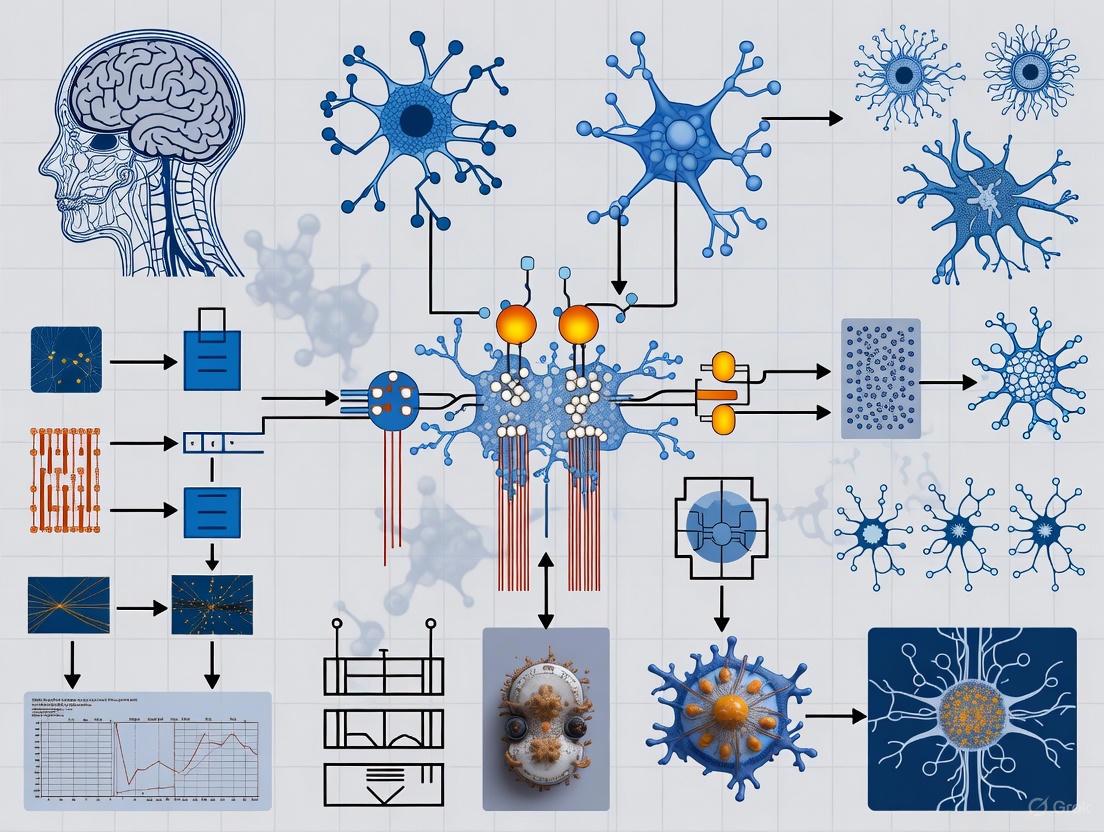
Figure 1: The complete BCI pipeline illustrates the sequential stages from neural signal acquisition to closed-loop execution, with feedback influencing subsequent neural activity.
Signal Acquisition Technologies
Signal acquisition constitutes the critical first stage of the BCI pipeline, with the detection and recording of cerebral signals directly determining system effectiveness [21]. The classification of signal acquisition technologies can be understood through a two-dimensional framework encompassing surgical invasiveness and sensor operating location [21].
Surgical Dimension: Invasiveness of Procedures
The surgical dimension classifies BCI approaches based on the invasiveness of the procedure required for signal acquisition, ranging from non-invasive to minimally invasive and fully invasive techniques [21].
Non-invasive Methods: These approaches require no surgical intervention and do not cause anatomically discernible trauma. Electroencephalography (EEG) is the most prominent example, recording electrical activity along the scalp. EEG provides a practical balance of signal quality, cost, and safety, though it offers limited spatial resolution and signal-to-noise ratio compared to more invasive methods [21] [23].
Minimally Invasive Methods: These techniques cause anatomical trauma that spares brain tissue itself. Examples include vascular stent electrodes that leverage naturally existing cavities like blood vessels, and electrocorticography (ECoG), which involves placing a thin plastic pad of electrodes right above the brain's cortex [21] [23].
Invasive Methods: These approaches cause anatomically discernible trauma at the micron scale or larger to brain tissue. Microelectrode arrays implanted directly into gray matter fall into this category. While offering the highest signal quality, they present greater surgical risks and ethical considerations [21].
Detection Dimension: Sensor Operating Location
The detection dimension classifies BCIs based on where sensors operate during signal acquisition, which directly influences the theoretical upper limit of signal quality [21].
Non-implantation: Sensors remain on the body surface, as with EEG. This approach is analogous to "listening to a chorus from outside the building," where only large-scale sums of neuronal activity can be detected amid noise [21].
Intervention: Sensors leverage naturally existing cavities within the body, such as blood vessels, without harming the integrity of original tissue. Stent-based electrodes are an emerging technology in this category [21].
Implantation: Sensors are placed within human tissue, as with intracortical microelectrodes. These typically provide the highest signal quality but raise biocompatibility concerns and may become integrated with tissue over time, complicating removal [21].
Table 2: Comparison of BCI Signal Acquisition Technologies
| Technology | Surgical Dimension | Detection Dimension | Spatial Resolution | Temporal Resolution | Signal Quality | Clinical Risk |
|---|---|---|---|---|---|---|
| EEG | Non-invasive | Non-implantation | Low (cm) | High (ms) | Low | Minimal |
| MEG | Non-invasive | Non-implantation | Medium | High | Medium | Minimal |
| ECoG | Minimally Invasive | Implantation | High (mm) | High | Medium-High | Moderate |
| Strode Arrays | Invasive | Implantation | High (μm) | High | High | Significant |
| Stent Electrodes | Minimally Invasive | Intervention | Medium-High | High | Medium-High | Moderate |
Signal Preprocessing and Cleaning
Raw neural signals, particularly from non-invasive approaches like EEG, contain substantial noise and artifacts that must be addressed before meaningful analysis can occur. Preprocessing transforms raw data into cleaned signals suitable for feature extraction [24].
Experimental Protocol: EEG Preprocessing Pipeline
A standardized preprocessing protocol for EEG data involves sequential steps to address different types of noise and artifacts:
Data Importation: EEG data is typically acquired in standardized formats such as FIF (Functional Imaging File Format) or EDF (European Data Format), which can be processed using libraries like MNE-Python [24].
Bad Channel Identification and Interpolation: Visual inspection or automated algorithms identify malfunctioning electrodes. Spherical spline interpolation estimates missing data based on surrounding good channels [24].
Filtering: Multiple filtering techniques isolate signals of interest:
- High-pass filtering (cutoff ~0.1 Hz) removes slow drifts and DC offsets
- Low-pass filtering (cutoff ~30 Hz for cognitive tasks) removes high-frequency noise
- Notch filtering (50/60 Hz) eliminates power line interference [24]
Downsampling: Data sampling rates are reduced to decrease computational load while maintaining signal integrity according to the Nyquist-Shannon theorem [24].
Re-referencing: Electrode signals are recomputed against different reference schemes (e.g., linked mastoids, average reference) to improve signal interpretation [24].
Optimization of Preprocessing Parameters
Preprocessing stage optimization must balance accuracy and computational timing costs, particularly for real-time BCI applications. Research demonstrates that parameter optimization in the preprocessing stage can significantly impact final classification performance [25].
Table 3: Preprocessing Stage Optimization Results for Motor Imagery BCI
| Preprocessing Parameter | Options | Optimal for Accuracy | Optimal for Timing Cost |
|---|---|---|---|
| Time Interval | 0-2s, 0-3s, 0-4s, 0-5s | 0-4s | 0-2s |
| Time Window - Step Size | 2s-0.125s, 1s-0.125s, 0.5s-0.125s, 2s-0.5s | 2s-0.125s | 0.5s-0.125s |
| Theta Band (4-7 Hz) | Included, Not Included | Included | Not Included |
| Mu/Beta Band (8-30 Hz) | Included, Not Included | Included | Not Included |
Taguchi method optimization with Grey relational analysis has demonstrated that the highest accuracy performance is obtained with a 0-4s time interval, 2s window with 0.125s step size, and inclusion of both theta and mu/beta frequency bands [25].
Feature Extraction and Selection
Feature extraction transforms preprocessed signals into representative characteristics that capture essential patterns of underlying brain activity, reducing data dimensionality while highlighting relevant information [24].
Feature Extraction Methodologies
Time-Domain Features: These capture temporal signal characteristics, including amplitude measurements (peak-to-peak, mean, variance), latency metrics (onset latency, peak latency), and time-series analysis techniques (autoregressive models, moving averages) [24].
Frequency-Domain Features: These analyze power distribution across frequency bands, with Power Spectral Density (PSD) calculated using Fast Fourier Transform (FFT) and band power computations for standard frequency bands (delta, theta, alpha, beta, gamma) [24].
Time-Frequency Features: Techniques like Wavelet Transform and Short-Time Fourier Transform (STFT) capture dynamic changes in frequency content over time, particularly useful for analyzing non-stationary signals [24].
Feature Selection Algorithms
Feature selection identifies the most informative characteristics from extracted features to improve model performance and avoid overfitting [26].
Filter Methods: Select features based on intrinsic characteristics independent of the machine learning algorithm. Variance Thresholding removes low-variance features, while SelectKBest chooses top features based on statistical tests like ANOVA F-value [26].
Wrapper Methods: Evaluate feature subsets by training and testing models with each subset. Recursive Feature Elimination (RFE) iteratively removes less important features based on classifier performance [26].
Embedded Methods: Incorporate feature selection during model training. L1 Regularization (LASSO) adds a penalty term that drives weights of less important features toward zero [26].
Advanced optimization algorithms like the Whale Optimization Algorithm (WOA) have demonstrated exceptional performance for feature selection in BCI systems, achieving accuracy up to 98.6% when combined with k-NN classifiers for motor imagery tasks [23].
Classification Algorithms and Translation
Classification algorithms translate selected features into device commands, serving as the final decoding stage in the BCI pipeline [26].
Machine Learning Approaches
Linear Discriminant Analysis (LDA): A classic linear classification method that finds projections maximizing separation between classes. LDA offers simplicity, speed, and effectiveness for high-dimensional BCI data [26] [23].
Support Vector Machines (SVM): Construct optimal hyperplanes to separate different classes in feature space. Kernel functions (linear, polynomial, radial basis function) enable handling of non-linear decision boundaries [26] [23].
Other Classifiers: Various algorithms offer different trade-offs, including Logistic Regression, Decision Trees, Random Forests, and k-Nearest Neighbors (k-NN) [26] [23].
Deep Learning and Optimization
Deep learning approaches have shown increasing promise for BCI applications, with optimized architectures demonstrating significant performance improvements. Bayesian optimization for hyperparameter tuning in deep learning models has achieved 4-9% accuracy improvements over conventional classifiers [27].
Table 4: Performance Comparison of Classification Algorithms for Motor Imagery BCI
| Classification Algorithm | Average Accuracy | Advantages | Limitations |
|---|---|---|---|
| Linear Discriminant Analysis (LDA) | 70-85% | Fast, simple, works well with high-dimensional data | Limited to linear decision boundaries |
| Support Vector Machine (SVM) | 75-90% | Handles non-linearity via kernels, effective in high dimensions | Sensitivity to parameter tuning |
| k-Nearest Neighbors (k-NN) | 80-92% | Simple, no training phase, naturally handles multi-class | Computationally intensive during execution |
| Random Forest | 78-90% | Handles non-linearity, robust to overfitting | Less interpretable, more parameters to tune |
| Optimized Deep Learning | 85-98% | Automatic feature learning, high performance | Computationally intensive, requires large data |
Closed-Loop Systems and Output
The closed-loop aspect represents the culmination of the BCI pipeline, where system outputs provide feedback to users, creating an adaptive interface that responds to brain activity in real time [22].
Feedback Mechanisms
Feedback components inform users about the computer's interpretation of their intended actions, typically conveyed through visual, auditory, or tactile modalities. This feedback enables adjustments and supports the closed-loop design essential for effective BCI operation [21].
Real-World Applications
Neurorehabilitation: BCI closed-loop systems facilitate recovery for patients with strokes, head trauma, and other neurological disorders by promoting neuroplasticity through real-time feedback [22] [3].
Assistive Technologies: These systems enable control of external devices such as prosthetic limbs, wheelchairs, or communication interfaces for individuals with motor impairments [23].
Cognitive Monitoring: BCIs integrated with AI allow for real-time monitoring of cognitive states, with particular relevance for conditions like Alzheimer's disease and related dementias [22].
Neuromorphic Engineering Approaches
Emerging neuromorphic engineering approaches implement closed-loop BCI systems using brain-inspired computing architectures. These systems offer potential advantages in power efficiency and real-time processing for adaptive brain interfaces [28].
Figure 2: Closed-loop BCI system with adaptive algorithms that adjust acquisition and processing parameters based on feedback, optimizing system performance over time.
The Scientist's Toolkit: Research Reagents and Materials
Table 5: Essential Research Materials for BCI Development and experimentation
| Item | Specification/Example | Primary Function |
|---|---|---|
| EEG Recording System | Biosemi ActiveTwo, BrainVision, g.tec systems | Multi-channel electrophysiological data acquisition |
| Electrodes/Caps | Ag/AgCl electrodes, electrode caps with 32-256 channels | Signal transduction from scalp to recording system |
| Electrode Gel | Electro-Gel, Signa Gel, Ten20 conductive paste | Maintains conductivity between scalp and electrodes |
| Data Processing Software | MNE-Python, EEGLAB, BCILAB | Signal preprocessing, analysis, and visualization |
| Machine Learning Frameworks | Scikit-learn, TensorFlow, PyTorch | Implementation of classification algorithms |
| Neuromodulation Devices | TMS, tDCS, neurostimulation systems | Investigate causal relationships in neural circuits |
| Implantable Electrodes | Microelectrode arrays, ECoG grids, Utah arrays | Invasive and minimally invasive signal acquisition |
| Bioamplifiers | g.tec amplifiers, Blackrock Microsystems | Signal conditioning and amplification |
The BCI pipeline represents a mature framework for creating direct interfaces between the brain and external devices, with clearly defined stages from signal acquisition to closed-loop execution. Advances in neural engineering have progressively enhanced each component of this pipeline, with current research focusing on optimizing preprocessing parameters, developing sophisticated feature selection algorithms, and implementing adaptive classification approaches. The integration of artificial intelligence and machine learning has been particularly transformative, enabling more accurate decoding of neural signals and creating more responsive closed-loop systems.
Future directions in BCI research include the development of more sophisticated neuromorphic engineering approaches, improved minimally invasive acquisition technologies, and enhanced adaptive algorithms that optimize system parameters in real time. As these technologies mature, they offer significant potential for clinical applications in neurorehabilitation, cognitive assessment, and assistive devices, ultimately advancing how neural engineering interfaces with the nervous system for both therapeutic and enhancement purposes.
Neural engineering represents an interdisciplinary frontier that combines principles from neuroscience, engineering, and computer science to study, repair, replace, or enhance neural systems [3]. This field aims to understand the nervous system's function and develop technologies that can interact with it, creating devices that restore or improve neural function lost to disease or injury. The core of this interaction lies in establishing communication pathways between biological neural systems and external devices, forming what are known as neural-computer interfaces (NCIs) [29]. These interfaces offer unprecedented potential for restoring lost functions by decoding neural signals associated with movement and speech and by providing artificial sensory feedback, effectively bypassing damaged neural pathways.
The fundamental principle governing NCIs involves two complementary processes: decoding neural activity to understand user intent and encoding information into the nervous system to restore sensation. From a mathematical perspective, decoding involves interpreting neural responses K to predict a stimulus or intent x, formalized as P(x|K) – the probability of x given observed neural activity K [30]. This process enables the translation of thought into action, whether for controlling prosthetic limbs or generating speech. Conversely, encoding involves delivering precisely timed stimuli to the nervous system to generate percepts that substitute for lost sensory functions. Together, these processes form a bidirectional communication channel that can restore functional loops disrupted by neurological damage.
Neural-Computer Interfaces: Bridging the Gap
Classification and Fundamental Principles
Neural-computer interfaces can be systematically classified along several dimensions, each with distinct implications for function restoration [29]:
- Degree of Intervention: Invasive interfaces involve implantation into neural tissue (e.g., intracortical arrays), offering high signal resolution but requiring neurosurgery. Semi-invasive interfaces are placed within the skull but not within brain tissue (e.g., ECoG). Non-invasive interfaces (e.g., EEG, sEMG) operate entirely outside the body, sacrificing some signal quality for safety and accessibility.
- Direction of Communication: Brain-Computer Interfaces (BCIs) convert neural activity into commands for external devices. Computer-Brain Interfaces (CBIs) translate artificial signals into stimuli for the central nervous system. Bidirectional BCIs (BBCIs) combine both directions to create closed-loop systems.
- Mode of Interaction: Active NCIs generate outputs from consciously controlled brain activity independent of external events. Reactive NCIs use brain activity modulated in response to external stimuli. Passive NCIs monitor brain states not directly tied to volitional control.
The Bidirectional Closed-Loop System
The most advanced approach for restoring function involves bidirectional closed-loop systems that both decode motor intent and encode sensory feedback. This creates an artificial nervous pathway that mimics natural sensorimotor integration. For example, a system designed to restore arm and hand function might decode movement intentions from the motor cortex and simultaneously deliver tactile feedback from sensors on a prosthetic hand to the somatosensory cortex [29]. This continuous loop allows for real-time adjustment of movement based on artificial sensation, dramatically improving the precision and naturalness of control. The brain gradually integrates this artificial pathway through neuroplasticity, ultimately treating the external device as an extension of the body schema.
Advancing Motor Control Restoration
Invasive Approaches for Paralysis
Groundbreaking work in invasive BCIs has demonstrated the feasibility of restoring volitional movement to paralyzed limbs. The ReHAB (Reconnecting Hand and Arm to Brain) system represents a state-of-the-art example [31]. This system employs intracortical electrodes implanted in motor areas to decode movement intention, which is then translated into stimulation patterns delivered to peripheral nerves or muscles via implanted functional electrical stimulation (FES) systems. In one clinical application, this approach allowed a patient who had not moved his limb in a decade to restore arm and hand function, effectively bypassing a spinal cord injury [31].
The methodology involves precise surgical planning and multidisciplinary collaboration. Electrodes are placed in both the motor strip (for movement execution) and areas involved with the intent to perform actions like hand grasp or arm reach [31]. The neural signals are recorded, decoded using sophisticated algorithms, and translated into commands that reanimate the paralyzed limb through electrical stimulation of peripheral nerves. This creates a direct brain-to-muscle pathway that circumvents the spinal lesion.
Table 1: Quantitative Performance of Non-Invasive Neuromotor Interfaces for Hand Gestures [32]
| Task Type | Performance Metric | Performance Value | Context |
|---|---|---|---|
| Continuous Navigation | Target Acquisitions/Second | 0.66 | Wrist-based continuous control |
| Discrete Gesture Detection | Gesture Detections/Second | 0.88 | Nine distinct finger gestures |
| Handwriting Transcription | Words Per Minute | 20.9 | Writing with imaginary implement |
Non-Invasive Alternatives
Recent advances have also demonstrated the potential of non-invasive approaches. A 2025 study published in Nature described a generic non-invasive neuromotor interface based on surface electromyography (sEMG) [32]. The system uses a high-density, dry-electrode wristband that records muscle electrical signals representing motor commands from the central nervous system. Through data collection from thousands of participants and deep learning models, the system can decode gesture intent with high accuracy across users without individual calibration. Test users achieved a handwriting rate of 20.9 words per minute and 0.88 gesture detections per second in discrete gesture tasks [32].
The experimental protocol for developing this system involved collecting sEMG data from a demographically diverse participant group performing three tasks: wrist control, discrete gesture detection, and handwriting. A specialized data collection system recorded both sEMG activity and precise label timestamps using a real-time processing engine to minimize online-offline shift. A time-alignment algorithm was developed to infer actual gesture event times, accounting for participant reaction time and compliance variations [32].
Decoding Speech from Neural Signals
Technical Approaches and Challenges
Speech decoding from neural signals represents a particularly challenging frontier in neural engineering, with profound implications for individuals who have lost the ability to speak due to conditions like amyotrophic lateral sclerosis (ALS), stroke, or brainstem injury [33]. The primary technical approaches can be categorized by the speech modality they target:
- Overt Speech: Actual vocalized speech with audible output
- Whispered Speech: Vocalized but quiet speech with reduced distinctness
- Silent Speech: Articulated without vocalization
- Imagined/Covert Speech: Internally generated without vocalization or articulation
The most clinically relevant yet most challenging paradigm is imagined speech decoding, as it requires no muscular engagement and is thus suitable for completely locked-in patients [33]. However, the absence of sensory feedback and the subtle nature of the associated neural signals make this modality particularly difficult to decode accurately.
Experimental Protocols and Performance
A 2025 research study systematically investigated whether incorporating EEG data from overt speech could enhance imagined speech classification [33]. The experimental protocol involved 24 healthy participants who pronounced and imagined five Spanish words used in assistive communication devices. Researchers compared four classification scenarios by modifying the training dataset composition using EEGNet, a convolutional neural network designed for EEG-based BCIs.
The results demonstrated that in binary word-pair classifications, combining overt and imagined speech data during training led to accuracy improvements of 3%–5.17% in four out of ten word pairs compared to using imagined speech alone [33]. While the highest individual accuracy (95%) was achieved with imagined speech exclusively, incorporating overt speech data allowed more participants to surpass the 70% accuracy threshold (increasing from 10 to 15 participants). The study also found that word characteristics like length, phonological complexity, and frequency of use contributed to higher discriminability between certain imagined word pairs.
EEG-based Speech Decoding Workflow
Restoring Sensory Feedback
Principles of Sensory Encoding
While decoding neural signals enables motor output, encoding artificial sensory information is equally crucial for functional restoration. Sensory feedback systems operate as computer-to-brain interfaces (CBIs) that deliver precisely controlled stimuli to neural tissue to generate perceptual experiences [29]. These systems typically employ electrical, tactile, or magnetic stimulation of peripheral nerves, spinal pathways, or specific brain regions to evoke sensations that substitute for lost natural senses.
The fundamental challenge in sensory encoding lies in mapping sensor data to stimulation patterns that produce meaningful, discriminable percepts. For example, a prosthetic hand with tactile sensors might translate pressure readings into patterns of electrical stimulation delivered to either the residual peripheral nerves in the limb or directly to the somatosensory cortex. The intensity, location, and timing of stimulation must correlate intuitively with the sensory input to create a usable feedback loop.
Clinical Applications and Neuromodulation
Beyond prosthetic sensation, sensory encoding principles underpin numerous clinical neuromodulation therapies. Deep brain stimulation (DBS) delivers electrical pulses to specific brain structures to modulate pathological neural activity in conditions like Parkinson's disease, essential tremor, and obsessive-compulsive disorder [3]. Similarly, transcranial magnetic stimulation (TMS) uses magnetic fields to non-invasively modulate cortical excitability, with FDA-approved applications for treatment-resistant depression [3].
These approaches represent a form of "macro-level" sensory encoding where the stimulation doesn't convey specific sensory information but rather modulates overall neural circuit function to alleviate symptoms or restore more normal processing of natural sensory inputs.
The Scientist's Toolkit: Research Reagent Solutions
Table 2: Essential Research Tools in Neural Interface Development
| Research Tool | Type/Technology | Primary Function in Research |
|---|---|---|
| sEMG Research Device (sEMG-RD) [32] | Dry-electrode, multichannel wristband | Records high-quality surface EMG signals for non-invasive neuromotor interfacing |
| Multielectrode Arrays [34] | Invasive neural implants | Records spiking activity from populations of neurons during learning and behavior |
| EEGNet [33] | Convolutional Neural Network | Classifies EEG signals for imagined speech decoding and other BCI applications |
| Real-time Processing Engine [32] | Software infrastructure | Aligns neural data with behavioral labels and minimizes online-offline shift |
| Population-level Synaptic Plasticity Model [34] | Computational modeling | Identifies long-term synaptic plasticity changes from spiking activity during learning |
Signaling Pathways in Neural Interfaces
Bidirectional Neural Interface Pathway
Future Directions and Ethical Considerations
The field of neural engineering is rapidly advancing toward more sophisticated and integrated systems. Current research aims to develop adaptive BCIs that can learn and evolve with the user over time. The DARPA INSPIRE project, for example, seeks to unravel how long-term synaptic plasticity (LTSP) governs learning and memory formation by developing computational tools to measure synaptic strength changes directly from spiking activity during behavior [34]. This understanding could lead to BCIs that not only restore function but also adapt to the user's changing neural architecture and even facilitate relearning of lost skills.
Other promising directions include the development of electrochemical interfaces that can record and stimulate at the molecular level, biomimetic hierarchical systems that more naturally integrate with the nervous system's organization, and energy-efficient technologies capable of long-term implantation without external power sources [29].
These technological advances raise important ethical considerations regarding mental privacy, agency, and the potential for unintended consequences of neural augmentation. As noted in recent scholarship, the ability to infer sensitive information about individuals from neural data necessitates robust frameworks for protecting "cognitive biometrics" [35]. Ensuring that users maintain ultimate agency over neural interfaces, particularly in bidirectional systems, represents a critical challenge for the field's responsible development.
Neural engineering interfaces represent a transformative approach to restoring function lost to neurological disease or injury. Through sophisticated decoding of motor and speech signals and encoding of sensory information, these systems can bypass damaged neural pathways to re-establish communication between the brain and the external world. Current research demonstrates impressive progress, from enabling paralyzed individuals to control their limbs again [31] to achieving practical typing speeds through non-invasive gesture detection [32]. As the field advances toward more adaptive, bidirectional, and biomimetic interfaces, the potential for restoring increasingly complex functions grows accordingly. The integration of neural interfaces with our understanding of synaptic plasticity and learning [34] promises not only to replace lost functions but to actively promote neural repair and rehabilitation, ultimately offering more natural and integrated solutions for those with neurological impairments.
The convergence of neural engineering and psychiatry represents a paradigm shift in how we understand and treat mental health disorders. Neuroengineering is an interdisciplinary field that applies engineering principles to understand, repair, replace, or improve neural systems [36] [37]. When interfaced with psychiatry, it enables the development of technologies that can directly monitor and modulate the neural circuits underlying psychiatric conditions. This approach moves beyond traditional pharmacological methods toward precision treatments that target specific neural pathways and networks.
The fundamental thesis of this integration posits that engineering approaches can provide unprecedented access to the neural substrates of psychiatric disorders, enabling both novel interventions and deeper mechanistic understanding. This interface is built upon a foundation of quantitative approaches to measuring and manipulating nervous system function, creating a feedback loop between technological innovation and neuroscientific discovery [37] [38]. The field leverages multiple interface modalities—from fully implantable devices to non-invasive systems—to establish bidirectional communication with the nervous system, allowing for both recording pathological neural signatures and delivering targeted neuromodulation to restore normal function.
Technical Approaches for Neural Interfacing in Psychiatry
Invasive and Non-Invasive Interface Modalities
Neuroengineering solutions for psychiatric applications span a spectrum of invasiveness, each with distinct trade-offs between spatial resolution, temporal resolution, and risk profiles. Each modality provides unique advantages for specific psychiatric applications and research contexts.
Table 1: Neural Interface Modalities for Psychiatric Applications
| Modality Type | Technologies | Spatial Resolution | Temporal Resolution | Key Psychiatric Applications |
|---|---|---|---|---|
| Invasive | Deep Brain Stimulation (DBS), Intracortical Recording, Responsive Neurostimulation | Single neuron to local field potentials | Millisecond precision | Treatment-resistant depression, OCD, severe mood disorders |
| Minimally Invasive | Endovascular stents, Epidural electrodes | Mesoscale network activity | High (milliseconds) | Emerging applications for depression monitoring |
| Non-Invasive | EEG, tES (tDCS/tACS), TMS, tFUS | Centimeter scale | Variable (milliseconds for EEG, seconds for fMRI) | Depression, anxiety, cognitive enhancement |
Invasive approaches, particularly Deep Brain Stimulation (DBS), represent the most targeted interface strategy. DBS involves the surgical implantation of electrodes into specific deep brain structures, enabling precise electrical modulation of circuits implicated in psychiatric disorders [37]. For treatment-resistant depression, targets have included the subcallosal cingulate cortex, ventral capsule/ventral striatum, and medial forebrain bundle. The Responsive Neurostimulation System (RNS) from NeuroPace represents an advanced closed-loop approach that continuously monitors neural activity and delivers stimulation only when pathological patterns are detected [37]. This responsive approach is particularly relevant for psychiatric conditions characterized by episodic symptomatology.
Non-invasive interfaces provide access to neural function without surgical intervention. Electroencephalography (EEG) captures electrical activity from scalp electrodes, allowing researchers to identify neural signatures associated with psychiatric states [36] [39]. Transcranial Electrical Stimulation (tES), including tDCS and tACS, applies weak electrical currents through the scalp to modulate cortical excitability [40]. Emerging techniques like transcranial Focused Ultrasound (tFUS) offer the potential for non-invasive neuromodulation of deeper brain structures with higher spatial precision than other non-invasive methods [36]. These non-invasive approaches enable repeated measurements and interventions across the illness trajectory, facilitating longitudinal studies of disease progression and treatment response.
Signal Processing and Decoding Approaches
Advanced signal processing is essential for extracting meaningful information from neural data in psychiatric applications. The complex, high-dimensional nature of neural recordings requires sophisticated analytical approaches to identify features relevant to psychiatric states.
Table 2: Neural Signal Processing Methods for Psychiatric Applications
| Processing Stage | Methods | Psychiatric Application Examples |
|---|---|---|
| Feature Extraction | Time-frequency analysis, Graph theory networks, Event-related potentials | Identifying biomarkers of depression from EEG oscillations, Mapping functional connectivity in OCD |
| Dimensionality Reduction | Principal Component Analysis, Independent Component Analysis, Autoencoders | Reducing high-dimensional ECoG data to essential features for mood decoding |
| Classification/Decoding | Support Vector Machines, Random Forests, Convolutional Neural Networks, Recurrent Neural Networks | Classifying depressive states from neural activity, Predicting treatment response from baseline neural features |
| Closed-loop Control | Kalman filters, Proportional-Integral-Derivative controllers, Reinforcement learning | Adaptive DBS for depression based on neural biomarkers |
Recent advances in machine learning, particularly deep learning, have dramatically improved our ability to decode complex mental states from neural signals [36]. Convolutional neural networks can identify spatial patterns in neural data, while recurrent architectures can model temporal dynamics of psychiatric symptoms. For example, convolutional neural networks have been used to map electrical signals from brain-computer interfaces to neural features, significantly improving performance on tasks relevant to psychiatric assessment [41]. These approaches enable the development of biomarker-driven interventions that can be personalized to an individual's unique neural signature of illness.
Experimental Protocols for Psychiatric Neuroengineering
Protocol 1: Closed-Loop DBS for Treatment-Resistant Depression
This protocol outlines a methodology for implementing closed-loop deep brain stimulation for treatment-resistant depression, adapting approaches from recent clinical trials and research initiatives [37].
Objectives:
- To identify neural biomarkers of depressive states from implanted recording electrodes
- To implement a closed-loop stimulation system that automatically adjusts stimulation parameters based on these biomarkers
- To evaluate clinical efficacy using standardized depression rating scales
Materials and Equipment:
- Implantable pulse generator with sensing capabilities (e.g., NeuroPace RNS System, Medtronic Summit RC+S)
- Depth electrodes for targeted brain regions (e.g., subcallosal cingulate cortex, ventral capsule/ventral striatum)
- External programming interface with real-time data processing capabilities
- Clinical assessment tools (MADRS, HAM-D, self-report measures)
Procedure:
- Preoperative Planning: Identify stimulation targets using structural (T1-weighted) and functional MRI (resting-state connectivity).
- Surgical Implantation: Stereotactically implant depth electrodes in target regions using standard neurosurgical techniques under general anesthesia.
- Biomarker Identification Phase (2 weeks): Record continuous neural data (local field potentials, single-unit activity) during various states (sleep, rest, emotional tasks). Use machine learning approaches to identify neural features (specific frequency band power, cross-region coherence) that correlate with depressive symptom severity.
- Stimulation Parameter Optimization (1 week): Systematically test stimulation parameters (frequency, amplitude, pulse width) in double-blind fashion to identify optimal settings for symptom reduction while minimizing side effects.
- Closed-Loop Operation: Implement detection algorithms for state-specific neural features identified in Phase 3. Configure stimulator to automatically adjust stimulation when these features are detected.
- Assessment: Conduct weekly clinical assessments for 12 weeks, then monthly. Continuously record neural data and stimulation parameters for offline analysis.
Data Analysis:
- Compare depression rating scale scores pre- and post-implementation
- Analyze relationship between neural biomarker occurrence and symptom fluctuations
- Quantify percentage of time stimulation is active in responsive mode versus continuous mode
Protocol 2: Multimodal Non-Invasive Neuromodulation for Depression
This protocol integrates multiple non-invasive neuromodulation approaches with EEG monitoring for treatment of major depressive disorder, leveraging recent technological advances [36] [40].
Objectives:
- To evaluate the efficacy of combined tFUS and tACS for modulating depressive circuit pathology
- To identify EEG biomarkers predictive of treatment response
- To assess durability of effects through follow-up period
Materials and Equipment:
- High-density EEG system (64+ channels) with compatible stimulation equipment
- Transcranial focused ultrasound system with neuronavigation
- Transcranial alternating current stimulation device
- MRI for target localization and circuit mapping
- Neuropsychological assessment battery
Procedure:
- Baseline Assessment: Conduct clinical interviews, self-report measures, and cognitive testing. Acquire structural and resting-state fMRI for target identification.
- EEG Biomarker Acquisition: Record 10 minutes of resting-state EEG followed by task-based EEG during emotional processing tasks.
- Neuronavigation: Co-register MRI data with subject's head to ensure precise targeting of tFUS.
- Stimulation Protocol:
- Apply tFUS to dorsolateral prefrontal cortex (2 MHz, 30-second duration, 500 Hz PRF, 0-500 kPa pressure)
- Immediately follow with tACS to same region (6 Hz, 1 mA, 20 minutes)
- Administer 15 sessions over 5 weeks (3 sessions per week)
- Monitoring: Record EEG before, during, and after each stimulation session to assess immediate effects on brain activity.
- Follow-up: Conduct clinical and EEG assessments at 1, 3, and 6 months post-treatment.
Data Analysis:
- Compute changes in frontal alpha asymmetry from pre- to post-treatment
- Analyze phase-amplitude coupling between theta and gamma oscillations
- Correlate EEG changes with clinical improvement on depression rating scales
- Use machine learning to identify baseline EEG features predictive of treatment response
Signaling Pathways and Neural Circuits in Psychiatric Neuroengineering
Understanding the neural circuits and signaling pathways modulated by neuroengineering approaches is essential for advancing psychiatric applications. The following diagram illustrates key circuits and their modifications through neural interfaces.
The fronto-striatal circuit (blue) is implicated in cognitive control and reward processing, functions frequently disrupted across psychiatric disorders. The limbic circuit (red) mediates emotional processing and is hyperactive in conditions like depression and anxiety [1]. Neuroengineering interventions target specific nodes within these circuits: DBS primarily modulates the subgenual anterior cingulate cortex (sgACC) in the limbic circuit [37], while non-invasive approaches like tFUS and tACS typically target dorsolateral prefrontal cortex (DLPFC) in the fronto-striatal circuit [36]. These interventions work to restore normal circuit function by modulating oscillatory activity, synaptic plasticity, and network connectivity.
At the cellular and molecular level, these interventions engage multiple signaling pathways. Electrical stimulation affects voltage-gated ion channels, leading to changes in neuronal firing patterns. Repeated stimulation induces neuroplasticity through mechanisms involving brain-derived neurotrophic factor (BDNF) and NMDA receptor-mediated synaptic plasticity. There is also evidence that neuromodulation influences neurotransmitter systems including serotonin, dopamine, and glutamate, which are known to be dysregulated in psychiatric disorders [37]. The convergence of these effects across molecular, cellular, and circuit levels enables the restoration of normal neural computation and alleviation of psychiatric symptoms.
Research Reagent Solutions for Psychiatric Neuroengineering
Table 3: Essential Research Reagents and Materials for Psychiatric Neuroengineering
| Reagent/Material | Function | Example Applications | Technical Considerations |
|---|---|---|---|
| High-density EEG systems | Non-invasive recording of electrical brain activity | Monitoring neural correlates of depression, anxiety, psychosis | 64+ channels recommended for source localization; impedance <10 kΩ |
| tDCS/tACS devices | Non-invasive neuromodulation via scalp electrodes | Modulating cortical excitability in depression; entraining neural oscillations | Electrode placement per 10-20 system; typically 1-2 mA intensity |
| Implantable microelectrode arrays | Chronic recording and stimulation of neural ensembles | Investigating circuit mechanisms in animal models; human DBS | Biocompatible materials (e.g., platinum-iridium, polyimide); high electrode density |
| Calcium indicators (GCaMP) | Optical monitoring of neural activity in animal models | Recording neural population dynamics during behavior | Viral delivery methods; compatible with fiber photometry or 2-photon imaging |
| Optogenetic actuators (Channelrhodopsin) | Precise control of specific neural populations | Causally testing circuit hypotheses in animal models | Cell-type specific promotors (e.g., CaMKII for excitatory neurons) |
| Neurotransmitter sensors (dLight, GRAB) | Real-time monitoring of neurotransmitter release | Measuring dopamine, serotonin dynamics in reward processing | Genetically encoded; requires viral delivery and optical detection |
| Behavioral testing apparatus | Quantifying disease-relevant behaviors in models | Depression (forced swim test), anxiety (elevated plus maze) | Automated tracking software; controlled testing environment |
| Neural data analysis software | Processing high-dimensional neural datasets | Spike sorting, LFP analysis, connectivity mapping | Open-source options (e.g., MATLAB, Python); cloud-compatible |
The selection of appropriate research reagents should be guided by the specific research question and model system. For human studies, non-invasive approaches like EEG and tES provide translational pathways, while animal models enable cellular and circuit-level interrogation using optogenetics and fiber photometry. The GX Dataset represents an example of open-source data incorporating EEG, physiological measures, and behavior during transcranial electrical stimulation, providing a valuable resource for method development [40]. Integration across these levels through convergent approaches is accelerating progress in psychiatric neuroengineering.
The interface between neuroengineering and psychiatry is producing transformative approaches for understanding and treating mental health disorders. By applying engineering principles to measure and modulate neural circuits, this interdisciplinary field enables unprecedented precision in targeting the neural substrates of psychiatric conditions. Current technologies span from invasive deep brain stimulation to non-invasive neuromodulation, each with distinct advantages for different applications and patient populations.
Future directions include developing closed-loop systems that automatically adjust stimulation parameters based on neural biomarkers, creating miniaturized and wireless devices for chronic use, and advancing personalized approaches that tailor interventions to individual circuit dysfunction [11] [37]. There is also growing recognition of the importance of centering disabled users in neurotechnology development to ensure solutions are practical, effective, and ethically grounded [11]. As the field progresses, integration with other emerging technologies like artificial intelligence and stem cell therapies will likely open new frontiers for restoring neural circuit function in psychiatric disorders [42].
The continued advancement of psychiatric neuroengineering requires sustained collaboration across disciplines—engineering, neuroscience, clinical psychiatry, and computer science—and active engagement with patient communities to ensure technologies address real-world needs. Through these concerted efforts, neuroengineering approaches promise to revolutionize how we understand, diagnose, and treat mental health disorders, moving toward a future of precision psychiatry based on direct neural circuit interrogation and modulation.
Neural engineering is an interdisciplinary field that combines principles from neuroscience, engineering, and computer science to study, repair, replace, or enhance neural systems [3]. Its primary goal is to create a functional interface between the nervous system and the outside world. This interface operates in two primary directions: output interfaces, which record and decode neural signals to understand intention and brain function, and input interfaces, which stimulate neural tissue to restore sensory processing or modulate neural activity for treating disease [2]. The architecture of a closed-loop neural interface system exemplifies this bridge, typically comprising four components: a multielectrode recording array, a mapping or decoding algorithm, an output device, and a sensory feedback channel [2]. This foundational engineering framework is now being radically transformed by the integration of artificial intelligence (AI) and high-throughput data, enabling unprecedented scale and precision in neuro-discovery.
High-Throughput Screening: Accelerating Target and Therapeutic Discovery
The identification of new therapeutic targets and treatments for neurodegenerative diseases (NDDs) is a major challenge, attributed to the complexity of neural circuits, limited tissue regeneration, and incomplete understanding of pathological processes [43]. High-throughput screening (HTS) has emerged as a critical tool to address this, replacing traditional "trial and error" approaches by allowing the rapid investigation of hundreds of thousands of compounds per day [43].
HTS comprises several steps, including target recognition, compound management, reagent preparation, assay development, and the screening process itself [43]. The active compounds identified, or "hits," serve as prototypes from which drug "leads" are formed through additional chemistry and optimization for drug metabolism and pharmacokinetic (DMPK) properties [43]. The workflow for CNS drug discovery can be subdivided into four main areas:
- Receptor and target engagement
- Drug "hit" identification
- Lead identification
- Drug lead optimization [43]
A critical first step in advancing NDD treatments is developing accurate assays for investigating neurodegeneration. Cell-based assays are particularly valuable as they allow for the investigation of whole pathways and provide data on intracellular targets that cannot be obtained from biochemical assays alone [43]. These assays are often conducted in scaled-down formats using 96- or 384-well microtiter plates with 2D cell monolayers [43]. The creation of successful assays includes the ability to identify events that trigger cell death, such as metabolic fluctuation, energy metabolism, and DNA fragmentation [43].
Table 1: Key Assay Types and Applications in Neuro-discovery
| Assay Type | Description | Key Applications in Neuroscience |
|---|---|---|
| Cytoprotective Assays | Utilize dyes or fluorescent markers to classify therapeutics causing neuronal death. | Investigation of neurotoxicity and side effects of drug candidates [43]. |
| Phenotypic Screening | Detects complex disease-related signatures and functional impairments prior to cell death. | Early detection of neuronal overactivity in ALS and Alzheimer's disease [43]. |
| Primary Neuron HTS | Uses primary neurons in screening, despite complex culture protocols. | Offers high biological and clinical relevance for large-scale compound or RNAi testing [43]. |
A major challenge remains the development of reliable screening phenotypes that can detect complex, predictive disease signatures. HTS, particularly when combined with multi-well cell-based platforms, allows for the identification of small molecule modulators of biochemical and signal transduction pathways, thereby accelerating the entire discovery pipeline [43].
The Rise of Large-Scale Data and AI in Neurology
The increasing availability of large-scale, real-world data (RWD) offers a powerful tool for advancing the understanding of neurological diseases. The NeuroDiscovery AI database is one such comprehensive repository, containing de-identified electronic health record (EHR) data from U.S.-based neurology outpatient clinics [44]. This dataset includes sociodemographic details, clinical examinations, social and medical histories, ICD diagnoses, prescribed medications, neuroimaging data, and laboratory results [44].
As of October 2024, this dataset includes EHR data from 355,791 patients, with over 40% aged 60 or older and spanning 14,797 distinct diagnosis codes [44]. It represents over 15 years of longitudinal patient information, providing an invaluable resource for studying disease progression, treatment responses, and long-term outcomes in neurology [44]. The scale of this data is crucial for addressing the growing burden of neurological disorders; the disability-adjusted life years (DALYs) due to neurological disorders were 113.5 million in 2022 and are projected to rise to 191.07 million by 2050 [44].
Table 2: Neurological Disease Burden and Projections in the United States
| Condition | Current U.S. Prevalence (Approx.) | Projected U.S. Prevalence | Economic Burden |
|---|---|---|---|
| Alzheimer's Disease | 6.7 million (aged 65+, 2023) | 13.8 million by 2060 | Projected to rise from $305B (2020) to $2.2T (2060) [44] |
| Parkinson's Disease | 1 million (2020) | 1.2 million by 2030 | $52B annually, rising to $80B by 2037 [44] |
| Migraine | 3.8 million cases (2019) | Data not specified in source | Data not specified in source [44] |
| Multiple Sclerosis | Nearly 1 million (2019) | Data not specified in source | Data not specified in source [44] |
The analysis of such large-scale RWD generates real-world evidence (RWE) that complements clinical trial data and can be used to uncover disease mechanisms, identify patient subgroups in highly heterogeneous diseases like Alzheimer's, assess drug responses, and inform personalized therapeutic strategies [44]. The integration of AI is key to interrogating these vast datasets to identify subtle trends and patterns that would be impossible to discern through manual analysis.
Visualizing Complex Neuroscience Data
As neuroscience datasets become larger and more complex, effective data visualization becomes critical for both analysis and communication. Visualizations are vital for revealing relationships in large datasets and communicating information to a broad audience [45]. However, with this power comes a responsibility to ensure that graphs are clear and honest, not misleading [45].
A survey of 1451 figures from leading neuroscience journals revealed that graphical displays often become less informative as the dimensions and complexity of datasets increase [45]. For example, only 43% of 3D graphics labeled the dependent variable, and only 20% portrayed the uncertainty of reported effects [45]. Even for 2D data, nearly 30% of figures that included error bars failed to define the type of uncertainty (e.g., standard deviation vs. standard error of the mean) being portrayed, which can lead to misinterpretation [45].
To create more informative displays, design choices should reveal data rather than hide it. For instance, while bar plots are common, they often have low data-density. Alternative designs like box plots or violin plots can reveal the full shape of distributions, including skewness or bimodality, which might be hidden in a bar plot [45]. This distributional information can be crucial for assessing the assumptions of statistical models and for understanding the most interesting quantities to investigate.
Furthermore, all visualizations must adhere to accessibility standards. The Web Content Accessibility Guidelines (WCAG) require a minimum contrast ratio of 4.5:1 for standard text and 3:1 for large text to ensure that people with low vision or color deficiencies can read the content [46]. This principle applies not only to published papers but also to the design of software and tools used by researchers.
Experimental Protocols and the Scientist's Toolkit
Detailed Methodology for High-Throughput Screening in Primary Neurons
The following protocol, adapted from Sharma et al. (2013), provides a scalable method for large-scale testing, ranging from compound libraries to whole-genome RNA interference (RNAi) [43].
Protocol: HTS for Neurodegeneration in Primary Neurons
Primary Neuron Culture:
- Isolate primary neurons from relevant brain regions of rodent models (e.g., E18 rat hippocampi or cortices).
- Plate neurons in specialized 384-well microtiter plates pre-coated with poly-D-lysine (0.1 mg/mL) to promote adhesion.
- Maintain cultures in neuronal growth medium (e.g., Neurobasal medium supplemented with B-27, glutamax, and penicillin/streptomycin) for 7-14 days in vitro (DIV) to allow for maturation and synapse formation before screening.
Compound Library and Reagent Preparation:
- Prepare compound libraries in DMSO at a standardized concentration (e.g., 10 mM). Using an automated liquid handler, transfer nanoliter volumes of compounds or RNAi vectors into the assay plates, creating a copy of the library for the screen.
- Dilute compounds in the culture medium to the desired final testing concentration (e.g., 1-10 µM), ensuring the final DMSO concentration is ≤0.1% to minimize solvent toxicity.
Assay Execution and Incubation:
- Apply the compound-containing medium to the mature primary neuronal cultures. Include control wells on every plate: vehicle controls (DMSO only), positive controls (e.g., a known neurotoxin like staurosporine to induce maximal cell death), and negative controls (healthy neurons).
- Inculture the plates for a defined period (e.g., 24-72 hours) in a humidified incubator at 37°C and 5% CO₂.
Viability and Phenotypic Readout:
- After incubation, assess neuronal health using a multiplexed approach. Add fluorescent dyes or probes to the medium, such as:
- Cell Viability Indicator: Propidium iodide or TO-PRO-3 to label dead/dying cells with compromised membranes.
- Neuronal Health Marker: A fluorescently conjugated antibody against a neuronal-specific protein like Microtubule-Associated Protein 2 (MAP2) to quantify neuronal mass and integrity.
- Incubate with dyes and antibodies for 3-6 hours, then fix cells with a paraformaldehyde solution (e.g., 4% in PBS).
- After incubation, assess neuronal health using a multiplexed approach. Add fluorescent dyes or probes to the medium, such as:
High-Content Imaging and Analysis:
- Image each well of the assay plate using a high-content imaging system (e.g., a confocal or widefield automated microscope) with a 10x or 20x objective.
- Acquire multiple images per well to ensure a representative sample of thousands of neurons.
- Use automated image analysis software to quantify:
- The total number of nuclei.
- The number of nuclei positive for the cell death marker.
- The total neuronal area and fluorescence intensity of the MAP2 stain.
Hit Identification and Data Analysis:
- Normalize data from each well to the plate-level positive and negative controls.
- Calculate a percent protection or percent viability score for each compound.
- Apply a statistical threshold for hit selection. A common method is to select compounds whose values exceed three standard deviations from the mean of the DMSO-treated control wells, which offers a manageable false-positive statistical hit rate of about 0.15% [43]. Alternatively, if performed in triplicate, the median can be used to protect against outlier results [43].
Research Reagent Solutions for HTS in Neuro-discovery
Table 3: Essential Materials and Reagents for Neuronal HTS
| Item | Function/Description | Example Application |
|---|---|---|
| Primary Neurons | Biologically relevant cells capturing critical events in disease states. | Modeling neurodegenerative processes like those in Alzheimer's and ALS [43]. |
| 384-well Microtiter Plates | Standardized format for scaled-down, high-density cell culture and screening. | Enables testing of hundreds of compounds per plate in a miniaturized format [43]. |
| Fluorescent Dyes/Probes | Markers for cell viability, death, and specific neuronal components. | Propidium iodide for dead cells; MAP2 antibodies for neuronal integrity [43]. |
| High-Content Imaging System | Automated microscope for quantitative analysis of cellular phenotypes. | Captures thousands of images for automated analysis of neuronal count, health, and morphology [43]. |
| Compound/RNAi Library | Large collections of small molecules or gene silencing constructs. | Systematic perturbation of biological systems to identify novel targets or therapeutics [43]. |
Integrated Workflows and Future Outlook
The convergence of neural engineering, high-throughput data, and AI defines the future of neuro-discovery. Integrated workflows are emerging where clinical data from neural interfaces informs the design of in vitro assays, the results of which are then validated against large-scale EHR datasets, creating a virtuous cycle of discovery. This integrated approach is critical for addressing the technical and scientific challenges faced by these systems before they are widely adopted.
Future efforts will focus on leveraging these integrated workflows to overcome the current limitations in understanding neurodegenerative disease pathogenesis. The continued expansion and diversification of large-scale neurology datasets, coupled with more sophisticated AI models and high-content phenotypic assays, will be essential for identifying robust biomarkers, deconvoluting disease heterogeneity, and delivering on the promise of personalized therapeutic strategies for neurological disorders [44].
Navigating the Neuro-frontier: Troubleshooting Technical, Ethical, and Translational Hurdles
Neural engineering stands at the intersection of neuroscience, engineering, and computational sciences, striving to create bidirectional interfaces with the nervous system. These interfaces are crucial for both decoding neural commands to control external devices and encoding sensory information by modulating neural activity. The core of this interdisciplinary field faces three persistent technical barriers: device longevity, concerning the stable and chronic integration of bioelectronic interfaces with biological tissues; signal fidelity, which involves the accurate acquisition and interpretation of complex neural signals; and computational efficiency, essential for processing high-dimensional neural data in real-time. This whitepaper provides an in-depth analysis of these challenges, framed within the context of foundational nervous system research, and details current experimental methodologies and material solutions aimed at overcoming these hurdles to advance neuroprosthetics, brain-computer interfaces (BCIs), and our fundamental understanding of neural circuits.
Device Longevity
Device longevity refers to the ability of an implanted neural interface to maintain its structural integrity and functional performance over extended periods within the biological environment. The primary obstacle to longevity is the foreign body response (FBR), a chronic inflammatory reaction that ensues following device implantation [47]. The FBR is triggered by the mechanical mismatch between typically rigid, inorganic neural electrodes and the soft, dynamic neural tissue, which has a Young's modulus in the range of 1 to 10 kPa [47]. This mismatch leads to continuous micromotion, causing acute injury and chronic inflammation. The biological cascade involves protein adsorption, activation of microglia and astrocytes, and the eventual formation of a glial scar—a dense layer of astrocytes and extracellular matrix that electrically insulates the electrode from its target neurons [47]. This scar formation increases impedance at the electrode-tissue interface, degrading recording quality and stimulation efficacy, and ultimately leads to device failure.
Key Research Reagents and Material Solutions
Addressing device longevity requires a materials science approach focused on enhancing biocompatibility and mitigating the FBR. The table below catalogs key material strategies and their functions.
Table 1: Research Reagent Solutions for Enhancing Device Longevity
| Material/Strategy | Function and Mechanism of Action |
|---|---|
| Soft Conductive Polymers | Reduces mechanical mismatch with neural tissue (Young's modulus ~ kPa range), minimizing micromotion-induced damage and chronic inflammation [47]. |
| Carbon Fiber Electrodes | Provides high stiffness for insertion despite fine diameter (e.g., 7 µm), enabling high-density arrays with minimal tissue displacement [47]. |
| Biocompatible Coatings | Serves as an interfacial layer to suppress the foreign body response; can be functionalized with immunosuppressive or bioactive molecules to promote integration [47]. |
| Fiber-Based Electronic Devices (FEDs) | Offers mechanical flexibility, minimal volume, and conformability for chronic implantation with reduced immune reaction, useful as smart sutures or deep-brain interfaces [48]. |
Experimental Protocols for Longevity Assessment
Protocol 1: Chronic In Vivo Safety and Efficacy Profiling This protocol is designed to evaluate the long-term functional stability and biological impact of implanted neural interfaces, such as intracortical microelectrode arrays [49].
- Device Implantation: Aseptically implant the neural interface (e.g., microelectrode arrays) into the target brain region (e.g., somatosensory cortex for touch restoration, ventral precentral gyrus for speech decoding) of an animal model or human clinical trial participant.
- Chronic Stimulation and Monitoring: Apply periodic intracortical microstimulation (ICMS) protocols over months to years. In a representative human study, participants received millions of electrical stimulation pulses over a combined 24 years [49].
- Endpoint Analysis:
- Functional Assessment: Quantify the percentage of electrodes that continue to evoke stable, high-quality tactile sensations or record high-fidelity signals over time. A key metric is more than half of electrodes functioning reliably after a decade [49].
- Safety Profiling: Monitor for serious adverse effects (SAEs) related to the implant or stimulation, such as persistent inflammation, infection, or tissue necrosis. Histological analysis of explanted tissue (in animal studies) can quantify glial scarring and neuronal loss.
Protocol 2: Accelerated Mechanical Fatigue Testing for Fiber-Based Devices This protocol assesses the durability of flexible fiber-based electronic devices (FEDs) under cyclic loading [48].
- Device Fabrication: Fabricate the fiber device (e.g., conductive Ag nanoparticle-based fibers, CNT-coated polymer fibers) using methods like dip-coating or melt extrusion.
- Cyclic Deformation: Subject the fiber device to repeated tensile, compressive, or bending strains using a mechanical tester, simulating dynamic in vivo conditions (e.g., movements in wearable garments or implantable tissues).
- Performance Monitoring: Continuously measure electrical properties (e.g., resistance, capacitance) throughout the test cycles. The primary failure modes to monitor are the emergence of microcracks or an exponential increase in electrical resistance, indicating mechanical fatigue and functional failure [48].
Diagram 1: Device Failure Pathway
Signal Fidelity
Signal fidelity encompasses the accuracy and reliability with which a neural interface captures neural activity (recording) and delivers patterned input (stimulation). High-fidelity signals are characterized by a high signal-to-noise ratio (SNR) and minimal distortion. The challenge is multifaceted: for recording, the insulating glial scar increases electrical impedance, attenuating neural signals [47]. Furthermore, non-stationarity of neural signals and environmental noise complicate decoding. For stimulation, achieving precise, localized activation of neurons without causing tissue damage or charge injection limits remains difficult. The emergence of high-density electrode arrays, while increasing spatial resolution, further exacerbates the data volume and complexity problem.
Quantitative Data on Performance
Recent clinical advances demonstrate significant progress in achieving high signal fidelity over long durations.
Table 2: Signal Fidelity Performance in Recent Clinical BCI Studies
| Application / Study | Key Fidelity Metric | Performance Outcome |
|---|---|---|
| Speech Decoding [49] | Word output accuracy in controlled tests | Up to 99% accuracy |
| Chronic Intracortical BCI [49] | Long-term stability of decoding performance | Stable performance over 2+ years without daily recalibration |
| Somatosensory Stimulation [49] | Stability of evoked sensation quality | High-quality, stable tactile sensations over years (up to 10 years in one participant) |
Experimental Protocols for Fidelity Assessment
Protocol 1: Chronic Speech and Motor Decoding in Humans This protocol is used in clinical trials like BrainGate2 to validate the long-term fidelity of intracortical BCIs for communication and control [49].
- Participant and Setup: A participant with tetraplegia (e.g., from ALS) is implanted with microelectrode arrays (e.g., 256 electrodes) in the ventral precentral gyrus. The BCI system is connected for independent home use.
- Task Paradigm:
- Speech Decoding: The participant attempts to speak words or sentences. The BCI decodes neural activity into text in real-time.
- Cursor Control: The participant attempts to make hand movements to control a computer cursor.
- Data Collection and Analysis: Over thousands of hours of use, record the number of communicated sentences and words per minute. In controlled tests, calculate the word error rate to establish accuracy (e.g., 99%) [49]. Monitor system performance to determine the necessity for recalibration.
Protocol 2: Validation of Artificial Somatosensation via ICMS This protocol assesses the fidelity of input signals by measuring the quality and stability of evoked sensations [49] [11].
- Stimulation Setup: Implant microelectrode arrays in the somatosensory cortex of human participants with spinal cord injury.
- Stimulation and Psychophysical Testing: Deliver controlled ICMS pulses through individual electrodes. Ask participants to describe the location, quality (e.g., "tingling," "pressure"), and intensity of the evoked sensation.
- Long-Term Tracking: Repeat the testing at regular intervals over months and years. The key fidelity metric is the consistency of the perceived location and quality of sensations, and the proportion of electrodes that remain functional over time [49].
Computational Efficiency
Computational efficiency is critical for the real-time operation of closed-loop neural interfaces, which require ultra-low latency between signal acquisition, decoding, and output. The core challenge lies in the immense data bandwidth generated by high-electrode-count arrays and the computational complexity of decoding algorithms, especially those powered by artificial intelligence (AI). For instance, a single 256-electrode array sampling at 30 kHz generates a massive data stream that must be processed to decode intended speech or movement often within tens of milliseconds to enable naturalistic interaction [49]. Traditional algorithms and hardware struggle with this burden, creating a bottleneck for the development of more complex and responsive BCIs.
AI and Novel Model Architectures
The integration of AI is transformative for computational neuroscience and BCI. AI techniques, particularly deep learning, have demonstrated remarkable effectiveness in decoding complex neural activity patterns for prosthetic control and communication [47]. A key advancement is the development of state-space models designed for efficient sequential data processing. Researchers from MIT's CSAIL recently developed the Linear Oscillatory State-Space Model (LinOSS), a novel AI model inspired by neural oscillations and harmonic oscillators in physics [50]. LinOSS addresses the instability and computational inefficiency of prior models when handling extremely long data sequences. It has been proven to possess universal approximation capability and in empirical tests, it outperformed the widely-used Mamba model by nearly two times in tasks involving sequences of extreme length [50]. This model provides a powerful tool for accurate and efficient long-horizon forecasting and classification of neural data.
Experimental Protocols for Computational Workflow
Protocol: Benchmarking Computational Models for Neural Decoding This protocol outlines the steps for evaluating the performance and efficiency of different computational models, such as LinOSS, on neural decoding tasks.
- Data Acquisition and Preprocessing: Collect neural recording datasets (e.g., spike trains, local field potentials) from chronic implants during well-defined behavioral tasks. Preprocess the data to remove noise and extract relevant features.
- Model Training and Comparison: Train multiple models (e.g., LinOSS [50], Mamba, LSTM networks [48]) to map the neural data to an output variable (e.g., kinematic parameters, phonemes, intended words).
- Benchmarking Metrics: Evaluate and compare models based on:
- Accuracy: Prediction accuracy (e.g., word error rate, correlation coefficient with actual movement).
- Computational Efficiency: Inference latency, memory footprint, and power consumption.
- Stability and Scalability: Performance on very long sequences (hundreds of thousands of data points) and stability over time [50].
Diagram 2: Neural Data Processing
The frontiers of neural engineering are being shaped by the concerted effort to overcome the intertwined challenges of device longevity, signal fidelity, and computational efficiency. Progress is evident: the demonstration of decade-long safety profiles for intracortical microstimulation and year-scale high-fidelity speech decoding marks a paradigm shift from proof-of-concept to clinically viable technology [49]. The future direction points toward the deep integration of biocompatible, soft material sciences [47] [48] with computationally efficient, brain-inspired AI models [50] to create closed-loop systems that seamlessly interact with the nervous system. This co-advancement will not only enable powerful new therapies for neurological disorders but also provide unprecedented tools for fundamental neuroscience research, allowing scientists to interrogate and interface with neural circuits with greater precision and over longer timescales than ever before.
The "Translation Valley of Death" is a pervasive metaphor in biomedical research describing the critical gap where promising scientific discoveries from academic laboratories fail to become commercialized therapies for patients [51] [52]. This chasm represents the failure to translate basic research findings into practical clinical applications, particularly affecting the development of novel treatments for nervous system disorders [51]. In the specific context of neural engineering, this valley manifests when fundamental discoveries about neural interface mechanisms struggle to advance into approved neuromodulation devices, neural repair therapies, or diagnostic technologies.
The magnitude of this challenge is substantial, with an estimated 90% of research projects failing before human testing and over 95% of drugs entering clinical trials ultimately not gaining approval [52]. For central nervous system (CNS) targeted therapies, the failure rate is notably higher than in other therapeutic areas, leading to a more than 50% decline in CNS-focused discovery programs within the pharmaceutical industry in recent years [51]. This translation gap persists despite significant investment, with approximately $58 billion spent annually on mental health in the United States alone [51].
Quantifying the Challenge: The Valley of Death in Numbers
The translational pathway from basic discovery to approved product is characterized by staggering timelines, costs, and attrition rates. The following tables summarize key quantitative challenges facing translational research, particularly in neural engineering and nervous system disorders.
Table 1: Attrition Rates and Timelines in Therapeutic Development
| Development Phase | Attrition Rate | Typical Timeline | Key Challenges in Neural Engineering |
|---|---|---|---|
| Basic Research | >80% of projects fail before human testing [52] | 3-7 years | Target identification, mechanism validation in complex neural systems |
| Preclinical Development | ~95% failure rate for drugs entering this stage [52] | 2-4 years | Species translation, disease model relevance, safety profiling for neural interfaces |
| Clinical Phase I | 30-40% failure [52] | 1-2 years | Safety assessment in humans, device-tissue compatibility |
| Clinical Phase II | 60-70% failure [52] | 2-3 years | Proof of concept, dosing optimization, patient stratification |
| Clinical Phase III | ~50% failure [52] | 3-5 years | Large-scale efficacy demonstration, risk-benefit assessment |
| Regulatory Review | Varies by application type | 1-2 years | Evidence standards, benefit-risk determination for novel neurotechnologies |
Table 2: Financial and Resource Challenges in Translation
| Cost Component | Estimated Investment | Notes |
|---|---|---|
| Total Development per Approved Drug | $1-2 billion [51] | Ranges from $1-2 billion according to different estimates |
| Alternative Cost Estimate | $2.6 billion [52] | 145% increase, correcting for inflation, over 2003 estimates |
| Development Timeline | 15-20 years [51] | From discovery to approved therapeutic |
| Return on R&D Investment | <$1 returned per $1 spent on average [52] | Demonstrating productivity challenges |
| NIH Annual Mental Health Expenditure | ~$58 billion [51] | United States expenditure |
Root Causes: Multifactorial Origins of the Translation Gap
Biological and Methodological Challenges
The complex biology of the nervous system presents unique translational challenges. The traditional categorical approach to diagnosing neurological and psychiatric disorders, such as the Diagnostic and Statistical Manual of Mental Disorders (DSM), primarily describes cross-sectional symptom clusters while prioritizing clinical reliability over biological validity [51]. This approach insufficiently addresses disease trajectory and underlying biology, leading to treatments that target symptoms rather than core pathophysiology [51].
Biological heterogeneity represents a fundamental barrier, manifesting as intrinsic variability in how neural systems respond to interventions [53]. This heterogeneity challenges conventional statistical approaches that struggle to account for the implications of such variability in experimental and clinical datasets [53]. Additionally, current preclinical models often lack predictive validity for human nervous system disorders, with poor correlation between animal model results and human clinical outcomes [52] [54].
Structural and Systemic Barriers
Translational efforts face significant structural barriers between academic and commercial sectors. Academic laboratories often lack sufficient knowledge or infrastructure to translate findings into commercial or clinical applications [51]. Meanwhile, the pharmaceutical industry has faced substantial business challenges, leading to downsizing of early discovery operations, particularly for CNS disorders [51].
The reproducibility crisis profoundly impacts translation, with one study demonstrating that replications of more than 100 promising drug candidates in an established mouse model of ALS fell well short of the original published findings [54]. Similarly, the Reproducibility Project: Cancer Biology found replication effect sizes were on average 85% smaller than original effect sizes [54]. Driving factors include shortcomings in research design, conduct, and communication, such as small sample sizes, lack of blinding and randomization, reagent quality control issues, and insufficient transparency of methods and results [54].
Bridging Strategies: Constructing Reproducible Pathways
Educational and Training Initiatives
Restructuring education and academic research is essential to cultivate the interface between academia and industry [51]. Effective strategies include educating young trainees in the entire drug development process, encompassing target identification and validation, high-throughput screening, medicinal chemistry, pharmacokinetics and pharmacodynamics analyses, animal model assessment, preclinical safety assessment, clinical trials, and regulatory approval [51].
Innovative programs are emerging worldwide to address these needs:
- The Johns Hopkins Drug Discovery Program offers a graduate course on drug discovery case studies with lecturers from the pharmaceutical industry [51]
- The Department of Drug Discovery Medicine at Kyoto University's Medical Innovation Center aims to cultivate academic researchers who enhance all aspects of drug discovery and development [51]
- Industrial Ph.D. programs in Denmark allow trainees to conduct translational research in the pharmaceutical industry guided by both academic and industry mentors [51]
- The Neurotherapeutics Course administered by NINDS is taught by experts from industry, government, and academia to educate researchers in drug development processes [51]
Research Framework Innovations
The Research Domain Criteria (RDoC) initiative from NIMH provides a biology-based framework that dissects mental disorders according to a matrix of dimensions or phenotypes with defined biological etiology [51]. This approach facilitates translation of comparable phenotypes between animal models and humans during research and clinical trials.
Translational Systems Biology represents another innovative framework emphasizing dynamic computational modeling to accelerate the preclinical scientific cycle and increase therapy development efficiency [53]. This approach utilizes simulations of clinical implementation via in silico clinical trials and personalized simulations to increase efficiency in the terminal phase of therapy development [53].
Operational and Process Improvements
Enhancing replicability of preclinical research requires improved methods of conducting research and sharing findings [54]. Key strategies include:
- Increasing sample sizes and implementing bias-reducing mechanisms like blinding and randomization
- Enhancing descriptions and sharing of data, code, protocols, and materials
- Incorporating preregistration of study designs
- Implementing structured preclinical phases with independent replication requirements
The NIH Somatic Cell Genome Editing (SCGE) Consortium demonstrates how additional funding can be contingent on replication success [54]. The Stroke Pre-Clinical Assessment Network (SPAN) exemplifies this approach through its investigation of six candidate therapies that underwent replication in multiple laboratories using different rodent models of ischemic stroke [54].
Diagram 1: Enhanced Translation Framework. This framework incorporates independent replication and multiple model testing as formal stages, with continuous clinical feedback.
Neural Engineering Applications: Crossing the Valley in Nervous System Research
Advanced Methodologies for Neural Interface Translation
Neural engineering faces unique translation challenges requiring specialized approaches. The implementation of medical digital twins—virtual representations of physical systems that are continuously updated with patient data—enables in silico testing and personalized simulation of neural interfaces [53]. Similarly, in silico clinical trials using computational models can augment traditional trials, potentially reducing costs and timelines while providing insights into heterogeneous treatment effects [53].
The principles of "True" Precision Medicine offer a framework for addressing variability in neural system responses [53]:
- Axiom 1: Patient A is not the same as Patient B (Personalization)
- Axiom 2: Patient A at Time X is not the same as Patient A at Time Y (Precision)
- Axiom 3: The goal of medicine is to treat; prognosis is not enough (Treatment)
- Axiom 4: Precision medicine should find effective therapies for every patient (Inclusiveness)
The Scientist's Toolkit: Essential Research Reagent Solutions
Table 3: Key Research Reagent Solutions for Neural Engineering Translation
| Reagent/Technology | Function in Translation | Application in Neural Engineering |
|---|---|---|
| Human iPSC-derived Neurons | Physiologically relevant human cell models for target validation | Disease modeling, toxicity screening, mechanism studies |
| Blood-Brain Barrier (BBB) Models | Assessment of compound penetrance into CNS | Predicting therapeutic candidate delivery to neural targets |
| Neural-specific Biomarker Assays | Objective measures of target engagement and pharmacodynamics | Demonstrating biological activity in preclinical and clinical studies |
| Multi-electrode Array Systems | Functional assessment of neural network activity | Evaluating effects of neuromodulation, neurotoxicity, therapeutic efficacy |
| Animal Models with Humanized Targets | Improved species translation for nervous system targets | Better prediction of human physiological responses |
| Target-specific Biologicals | Validation of candidate targets through pharmacological modulation | Establishing causal relationships between targets and functional outcomes |
Future Directions: Building Sustainable Bridges
The future of crossing the translation valley of death in neural engineering will require continued development of innovative approaches. Academic-Industry Partnerships through consortia like the Academic Drug Discovery Consortium (ADDC), which includes over 140 centers and 1,500 members worldwide, provide networking opportunities, collaborations with pharmaceutical companies, and resources to foster translational research [51].
Enhanced Computational Modeling approaches that incorporate clinical insights earlier in the preclinical pipeline can better inform how to design, measure, and model human disease [54] [53]. Rather than treating preclinical research as a single phase, implementing structured phases with go/no-go decision points based on independent replication can increase confidence in which promising leads warrant further investment [54].
The valley of death remains a significant challenge in neural engineering and nervous system research, but through coordinated efforts addressing educational gaps, methodological limitations, and structural barriers, sustainable bridges can be constructed to accelerate the translation of basic discoveries to clinical applications that improve patient lives.
Diagram 2: Multidimensional Strategy for Crossing the Valley of Death. Successful translation requires coordinated advances across education, research frameworks, and operational processes.
Neural engineering represents a revolutionary interdisciplinary field that combines neuroscience, engineering, computer science, and mathematics to study, repair, replace, or enhance neural systems [3]. This field aims to understand nervous system function and develop technologies to interact with it, creating devices that can restore or improve neural function through various interface modalities [3]. The rapid advancement of these technologies has enabled unprecedented capabilities to both "read" mental states by decoding neural activity patterns and "write" or modulate mental states by manipulating neural computation [55]. These developments are ethically motivated by the principle of beneficence, offering potential therapies for conditions such as dementia, chronic pain, depression, addiction, and autism [55]. However, these same capabilities have sparked an urgent debate about the need for ethical guardrails specifically designed to protect mental privacy and personal identity in the face of neurotechnological intervention.
The emerging concept of "neurorights" seeks to address these concerns by proposing specific protections for the inner dimensions of human experience. As neural engineering technologies increasingly interface with the nervous system—from brain-computer interfaces (BCIs) that enable direct communication between the brain and external devices to neuroprosthetics that replace or enhance damaged neural systems—they raise fundamental questions about who controls the contents of the mind [56] [3]. This whitepaper examines the core neurorights of mental privacy and personal identity within the context of neural engineering research, analyzing both the technical foundations and ethical implications of these emerging technologies.
Neural Engineering: Interfacing with the Nervous System
Fundamental Approaches and Technologies
Neural engineering employs convergent research approaches that integrate multiple disciplines to address specific challenges at the neural interface [37]. This methodology combines knowledge and methods from diverse fields, developing new communication frameworks to advance neurotechnology. The field encompasses several core technological approaches:
Table: Core Neural Engineering Technologies and Applications
| Technology | Interface Type | Primary Applications | Key Functions |
|---|---|---|---|
| Deep Brain Stimulation (DBS) | Invasive | Parkinson's disease, essential tremor, dystonia, OCD, depression [37] | Targeted electrical stimulation of specific brain areas to modulate neural circuits |
| Brain-Computer Interfaces (BCIs) | Invasive/Non-invasive | Paralysis restoration, prosthetic control, communication [3] | Direct communication pathway between brain and external devices |
| Neuroprosthetics | Invasive | Cochlear implants (hearing), retinal implants (vision), limb prosthetics [3] | Replaces or enhances function of damaged neural systems through artificial devices |
| Neuromodulation | Invasive/Non-invasive | Chronic pain, epilepsy, depression, migraine [3] | Therapeutic alteration of nerve activity through targeted delivery of stimuli |
| Neural Signal Processing | Non-invasive | Brain function mapping, neurofeedback, cognitive research [3] | Analysis and interpretation of signals from the nervous system |
These technologies operate across a spectrum of invasiveness, from externally worn devices to fully implanted systems, with varying implications for both therapeutic potential and ethical concern [37]. The mechanisms through which these technologies interface with the nervous system involve sophisticated engineering approaches that leverage advances in materials science, signal processing, and neural computation.
Experimental Methodologies and Research Protocols
Neural engineering research employs rigorous experimental protocols to ensure both scientific validity and ethical compliance. The following methodology outlines a standardized approach for neural interface development and validation:
Table: Neural Engineering Experimental Protocol Framework
| Research Phase | Key Activities | Methodological Considerations | Ethical Checkpoints |
|---|---|---|---|
| Neural Signal Acquisition | Electrophysiological recording (EEG, ECoG, LFP), neural data preprocessing, signal filtering and amplification [37] | Signal-to-noise ratio optimization, sampling rate selection, artifact removal | Informed consent for data collection, privacy safeguards for neural data |
| Feature Extraction & Decoding | Time-frequency analysis, spike sorting, machine learning pattern recognition [37] | Algorithm selection, feature selection, dimensionality reduction | Bias mitigation in algorithms, transparency in decoding methodologies |
| Neural Modulation | Electrical stimulation, magnetic stimulation, optogenetic manipulation [37] | Parameter optimization (amplitude, frequency, pulse width), closed-loop control systems | Safety thresholds, monitoring for adverse effects, identity impact assessment |
| System Validation | Performance metrics calculation (accuracy, latency), biocompatibility testing, long-term stability assessment [37] | Controlled experimental paradigms, statistical power analysis, replication across subjects | Assessment of cognitive and emotional side effects, long-term impact evaluation |
The development of these experimental protocols represents a crucial intersection between technical feasibility and ethical consideration, particularly as neural engineering technologies advance toward more sophisticated applications with greater potential for impacting personal identity and mental privacy.
The Neurorights Framework: Mental Privacy and Personal Identity
Defining Mental Privacy in the Neurotechnological Context
Mental privacy represents one of the most widely discussed neurorights, encompassing the idea that individuals should maintain control over access to their neural data and the information about their mental processes that can be obtained by analyzing it [55]. This right is predicated on the unique sensitivity of neural data, which can reveal information about intentions, emotional states, preferences, and thoughts that may not be accessible through other means. The Morningside Group's framework, which has influenced policy developments in countries like Chile, Brazil, and Spain, positions mental privacy as a fundamental protection requiring special legal treatment [55].
The threat to mental privacy emerges from "mind-reading" neurotechnologies that include diverse applications such as interpreting isolated neural activity patterns to determine thoughts, using neural responses to consciously perceived stimuli for identifying recognition experiences, and employing subliminal stimuli for detecting sexual preferences and empathic responses [55]. These technologies bypass the fundamental cognitive filtering process that typically defines privacy—the conscious consideration, reasoning about personal and social meaning, and selective sharing of information [55]. This direct access to mental content without cognitive mediation constitutes a unique threat that differentiates mental privacy violations from other forms of privacy infringement.
Conceptualized as a psychological capacity, mental privacy can be understood through Irwin Altman's boundary regulation process, which views privacy as the regulation of social interaction aimed at achieving an ideal level of interpersonal contact [55]. This process depends on control over both social inputs (accepting or rejecting others' opinions) and social outputs (controlling who listens to one's opinions), with its main function being the construction and maintenance of personal identity [55]. When neurotechnologies circumvent this boundary regulation process, they potentially undermine the very mechanisms through which individuals develop and maintain their self-understanding.
Personal Identity as a Neuroethical Concern
The neuroright to personal identity aims to establish boundaries that prohibit neurotechnologies from disrupting an individual's sense of self [57]. This protection responds to concerns that interventions in the brain may cause alterations in the mind that potentially threaten personal identity, particularly as neurotechnologies expand beyond therapeutic applications into enhancement domains [57]. The conceptual challenge lies in defining what constitutes the "self" that requires protection and determining how neurotechnologies might threaten this essential aspect of human experience.
A prominent approach in neuroethics conceptualizes personal identity through a narrative and relational framework, where identity is not discovered but rather created through the development of self-narratives [55]. These cognitive structures allow individuals to interpret personal histories and psychological traits while shaping intentions and plans. Critically, this self-creation process occurs relationally through ongoing negotiation between self-ascriptions of identity and the interpretation and recognition of these ascriptions by others [55]. The stability of self-narrative depends on interpersonal communication and temporary equilibrium between personal perspective and social recognition.
Within this framework, mental privacy as a boundary regulation process becomes constitutive of identity formation itself [55]. The inter-subjective equilibrium required for constructing a stable self-narrative depends on the individual's capacity to regulate communicative output (projected self-narrative) and input (perceived narrative from others). When violations of mental privacy disrupt cognitive control over what personal information is shared or received, they directly impact the communicative process underlying identity formation [55]. This explains why neural intrusions are psychologically distinct from other privacy breaches—they reach into the very processes that construct and maintain selfhood.
Technical Implementation and Research Tools
Research Reagent Solutions for Neural Interface Studies
The development of ethical neural technologies requires specific research tools and reagents that enable precise investigation while maintaining ethical standards. The following table details essential materials and their functions in neural engineering research:
Table: Essential Research Reagents and Materials for Neural Engineering Studies
| Research Reagent/Material | Function | Ethical Application Considerations |
|---|---|---|
| Implantable Electrode Arrays | Neural signal recording and electrical stimulation delivery [37] | Biocompatibility testing, long-term stability assessment, minimization of tissue damage |
| Signal Processing Algorithms | Extraction of meaningful patterns from neural data [37] | Transparency in decoding methodologies, bias mitigation, accuracy validation |
| Machine Learning Models | Decoding neural signals into commands or interpretations [37] | Representative training datasets, privacy-preserving techniques (federated learning), algorithmic fairness |
| Biocompatible Encapsulants | Protection of implanted electronics from biological environment [37] | Long-term durability testing, failure mode analysis, inflammatory response assessment |
| Calibration Stimuli Sets | System validation using controlled sensory or cognitive tasks [37] | Ecological validity, minimization of discomfort or risk, informed consent for novel stimuli |
These research tools enable the development of neural technologies while providing opportunities to implement ethical considerations at the fundamental level of technological design. The selection of appropriate reagents and materials directly influences both the performance and the ethical implementation of neural interfaces.
Neural Data Processing and Mental Privacy Protection
The processing of neural data presents unique challenges for mental privacy protection. Advanced computational approaches are being developed to balance the need for data utility in research with robust privacy safeguards:
This processing pipeline illustrates the integration of privacy protection mechanisms at critical stages of neural data handling. Federated learning approaches are particularly promising, as they enable model training across decentralized data sources without centralizing raw neural information, thereby maintaining privacy while allowing for algorithmic advancement [57]. These technical solutions must be developed in tandem with policy frameworks to ensure comprehensive mental privacy protection.
Ethical Analysis and Implementation Challenges
Critical Perspectives on Neurorights Implementation
While the conceptual foundation for neurorights is well-established, significant implementation challenges remain. Critical perspectives highlight conceptual, practical, and logical problems that must be addressed for effective neurorights governance:
Conceptual Tensions: The neuroright to personal identity may enter into an "antinomy" or contradiction with enhancement technologies, as any intervention in the brain potentially alters mental processes and thus could be seen as threatening identity [57]. Defining the boundaries of acceptable versus disruptive change to the self remains philosophically and practically challenging.
Privacy-Bias Tradeoffs: Strong mental privacy protections that restrict data sharing may inadvertently hinder the development of fair algorithms by limiting the availability of diverse, representative datasets needed to identify and mitigate biases [57]. This creates tension between individual privacy rights and collective interests in algorithmic fairness.
Implementation Practicality: The proposal to treat neural data as organic tissue—prohibiting commercial transfer regardless of consent status and requiring opt-in authorization—presents significant challenges for research and development ecosystems that often rely on data sharing and commercial incentives [55] [57].
Global Equity Concerns: Neurorights frameworks must address significant disparities between developed and developing nations in resources, regulatory capacity, and access to neurotechnologies [57]. Without deliberate equity measures, neurorights protections may become another source of technological inequality.
Governance Frameworks and Policy Recommendations
Effective governance of neurotechnologies requires a multi-layered approach that combines technical standards, ethical guidelines, and legal frameworks. The following principles emerge from current analyses of neurorights implementation:
Mental-Privacy-First Data Rules: Neural data should be treated as inherently sensitive, requiring explicit, revocable, and informed consent for collection, use, and sharing, with clear limits on secondary uses [56].
Procedural Safeguards in Research: Clinical trials for neural devices must meet rigorous safety, welfare, and informed-consent standards, with particular attention to protecting participants' psychological well-being and identity formation processes [56].
Transparency and Oversight Mechanisms: Companies and research institutions should disclose data flows, model-training practices, and commercial sharing of neural signals, with independent audits and enforceable penalties for misuse [56].
Protection Against Coercion: Specific protections should prohibit coercive uses of neural monitoring in employment, educational, or criminal-justice settings without robust legal protections and judicial oversight [56].
Equity and Access Provisions: Governance frameworks should avoid creating two-tier systems where only affluent groups receive safe, beneficial neurotechnologies while others face surveillance or low-quality interventions [56].
These governance principles recognize that while neurotechnologies offer significant benefits, their development and deployment must be guided by protections for fundamental human capacities, including mental privacy and identity formation.
The neurorights debate represents a critical frontier in neuroethics, addressing fundamental questions about human identity and privacy in an era of rapidly advancing neural engineering capabilities. As neurotechnologies increasingly interface with the nervous system—offering unprecedented opportunities to understand, repair, and enhance neural function—they simultaneously challenge essential aspects of human experience that have traditionally been considered inviolable. The frameworks for mental privacy and personal identity protection developed through this debate will shape not only the future of neural engineering but also the very definition of human selfhood in relation to technology.
The successful navigation of this ethical terrain requires ongoing collaboration between neural engineers, neuroscientists, ethicists, policymakers, and the public. Through thoughtful governance that balances innovation with protection, society can harness the benefits of neural engineering while safeguarding the mental privacy and personal identity that form the foundation of human dignity and autonomy. The neurorights framework establishes essential guardrails for this endeavor, creating a structure within which neural engineering can continue to advance while respecting the fundamental rights and freedoms that define our humanity.
The clinical advancement of neurotechnologies, such as brain-computer interfaces (BCIs) and intelligent neuroprostheses, is accelerating rapidly, driven by significant public and private investment and advances in artificial intelligence (AI) [58]. As these technologies begin to directly interface with the human central nervous system to treat conditions like Parkinson's disease, epilepsy, and major depressive disorder, a critical gap has emerged: the subjective experience of the user is often overlooked in favor of technical performance metrics [58]. A 2024 systematic review of qualitative studies on clinical neurotechnology revealed a pronounced focus on usability and technical aspects, paralleled by a relative neglect of considerations regarding agency, self-perception, and personal identity [58]. This whitepaper argues that for neural engineering to fulfill its promise, it must integrate disability perspectives and user-centered design principles at every stage, from basic research to clinical deployment. This approach ensures that neurotechnologies not only interface with the nervous system effectively but also align with the lived experiences and values of the individuals they are designed to serve.
The Current Landscape of Neurotechnology and Identified Gaps
Dominant Technical Focus and Its Limitations
The current development paradigm for neurotechnologies is heavily skewed towards quantitative performance and engineering benchmarks. A synthesis of existing qualitative research highlights this imbalance, showing that studies investigating user experience with neurotechnology predominantly focus on usability and technical aspects [58]. This focus often comes at the expense of understanding the profound impact these technologies can have on a person's sense of self and autonomy. Furthermore, the research landscape itself is geographically constrained, with studies exclusively conducted in Western countries, potentially limiting the diversity of perspectives incorporated into design [58].
A significant challenge in the BCI field is the phenomenon of "BCI inefficiency," where 15-30% of users cannot control the device effectively, even after extensive training [59]. This inefficiency limits the diffusion of BCIs beyond laboratory settings and points to a fundamental mismatch between technology and user. Efforts to address this have primarily focused on improving signal detection and classification algorithms, but a user-centered approach would also consider individual differences in neurophysiology, user strategy, and adaptive interface design.
The Ethical and Social Dimensions of Neurotechnology
The ethical implications of neurotechnology are profound. As these devices begin to read from and write to the human brain, they raise fundamental questions about personality, identity, autonomy, authenticity, and agency (PIAAAS) [58]. The concept of personhood itself, which has historically been used to exclude individuals with cognitive disabilities from full societal participation, is being challenged and potentially redefined by the capabilities of AI and neurotechnology [60].
Generative AI, in particular, presents both an opportunity and a challenge. It can function as a "social mirror," reflecting and sometimes amplifying societal biases about disability [60]. For instance, a study involving 56 participants with disabilities found that AI dialogue models frequently perpetuated harmful stereotypes, portraying people with disabilities as passive, sad, and lonely, and often fixating on physical disabilities while neglecting cognitive ones [60]. This mirroring effect is critical for developers to understand, as AI-driven neurotechnologies risk hard-coding these very biases into their operational fabric, thereby exacerbating existing forms of exclusion [60].
Table 1: Key Gaps in Current Neurotechnology Research and Development
| Domain | Current Focus | User-Centered Priority |
|---|---|---|
| Research Methodology | Quantitative metrics, technical performance | Qualitative understanding of lived experience, identity, and agency [58] |
| BCI Development | Overcoming "BCI inefficiency" via engineering | Understanding neurophysiological, strategic, and interface-based causes of inefficiency [59] |
| Ethical Consideration | Abstract ethical principles | Empirical investigation of PIAAAS (Personality, Identity, Autonomy, Authenticity, Agency) [58] |
| AI Integration | Functional optimization and capability | Mitigation of bias amplification and function as a "social mirror" [60] |
| Geographical Scope | Western-centric research and design | Inclusive, globally diverse user perspectives [58] |
Methodological Frameworks for Integrating User Perspectives
Qualitative and Participatory Research Protocols
To center the user, research and development processes must incorporate robust qualitative methodologies that capture the richness of first-person experiences. This involves moving beyond standardized questionnaires to methods that allow for free report and deep exploration.
- Protocol for Qualitative Interviewing of Neurotechnology Users:
- Objective: To understand the subjective experience of using a neurotechnology, including its impact on daily life, self-perception, and social relationships.
- Participant Recruitment: Purposive sampling of current or prospective users of the neurotechnology, ensuring diversity in type and duration of disability, age, gender, and technological proficiency.
- Data Collection: Conduct semi-structured interviews or focus groups, using open-ended questions. Example questions include: "Can you describe a typical day using the device?"; "How, if at all, has using the device affected how you see yourself?"; "What has been your experience when using the device in social situations?" [58].
- Data Analysis: Employ thematic analysis or interpretative phenomenological analysis to identify recurring themes and unique insights. Codes should explicitly cover domains such as usability, agency, self-perception, identity, social relations, and ethical concerns [58].
- Integration with Development: Findings should be translated into concrete design requirements and reviewed iteratively with user panels throughout the development lifecycle.
A Framework for Generative AI and Cognitive Partnership
When integrating generative AI into neurotechnology, a structured, ethical framework is necessary to ensure it acts as a supportive "cognitive copilot" rather than a source of bias.
- Protocol for Developing AI as a Cognitive Copilot:
- Objective: To create AI systems that provide personalized assistance to individuals with cognitive disabilities in daily tasks, social interactions, and environmental navigation [60].
- User Involvement: Embed co-design principles from the outset, involving individuals with cognitive disabilities and their caregivers in all stages—from defining needs and requirements to testing prototypes.
- Bias Mitigation: Actively audit training data and algorithms for biases related to disability. This includes techniques like data diversification and adversarial debiasing to prevent the perpetuation of stereotypes [60].
- Personalization: Utilize machine learning to dynamically respond to users' physiological and behavioral data, optimizing support for individual needs and contexts [61].
- Ethical Oversight: Establish clear boundaries for AI assistance to protect user autonomy and ensure the human remains the ultimate decision-maker.
Quantitative Data and Experimental Outcomes
Empirical evidence underscores the value of user-centered approaches. The following table synthesizes key quantitative findings from recent research, highlighting both the potential benefits and the challenges of neurotechnologies.
Table 2: Quantitative Data on Neurotechnology Applications and User Experience
| Neurotechnology Type | Primary Application | Key Performance Metric | Outcome / Challenge |
|---|---|---|---|
| Non-Invasive BCI | General translation of brain activity to commands | Intent Detection Failure Rate | 15-30% of subjects ("BCI inefficiency") cannot control the device effectively [59] |
| Mobility Assistive Devices | Improving autonomy in individuals with motor impairments | Impact on Independence & Quality of Life | Significant improvement in mobility and autonomy; reduction in risk of falls and pain [61] |
| Deep Brain Stimulation (DBS) | Treatment of Parkinson's Disease (PD) | Subjective Experience Focus | 19 out of 36 qualitative studies focused on DBS for PD, indicating a significant research focus on this patient group [58] |
| National AT Distribution Program | Providing assistive devices to elderly with limited mobility | Program Coverage & Follow-up | Only 25% of estimated need covered; only 2% of patients received adequate follow-up [61] |
The Scientist's Toolkit: Research Reagent Solutions
For researchers embarking on user-centered neurotechnology development, the following tools and resources are essential.
Table 3: Essential Research Reagents and Resources for User-Centered Neurotech
| Item / Resource | Function / Purpose | Example & Notes |
|---|---|---|
| Qualitative Analysis Software | To systematically code and analyze interview and focus group transcriptions. | Atlas.ti, NVivo; Facilitates thematic analysis and management of large qualitative datasets [58]. |
| Accessibility Color Palette | To ensure high visual contrast in user interfaces for individuals with low vision. | Use WCAG 2.1 AA guidelines: 3:1 contrast for non-text elements; tools like Color Contrast Analyzer can verify ratios [62]. |
| Bias Audit Framework | To identify and mitigate biases in AI models and training datasets. | Custom scripts or commercial tools to analyze data for stereotypes related to disability [60]. |
| User Experience (UX) Metrics Suite | To quantitatively assess usability, satisfaction, and cognitive load. | System Usability Scale (SUS), NASA-TLX; Should be supplemented with qualitative feedback. |
| Modular BCI Platform | For flexible prototyping and testing of BCI paradigms with diverse user groups. | Platforms like OpenBCI or custom research setups that allow for easy modification of signal processing and feedback parameters. |
Visualizing the User-Centered Neurotechnology Workflow
The following diagram illustrates an integrated workflow for developing neurotechnology that centers users and their experiences from basic research through to post-deployment monitoring.
Diagram 1: Integrated User-Centered Development Workflow for Neurotechnology. This flowchart depicts a cyclical and iterative process that begins and ends with user input, ensuring that the perspectives of individuals with disabilities inform basic research, design, ethical review, clinical testing, and post-market surveillance.
The integration of neural engineering with nervous system research holds immense promise for treating a wide range of neurological and psychiatric conditions. However, realizing this promise requires a fundamental shift from a purely technology-driven paradigm to a human-centered one. By systematically incorporating disability perspectives through qualitative research, participatory design, ethical AI frameworks, and continuous iteration, developers can create neurotechnologies that are not only technically sophisticated but also truly aligned with the needs, values, and identities of their users. This approach is not merely an ethical imperative but a technical necessity for overcoming challenges like BCI inefficiency and ensuring that the benefits of neurotechnology are accessible, effective, and empowering for all.
Regulatory Pathways and the Challenge of Predatory Data Agreements
Neural engineering is fundamentally a data-intensive discipline. Modern research interfaces with the nervous system using high-density electrophysiology, calcium imaging, and large-scale neural recordings, generating unprecedented volumes of highly sensitive biological and behavioral data [11] [1]. The field's rapid progression from basic research to therapeutic applications—such as implanted brain-computer interfaces (iBCIs) for restoring speech and motor control—creates critical data governance challenges at the intersection of regulatory compliance, research ethics, and scientific innovation [11]. The emerging era of "relative" personal data, as articulated in the CJEU's landmark EDPS v. SRB decision, introduces particular complexity. This judgment establishes that sufficiently strongly pseudonymized data may constitute personal data for the original controller but not for a recipient unable to reverse the pseudonymisation [63]. This legal framework creates potential vulnerabilities that predatory data agreements can exploit, particularly when neural data is shared across international research collaborations or with commercial partners. This whitepaper examines these regulatory pathways and provides a technical framework for securing neural data against exploitative agreements.
Regulatory Frameworks Governing Neural Data
The Evolving Definition of Personal Data in Neural Research
The CJEU's September 2025 ruling in EDPS v. SRB (C-413/23 P) represents a paradigm shift in how pseudonymized research data is classified. The court explicitly endorsed a "relative concept of personal data," meaning the same dataset can be considered personal data in the hands of one research institution (which holds the key) while being anonymous for a collaborative partner that cannot reverse the pseudonymisation [63]. For neural engineers, this creates a complex compliance landscape where data classification depends on the specific technical and organizational measures in place at each institution and the legal jurisdiction governing each party.
United States Regulatory Patchwork
The United States employs a sectoral approach to data protection with no single comprehensive federal law, creating a complex compliance environment for multi-institutional neural engineering research [64]. Key regulations impacting neural data include:
Table 1: Key U.S. Regulations Impacting Neural Data Research
| Regulation | Scope | Key Requirements | Relevance to Neural Engineering |
|---|---|---|---|
| Health Insurance Portability and Accountability Act (HIPAA) | Healthcare providers, health plans, healthcare clearinghouses | Privacy and Security Rules for protected health information | Protects identifiable neural data collected in clinical settings |
| California Consumer Privacy Act (CCPA/CPRA) | Businesses collecting California residents' personal information | Consumer rights to access, delete, and opt-out of sale of personal information | Applies to neural data collected from California participants |
| Illinois Biometric Privacy Act (BIPA) | Entities collecting biometric data in Illinois | Requires consent and establishes private right of action | Strictly regulates neural fingerprints and biometric patterns |
| Gramm-Leach-Bliley Act (GLBA) | Financial institutions | Privacy and safeguard rules for customer information | Potentially applies to financial data from neural stimulation studies |
| FDA Device Regulations | Medical devices | Premarket approval, 510(k) clearance, Quality System Regulation | Governs implanted neural devices and associated data streams |
Recent state-level developments are particularly significant. Updated CCPA regulations effective January 1, 2026, now mandate cybersecurity audits, risk assessments, and specific disclosures for automated decision-making technology (ADMT) [65]. This directly impacts neural engineers using algorithmic processing of neural signals for decoding intent or modulating brain activity.
International Transfer Challenges
The regulatory divergence between the EU's GDPR (with its relative data concept) and the U.S. sectoral approach creates significant challenges for international neural engineering collaborations. Data transfer mechanisms must account for differing classifications of what constitutes personal data, particularly for pseudonymized neural datasets. The Homebuyers Privacy Protection Act signed in October 2025 demonstrates the continuing trend toward sector-specific federal privacy laws in the U.S., which may foreshadow future neural-data-specific regulations [65].
Predatory Data Agreements: Identification and Mitigation
Characteristics of Predatory Terms
Predatory data agreements in neural engineering often contain clauses that exploit power asymmetries between research institutions, commercial partners, and individual participants. Common problematic provisions include:
- Overbroad Intellectual Property Claims: Agreements that claim ownership of all derivatives of shared neural data, including future research findings.
- Inadequate Security Specifications: Vague or minimal security requirements that fail to address the sensitive nature of neural data.
- Asymmetrical Re-identification Rights: Provisions that allow one party to reverse pseudonymization while restricting this capability from other parties.
- Burdensome Compliance Obligations: Requirements that impose disproportionate regulatory burdens on research institutions compared to commercial entities.
The Data Processing Agreement Dilemma
The CJEU's SRB judgment leaves open whether Article 28 GDPR applies to pseudonymised personal data in controller-processor relationships [63]. Processors might argue such agreements are unnecessary when they cannot identify data subjects, while controllers may insist on contracts to mitigate risks of re-identification. This legal uncertainty creates negotiation leverage that can be exploited in data sharing agreements.
Compliance Protocols for Neural Data Management
Technical Safeguards for Pseudonymized Neural Data
Implementing robust technical safeguards is essential for maintaining the "relative" anonymous status of neural data when shared with collaborators. The following workflow illustrates a compliant neural data pseudonymization and sharing process:
Diagram 1: Neural Data Pseudonymization Workflow
Organizational Governance Framework
Effective neural data governance requires clear organizational structures and processes. The following framework establishes accountability and oversight for data sharing agreements:
Diagram 2: Data Governance Organizational Structure
The Scientist's Toolkit: Research Reagent Solutions
Table 2: Essential Resources for Compliant Neural Data Research
| Tool/Resource | Function | Compliance Application |
|---|---|---|
| Design-Expert Software | Design of experiments (DOE) and multifactor testing | Optimizes data collection protocols to minimize unnecessary personal data collection [66] |
| NIST Cybersecurity Framework 2.0 | Structured cybersecurity assessment | Implements security controls for sensitive neural datasets [65] |
| Automated Cybersecurity Evaluation Tool (ACET) | Cybersecurity readiness assessment | Evaluates security posture for neural data systems, particularly in financial institutions [65] |
| WebAIM Contrast Checker | Color contrast verification | Ensures accessibility in data visualization interfaces [67] |
| Data Processing Agreement Templates | Standardized contractual terms | Establishes GDPR-compliant relationships with data processors [63] |
Implementation Roadmap for Research Institutions
Near-Term Actions (0-6 Months)
- Conduct a comprehensive neural data inventory classifying datasets by jurisdiction, identifiability level, and consent restrictions.
- Develop standardized data processing agreements that address the relative nature of pseudonymized neural data, specifying precisely which parties can re-identify data and under what circumstances.
- Implement technical safeguards for data pseudonymization and secure transfer, following the workflow in Diagram 1.
Medium-Term Initiatives (6-18 Months)
- Establish a neural data governance committee with representation from research, ethics, legal, and information security domains.
- Create standardized protocol templates for neural data sharing that incorporate jurisdiction-specific requirements.
- Develop researcher training programs focused on identifying predatory agreement terms and understanding regulatory obligations.
As neural engineering continues its rapid advancement toward clinical translation and commercial application, the regulatory landscape governing neural data will inevitably become more complex. The emerging concept of "relative" personal data creates both challenges and opportunities for the field. By implementing robust technical and organizational frameworks, neural engineers can protect research participants, maintain regulatory compliance, and prevent predatory data agreements from undermining scientific progress. The protocols and resources outlined in this whitepaper provide a foundation for responsible neural data stewardship that balances innovation with ethical obligation and legal compliance.
Proving Ground: Validating Neurotechnologies and Comparative Analysis for Clinical Use
The year 2025 represents a pivotal inflection point for brain-computer interface (BCI) technologies, marking their accelerated transition from laboratory research to clinical application. The current clinical trial landscape is characterized by unprecedented diversity in technological approaches, a rapidly expanding roster of participants, and a clear focus on restoring communication and motor function for patients with severe neurological disabilities. With over 90 active human trials underway globally and multiple companies advancing toward pivotal regulatory milestones, neural engineering is demonstrating tangible clinical impact by creating direct pathways between the nervous system and external devices [68]. This overview examines the key players, methodological approaches, and technical considerations shaping human BCI research in 2025, providing researchers and drug development professionals with critical insights into this rapidly evolving field.
Current Clinical Trial Landscape
The 2025 BCI clinical trial ecosystem reflects a vibrant competition between established medical device companies, well-funded startups, and academic research consortia. The table below summarizes the key active trials and their status as of mid-2025.
Table 1: Key BCI Clinical Trials and Status in 2025
| Company/Institution | Device Name/Platform | Primary Application | Trial Status (2025) | Key Details |
|---|---|---|---|---|
| Neuralink | N1 Implant | Severe paralysis; digital device control | Early human trials | 5 participants with severe paralysis using device to control digital/physical devices [68] |
| Synchron | Stentrode | Paralysis; computer control | Advanced trials; preparing for pivotal trial | Minimally invasive endovascular approach; partnered with Apple/NVIDIA [68] [69] |
| Paradromics | Connexus BCI | Speech restoration | First-in-human recording completed; full trial planned for late 2025 | 421-electrode array; temporary implant in epilepsy patient [68] [70] |
| Precision Neuroscience | Layer 7 Cortical Interface | Communication for ALS patients | FDA 510(k) cleared (April 2025) | Ultra-thin electrode array; minimally invasive; up to 30 days implantation [68] |
| Blackrock Neurotech | Neuralace | Paralysis; daily in-home use | Expanding trials with long-term implants | Longest-serving BCI patient (9+ years); developing new flexible lattice electrode [68] [69] |
| UC Davis/Stanford | Multiple research platforms | Speech restoration for ALS/paralysis | Active BrainGate2 trial | Up to 97% accuracy in speech-to-text conversion; award-winning research [71] [72] |
The geographic distribution of trials spans North America, Europe, Asia, and Australia, with an unprecedented number of human participants enrolled across approximately 90 active studies [68]. This expansion reflects growing confidence in the safety and efficacy of next-generation BCI systems. No fully implantable BCI has yet received general medical approval, but multiple companies are targeting regulatory submissions within the next 2-3 years, positioning the field similarly to gene therapy in the 2010s—on the cusp of transitioning from experimental to regulated clinical use [68] [69].
Technological Approaches and Methodologies
BCI systems fundamentally operate through a sequential pipeline of signal acquisition, processing, decoding, and output generation. The following diagram illustrates this core workflow:
Signal Acquisition Modalities
Clinical trials in 2025 employ three primary signal acquisition approaches, each with distinct trade-offs between signal fidelity and invasiveness:
Invasive Intracortical Interfaces: Neuralink, Paradromics, and Blackrock Neurotech utilize microelectrode arrays implanted directly into cortical tissue. These systems record from individual neurons, providing high spatial and temporal resolution signals essential for complex applications like speech decoding and fine motor control. Neuralink's approach uses 64 flexible polymer threads with 16 recording sites each, while Paradromics employs a modular array with 421 electrodes [68] [70].
Minimally Invasive Approaches: Synchron's Stentrode represents a novel endovascular approach, deploying a stent-based electrode array through blood vessels to position electrodes near the motor cortex without open brain surgery. Precision Neuroscience has developed an ultra-thin electrode array that sits on the cortical surface, requiring only a small dural incision [68] [69].
Electrocorticography (ECoG): Surface arrays placed directly on the brain (but not penetrating it) provide higher signal resolution than non-invasive EEG while avoiding some risks of intracortical recording. Precision Neuroscience's Layer 7 device exemplifies this approach, using a flexible "brain film" that conforms to the cortical surface [68].
Decoding Methodologies
Modern BCI systems employ sophisticated machine learning algorithms, primarily deep neural networks, to translate neural signals into intended commands. The decoding process varies significantly based on the target application:
Speech Decoding: Systems like those developed at Stanford and UC Davis identify phonemic components from neural activity patterns in the motor cortex, then stitch these units into words and sentences. The algorithms are typically trained on neural data collected while users attempt to speak or imagine speaking predefined texts [71] [72].
Motor Intent Decoding: For cursor control or prosthetic manipulation, systems typically decode movement direction, velocity, and target selection from motor cortex activity. The widely used Kalman filter and recurrent neural network approaches model the relationship between neural firing patterns and movement parameters [68].
Discrete Command Decoding: For simpler applications like menu selection, BCIs often classify distinct mental states (e.g., imagining hand squeezing versus foot movement) using support vector machines or linear discriminant analysis [68].
The following diagram contrasts the primary technological approaches being tested in 2025 clinical trials:
Detailed Experimental Protocols
Speech Restoration Protocol (UC Davis/Stanford)
The award-winning speech BCI research exemplifies rigorous clinical trial methodology for neural engineering applications:
Participant Profile: Adults with severe speech impairment due to ALS, brainstem stroke, or other neurological conditions, with preserved cognitive function [71] [72].
Surgical Implantation: Microelectrode arrays (smaller than a pea) are surgically implanted in regions of the motor cortex associated with speech production (lip, tongue, larynx areas). At UC Davis, the implantation is performed by neurosurgeons using standard stereotactic techniques [72].
Training Phase: Participants engage in supervised training sessions where they attempt to speak or imagine speaking words and sentences presented visually. Neural signals are recorded simultaneously with the target phrases to create a labeled dataset. At Stanford, researchers use phoneme-based training to build a dictionary of neural patterns corresponding to the smallest units of speech [71].
Decoding Model Development: Machine learning models (typically deep neural networks) are trained to map neural activity patterns to intended speech elements. The Stanford approach involves two-stage training: first at the phoneme level, then with language models to constrain word sequences [71].
Validation and Testing: Real-time closed-loop testing assesses decoding accuracy, typically measured as word error rate or character accuracy. The UC Davis system achieved up to 97% accuracy for limited vocabularies, while Stanford researchers reported effective decoding of inner speech with appropriate privacy safeguards [71] [72].
Motor Restoration Protocol (Multi-site Trials)
Trials focused on restoring motor function share common methodological elements:
Participant Selection: Individuals with cervical spinal cord injuries, amyotrophic lateral sclerosis, or stroke resulting in significant upper limb impairment [68] [69].
Implantation Strategy: Arrays are typically implanted in hand/arm areas of the primary motor cortex. Neuralink targets the hand knob region for tetraplegic participants, while Synchron places its Stentrode in veins draining the motor cortex [68] [69].
Calibration Paradigm: Participants imagine or attempt specific hand and arm movements while observing visual cues, building the decoder's movement repertoire [68].
Closed-Loop Control Training: Gradual progression from simple one-dimensional control to complex multi-dimensional tasks, with real-time visual feedback essential for user learning and system adaptation [68].
The Scientist's Toolkit: Essential Research Reagents and Materials
Successful BCI implementation requires specialized materials and instrumentation. The following table details key components currently employed in leading clinical trials.
Table 2: Essential Research Reagents and Materials for BCI Clinical Trials
| Component Category | Specific Examples | Function/Purpose | Representative Users |
|---|---|---|---|
| Microelectrode Arrays | Utah array (Blackrock), Flexible threads (Neuralink), Connexus array (Paradromics) | Neural signal acquisition from cortical neurons | Blackrock Neurotech, Neuralink, Paradromics [68] [70] |
| Minimally Invasive Electrodes | Stentrode (Synchron), Layer 7 array (Precision) | Signal acquisition without penetrating brain tissue | Synchron, Precision Neuroscience [68] [69] |
| Neural Signal Processors | N1 chip (Neuralink), Custom ASICs | Amplification, filtering, and wireless transmission of neural signals | Neuralink, Paradromics [68] [70] |
| Surgical Implantation Tools | Robotic inserter (Neuralink), Endovascular catheters (Synchron) | Precise, minimally traumatic device placement | Neuralink, Synchron [68] [69] |
| Decoding Algorithms | Deep neural networks, Kalman filters, Support vector machines | Translation of neural signals to intended commands | All major research groups [68] [71] [72] |
| Validation Software Suites | Custom MATLAB/Python toolkits, Real-time performance metrics | System calibration and performance assessment | Academic research centers [71] [72] |
Emerging Challenges and Research Directions
Despite rapid progress, the field continues to grapple with several significant challenges that shape current research priorities:
BCI Inefficiency: An estimated 15-30% of users cannot achieve effective control even after extensive training, a phenomenon termed "BCI inefficiency" that limits clinical adoption [59]. Research initiatives like the NxGenBCI 2025 workshop are addressing this through improved neurophysiological predictors, adaptive algorithms, and optimized training protocols [59].
Signal Stability: Chronic implantation presents challenges with signal degradation due to tissue response and encapsulation. Companies are addressing this through novel electrode materials, flexible substrates, and adaptive algorithms that compensate for signal evolution [68].
Privacy and Ethical Safeguards: The potential for decoding inner speech raises novel privacy concerns. Stanford researchers have implemented password-protection systems that prevent accidental decoding of private thoughts, requiring users to mentally "speak" a specific passphrase before enabling communication functions [71].
Regulatory Pathways: As BCIs transition toward commercial medical devices, regulatory frameworks are evolving. The FDA's Breakthrough Device Designation has been granted to multiple BCI systems, accelerating development paths for serious neurological conditions [69].
Global Research Expansion: China has emerged as a significant force in BCI research, with government-led initiatives targeting core technological breakthroughs by 2027. Recent demonstrations include a quadriplegic patient playing chess via BCI and the development of high-channel-count interface chips [73].
The convergence of neural engineering with nervous system research continues to accelerate, with 2025 representing a watershed year for clinical translation. As these technologies mature, they offer unprecedented opportunities to restore function for people with severe neurological disabilities while simultaneously advancing fundamental understanding of human neural coding.
Neural engineering represents a transformative, interdisciplinary field that combines principles from neuroscience, engineering, and computer science to study, repair, replace, or enhance neural systems [3]. A core focus within this domain is neural tissue engineering, which aims to create biological substitutes that can restore, maintain, or improve neural tissue function [3]. The development of synthetic brain tissue models emerges directly from this endeavor, creating a crucial interface for fundamental nervous system research.
These engineered models address a critical translational gap in neurology. Traditional research relying on animal models faces significant limitations due to the substantial genetic and physiological differences between rodent and human brains [74]. This discrepancy contributes to the staggering statistic that over 90% of drugs that appear safe and effective in animal studies ultimately fail once they reach human trials [75]. Synthetic brain tissue platforms offer a paradigm shift by providing human-relevant, reproducible systems that can better predict patient outcomes, thereby accelerating the development of treatments for neurological disorders while aligning with regulatory efforts to reduce animal testing [74] [76].
The Scientific Basis for Synthetic Brain Models
The human brain's complexity arises from its intricate 3D architecture, diverse cell types, and dynamic cell-to-cell communication [77]. Reproducing this environment in vitro has been a persistent challenge in neurobiology and neuropharmacology. The fundamental goal is to mimic not just the brain's structure but its functional capacity for brain plasticity—the ability to modify its structure or function in response to various stimuli [77].
Early in vitro models progressed from simple 2D cultures to more complex 3D systems, but these often failed to recapitulate the intercellular interactions essential for normal brain function and pathological processes [77] [78]. The emergence of brain organoids—3D cell culture models derived from human stem cells that self-organize to resemble the brain—marked a significant advancement [78]. However, many organoid platforms rely on poorly defined, animal-derived biological coatings such as Matrigel, which introduce variability and limit reproducibility for controlled drug testing [74] [78]. This limitation highlighted the need for fully synthetic, chemically defined alternatives that could provide a more standardized platform for pharmaceutical development.
Breakthroughs in Fully Synthetic Brain Tissue Engineering
The UC Riverside Synthetic Scaffold Model
A team at UC Riverside has engineered the first fully synthetic brain tissue model that functions without any animal-derived materials or added biological coatings [74]. This model uses a scaffold composed primarily of polyethylene glycol (PEG), a common polymer known for its chemical neutrality [74]. Typically, living cells do not attach to PEG without added proteins, but the research team reshaped it into a maze of textured, interconnected pores that cells recognize and colonize [74].
Key Innovation: The scaffold's interconnected porous structure allows oxygen and nutrients to circulate efficiently, essentially feeding donated stem cells and enabling them to grow, organize, and communicate in brain-like clusters [74]. The fabrication process involves a mixture of water, ethanol, and PEG flowing through nested glass capillaries, with a flash of light stabilizing the separation to lock in the porous structure [74]. This engineered scaffold's stability permits longer-term studies, which is especially important as mature brain cells better reflect real tissue function when investigating diseases or traumas [74].
The MIT "miBrain" Multicellular Model
Researchers at MIT have developed another advanced model dubbed "Multicellular Integrated Brains (miBrains)"—the first in vitro system to contain all six major cell types present in the human brain [79]. These include neurons, various glial cells, and vasculature, all grown from individual donors' induced pluripotent stem cells (iPSCs) [79].
Key Innovation: The miBrain platform features a customized hydrogel-based "neuromatrix" that mimics the brain's extracellular matrix (ECM) with a precise blend of polysaccharides, proteoglycans, and basement membrane components [79]. This provides an optimal scaffold that supports the development of functional neurons and the formation of neurovascular units with a blood-brain-barrier capable of gatekeeping which substances may enter the brain [79]. The highly modular design allows precise control over cellular inputs and genetic backgrounds, making it particularly valuable for disease modeling and drug testing applications [79].
Table 1: Comparison of Advanced Synthetic Brain Models
| Feature | UC Riverside Synthetic Scaffold | MIT miBrain Model |
|---|---|---|
| Core Material | Polyethylene glycol (PEG) polymer | Custom hydrogel "neuromatrix" blend |
| Key Structural Feature | Textured, interconnected pores | Biomimetic extracellular matrix |
| Cell Sourcing | Donor stem cells | Patient-derived induced pluripotent stem cells (iPSCs) |
| Cellular Complexity | Neural networks | All six major brain cell types |
| Defined Composition | Fully defined synthetic material | Defined synthetic matrix |
| Blood-Brain Barrier | Not specified | Included in neurovascular units |
| Primary Application | Drug testing, disease modeling | Disease mechanism research, personalized medicine |
Advantages Over Traditional Models and Methods
Enhanced Reproducibility and Defined Composition
The fully synthetic nature of these models eliminates a major source of variability in neurological research. Traditional platforms utilizing biological coatings are "poorly defined, which makes it difficult to recreate their exact composition for reliable testing" [74]. By replacing these variable components with chemically defined synthetic materials, researchers can achieve unprecedented reproducibility in their experimental systems, a critical requirement for robust drug screening and validation.
Improved Human Relevance and Translation
These human-derived models circumvent the species differences that frequently undermine the predictive value of animal studies. As noted in the search results, "There are significant genetic and physiological differences between rodent and human brains" [74], and "rodents have a lower percentage of white matter than humans" [78]. By using human stem cells in an environment that more closely mimics human brain tissue, these synthetic platforms provide more clinically relevant data for drug development.
Enabling Personalized Medicine Approaches
The modular design of platforms like miBrains, where "cell types are cultured separately, they can each be genetically edited so that the resulting model is tailored to replicate specific health and disease states" [79]. This capability enables researchers to create patient-specific models that reflect individual genetic backgrounds, including specific disease-associated variants like the APOE4 allele linked to Alzheimer's disease [79]. This personalization potential represents a significant step toward individualized therapeutic development.
Supporting Complex Disease Modeling
The integration of multiple cell types allows these synthetic models to capture complex cell-cell interactions that drive disease pathology. In a demonstration of this capability, MIT researchers used miBrains to discover how APOE4 astrocytes interact with microglia to produce tau pathology in Alzheimer's disease—a finding that required the multicellular environment [79]. This level of mechanistic insight is difficult to obtain from simpler models that lack critical cellular interactions.
Table 2: Quantitative Comparison of Brain Model Systems
| Characteristic | Traditional 2D Cultures | Animal Models | Synthetic Brain Tissue |
|---|---|---|---|
| Human Relevance | Low to Moderate | Moderate (species differences) | High (human cells) |
| Complexity | Low (limited cell types) | High (whole organism) | Moderate to High |
| Reproducibility | High | Variable (genetic, environmental factors) | Very High (defined components) |
| Throughput | High | Low | Moderate to High |
| Cost | Low | High | Moderate |
| Regulatory Alignment | N/A | Decreasing (FDA phasing out requirements) | Increasing (FDA encouraging alternatives) |
| Typical Study Duration | Days to weeks | Months to years | Weeks to months |
Detailed Experimental Protocols and Methodologies
Fabrication of UC Riverside PEG Scaffold
The UC Riverside team employed a sophisticated fabrication process to create their synthetic scaffold [74]:
Material Preparation: A mixture of water, ethanol, and polyethylene glycol (PEG) is prepared for processing.
Microfluidic Flow: The mixture is directed to flow through nested glass capillaries, creating controlled fluid dynamics.
Phase Separation: When the mixture reaches an outer water stream, its components begin to separate, forming an emulsion with the desired structural characteristics.
Photocrosslinking: A flash of light is applied to stabilize this separation, locking in the porous structure through photocrosslinking chemistry.
Cell Seeding: Donor-derived stem cells are introduced to the scaffold, where they colonize the porous structure and develop into functional neural networks.
The resulting scaffold is approximately two millimeters wide and supports long-term studies due to its stability, allowing cells to mature to a state more reflective of real tissue function [74].
Synthetic Brain Tissue Fabrication Workflow
Establishment of MIT miBrain Cultures
The MIT miBrain protocol involves a meticulously optimized process [79]:
Cell Type Generation: All six major brain cell types are independently differentiated from patient-derived induced pluripotent stem cells (iPSCs), with quality verification at each stage.
Optimized Cell Ratios: Researchers experimentally determined the optimal balance of cell types through iterative testing, ultimately identifying proportions that result in functional, properly structured neurovascular units.
Matrix Encapsulation: The carefully balanced cell mixture is encapsulated within the custom hydrogel neuromatrix designed to mimic the brain's natural extracellular environment.
Self-Organization: The embedded cells self-assemble into functioning units that develop key features including blood vessels, immune defenses, and nerve signal conduction capabilities.
Validation: Resulting miBrains are validated for the presence of a functional blood-brain-barrier and other critical characteristics through molecular and functional assays.
The modular nature of this system enables researchers to incorporate genetically modified cell types to model specific disease states or test experimental therapeutics.
Application in Disease Modeling and Drug Screening
Both platforms enable sophisticated experimental applications:
For Alzheimer's disease research using miBrains [79]:
- Isolation of Genetic Effects: APOE4 astrocytes are introduced into otherwise APOE3 miBrains to isolate the specific contribution of this risk variant.
- Cross-talk Investigation: Microglia are systematically omitted from cultures to determine their role in pathological cascades.
- Conditioned Media Testing: Media from different cell type combinations is applied to identify soluble factors driving pathology.
For general drug screening applications [74] [78]:
- Compound Administration: Drug candidates are introduced to the synthetic brain tissue models at controlled concentrations.
- Functional Assessment: Neural activity is monitored using electrophysiological or calcium imaging methods.
- Toxicity Evaluation: Cell viability, inflammatory responses, and tissue integrity are assessed following exposure.
- Barrier Permeability: For models with blood-brain-barrier components, compound penetration is quantified.
- Mechanistic Investigation: Molecular analyses identify pathways affected by drug treatments.
The Scientist's Toolkit: Essential Research Reagents
Table 3: Key Research Reagents for Synthetic Brain Tissue Engineering
| Reagent/Category | Function | Examples/Specifications |
|---|---|---|
| Polyethylene Glycol (PEG) | Synthetic polymer scaffold providing 3D structure without biological contaminants | Chemically neutral polymer modified with textured, interconnected pores [74] |
| Hydrogel Neuromatrix | Biomimetic extracellular matrix supporting multiple cell types | Custom blend of polysaccharides, proteoglycans, and basement membrane components [79] |
| Induced Pluripotent Stem Cells (iPSCs) | Patient-derived starting material for generating all neural cell types | Can be genetically edited to introduce disease-associated variants [79] |
| Microfluidic Systems | Precision fabrication of scaffold architecture with controlled porosity | Nested glass capillaries for emulsion templating [74] |
| Differentiation Factors | Direct stem cell fate toward specific neural lineages | Growth factors, small molecules patterning regional identity [78] [79] |
| Photocrosslinkers | Enable light-mediated stabilization of scaffold structure | UV-activated chemistry for locking in porous architecture [74] |
Signaling Pathways in Synthetic Brain Models
Synthetic brain tissues recapitulate critical signaling pathways essential for neural function and disease modeling. The following diagram illustrates key pathways involved in neural network formation and function within these engineered systems:
Key Signaling Pathways in Synthetic Brain Models
Future Directions and Research Applications
Scaling and System Integration
Current synthetic brain models, while advanced, remain limited in scale. The UC Riverside scaffold material is "only about two millimeters wide" [74], and scaling efforts are underway. The long-term vision includes developing "a suite of interconnected organ-level cultures that reflect how systems in the body interact" [74]. Such an integrated system would enable researchers to observe "how different tissues respond to the same treatment and how a problem in one organ may influence another" [74], representing a significant advancement for understanding systemic drug effects.
Regulatory Adoption and Industry Implementation
The regulatory landscape is increasingly supportive of these human-relevant testing platforms. The U.S. FDA has announced plans "to phase out animal testing requirements for monoclonal antibodies and other drugs" [76], actively encouraging the use of "cell lines and organoid toxicity testing in a laboratory setting" as part of New Approach Methodologies (NAMs) [76]. This regulatory shift, combined with the compelling economic rationale—billions of dollars currently wasted on failed clinical trials [75]—positions synthetic brain tissues to become central components of future drug development pipelines.
Personalized Medicine and Disease Modeling
The future of these technologies points toward increasingly personalized applications. As noted by MIT researchers, "I'm most excited by the possibility to create individualized miBrains for different individuals. This promises to pave the way for developing personalized medicine" [79]. This approach could revolutionize treatment development for neurological disorders by enabling patient-specific therapeutic testing before clinical trials, potentially dramatically improving success rates while reducing risks.
Synthetic brain tissue models represent a transformative advancement at the intersection of neural engineering and pharmaceutical development. By providing fully defined, reproducible, human-relevant platforms, these technologies address critical limitations of traditional animal models and biologically variable culture systems. The development of PEG-based scaffolds and multicellular hydrogel systems demonstrates how engineered neural tissues can recapitulate complex cellular interactions and disease processes while enabling standardized drug evaluation.
As these platforms continue to evolve through scaling efforts and integration with other organ systems, they promise to accelerate the development of effective neurological therapies while reducing reliance on animal testing. Within the broader context of neural engineering, synthetic brain tissues constitute a powerful interface for understanding and manipulating nervous system function, ultimately contributing to the fundamental goal of restoring and enhancing human neural health through scientifically advanced, ethically progressive means.
Neural engineering stands as a disciplinary bridge between neuroscience and engineering, dedicated to understanding, repairing, and enhancing neural systems [3]. This field employs engineering principles to develop technologies for diagnosing and treating neurological disorders, with brain-computer interfaces (BCIs) representing one of its most prominent applications [36]. A BCI is a system that measures central neural activity and converts it into real-time outputs to change the ongoing interactions between the brain and its environment [68]. At the heart of BCI development lies a fundamental trade-off: the conflict between the invasiveness of the interface and the fidelity of the neural data it can acquire. This trade-off represents a core engineering challenge that directly influences the safety, efficacy, and potential applications of neural interfaces for interfacing with the nervous system. As the field progresses, driven by initiatives like the U.S. BRAIN Initiative, the distinction between invasive, minimally invasive, and non-invasive approaches has become increasingly critical for guiding both research and clinical applications [80] [36].
Core BCI Architectures and Their Signal Acquisition Pathways
All BCIs, regardless of their implementation, share a common architectural backbone: a closed-loop design that acquires, decodes, and executes commands based on neural signals [68]. The initial stage—signal acquisition—is where the critical divergence between approaches occurs, fundamentally shaping the system's capabilities and limitations.
- Signal Acquisition: Electrodes or sensors detect the electrical activity of neurons. The physical proximity and integration of these sensors with neural tissue determine the signal's spatial resolution and frequency range.
- Processing and Decoding: Algorithms filter noise and interpret the user's intent from brainwave patterns. Advanced machine learning, particularly deep learning, has dramatically improved the accuracy and speed of this decoding process [68] [36].
- Output and Feedback: The decoded intent is translated into a command for an external device, such as a robotic limb or speech synthesizer. The user then perceives the result, creating a feedback loop that allows for mental strategy adjustment [68].
The following workflow diagram illustrates this fundamental BCI process and the key branching point that defines the invasiveness-fidelity trade-off.
Comparative Analysis of BCI Modalities
The core of the invasiveness-fidelity trade-off is manifested in three distinct BCI modalities. Each modality employs different technologies to interface with the nervous system, resulting in a characteristic profile of advantages and limitations.
Table 1: Comparative Analysis of BCI Modalities Based on Invasiveness
| Feature | Non-Invasive (e.g., EEG) | Minimally Invasive (e.g., Endovascular) | Fully Invasive (e.g., Cortical Implants) |
|---|---|---|---|
| Signal Fidelity | Low; records averaged signals from large neuron populations [36] | Medium; comparable to some invasive methods [81] | High; can record from individual neurons [82] |
| Spatial Resolution | Low (centimeter-scale) [83] | Medium (millimeter-scale) [81] | High (micrometer-scale) [68] |
| Clinical Risk | Low; no surgery required [3] | Medium; requires endovascular procedure [81] | High; requires craniotomy [82] |
| Butcher Ratio | Zero (no neurons killed) | Zero (no neurons killed) [82] | High (hundreds/thousands killed per neuron recorded) [82] |
| Long-Term Stability | Variable; subject to setup consistency | Promising; stable recordings in preclinical models [81] | Challenging; signal degradation from scarring [68] [82] |
| Primary Use Cases | Basic assistive control, research, wellness monitoring [36] | Digital communication for paralyzed patients [68] [81] | Complex motor control, speech decoding, high-DoF control [68] [83] |
The data fidelity of a BCI is not a monolithic concept but is composed of several quantifiable metrics that directly impact performance. These metrics are critical for sizing new BCI systems and understanding their fundamental limitations.
Table 2: Quantitative Metrics for BCI Data Fidelity and Performance
| Metric | Description | Impact on Performance | Representative Values |
|---|---|---|---|
| Information Transfer Rate (ITR) | The speed of information communication (bits/min) [83] | Determines the speed and complexity of possible commands | Varies by modality and algorithm |
| Spatial Resolution | The smallest distinguishable spatial detail in the signal | Higher resolution enables control of more degrees of freedom (e.g., individual finger movement) | EEG: ~1 cm; ECoG: ~1 mm; MEA: ~100 μm [83] [36] |
| Input Data Rate (IDR) | The rate of data flow from the sensors to the decoder [83] | A higher IDR is empirically correlated with achieving a higher classification rate; impacts power consumption | Ranges from kbps to Mbps depending on channel count [83] |
| Classification Accuracy | The accuracy of intent decoding from neural signals | Directly impacts the functional utility and user adoption | Modern speech BCIs can reach ~99% accuracy [68] |
Experimental Protocols for BCI Implementation
Translating BCI architectures from concept to functional system requires rigorous experimental protocols. The following methodologies detail the implementation of the primary BCI modalities, from non-invasive systems to advanced cortical interfaces.
Protocol for Non-Invasive BCI Using EEG
Objective: To establish a system for controlling an external device (e.g., a computer cursor) using non-invasively acquired electroencephalography (EEG) signals.
Equipment Setup:
- An EEG cap equipped with 16 to 128 electrodes, following the 10-20 international system.
- Biopotential amplifiers and an analog-to-digital converter.
- A computer with signal processing software (e.g., MATLAB with toolboxes, or custom BCI software).
Paradigm Design:
- Implement a motor imagery paradigm. Instruct the user to imagine moving either their left or right hand without executing the actual movement.
- Alternatively, use evoked potentials like P300, where the user focuses on a rare stimulus among frequent ones.
Signal Acquisition & Pre-processing:
- Apply conductive gel to lower electrode-scalp impedance.
- Record EEG signals at a sampling rate of 256 Hz or higher.
- Apply a band-pass filter (e.g., 0.5-40 Hz) and a notch filter (50/60 Hz) to remove line noise and artifacts.
- Use blind source separation algorithms (e.g., Independent Component Analysis) to identify and remove ocular and muscular artifacts.
Feature Extraction & Decoding:
- For motor imagery, compute the band power in specific frequency bands (e.g., Mu rhythm: 8-12 Hz) over the sensorimotor cortex.
- For P300, use time-locked averaging to detect the event-related potential.
- Train a classifier (e.g., Linear Discriminant Analysis or Support Vector Machine) on labeled training data to decode the user's intent.
Output & Feedback:
- Translate the classifier's output into a command for the external device.
- Provide real-time visual feedback to the user to facilitate learning and adjustment. This closed-loop operation is critical for effective BCI control [36].
Protocol for Minimally Invasive Endovascular BCI
Objective: To implant and validate a Stentrode device for enabling digital communication in a paralyzed patient.
Pre-operative Planning:
- Conduct MR Venography to map the patient's cerebral venous anatomy and confirm the suitability of the superior sagittal sinus as an implantation site [81].
Device Implantation:
- Perform a minimally invasive endovascular procedure under fluoroscopic guidance.
- Access the venous system via the jugular vein.
- Deploy the Stentrode—a stent-based electrode array—into the superior sagittal sinus, positioning it adjacent to the motor cortex. This method avoids open-brain surgery [68] [82].
Signal Recording & Validation:
- Record electrocorticography (ECoG)-like signals through the vessel wall after the device endothelializes.
- Post-operatively, instruct the patient to attempt specific motor imagery tasks (e.g., imagining ankle or knee movement).
- Confirm that the recorded signal patterns correspond to the attempted movements, validating the functional location of the device.
Device Operation:
Protocol for Fully Invasive BCI for Motor Control
Objective: To implant a microelectrode array (MEA) for high-fidelity recording and decoding of motor intention to control a multi-degree-of-freedom (DoF) prosthetic arm.
Surgical Implantation:
Neural Signal Acquisition:
- Record action potentials (spikes) and local field potentials (LFPs) from hundreds to thousands of individual neurons.
- Transmit this high-bandwidth data wirelessly from the implanted device.
Decoder Calibration:
- Ask the participant to observe and imagine performing various arm and hand movements while recording the corresponding neural population activity.
- Train a recurrent neural network (RNN) or a similar sophisticated decoder to map the complex neural firing patterns to the intended kinematics (velocity, position) of the prosthetic arm [31].
Closed-Loop Control:
Signaling Pathways in BCI Operation
The core function of a BCI depends on the electrophysiological signals generated by neurons. The relationship between signal type, recording technology, and the resulting data fidelity is fundamental. The following diagram maps the pathway from neural activity to recorded signal, highlighting how different BCI modalities access this information.
The Scientist's Toolkit: Essential Research Reagents and Materials
The advancement of BCI technology relies on a sophisticated toolkit of materials, devices, and computational resources. The following table details key components essential for research and development in this field.
Table 3: Key Research Reagents and Materials for BCI Development
| Item | Function/Description | Application Context |
|---|---|---|
| Utah Array | A "bed-of-nails" style microelectrode array with ~100 rigid needles for penetrating cortical tissue [82]. | The long-standing gold standard for invasive BCI research in humans; provides high-fidelity single-neuron recordings [82]. |
| Stentrode | A stent-based electrode array deployed via the blood vessels to record brain signals from within a vein [68] [81]. | A minimally invasive BCI approach; has enabled paralyzed patients to control digital devices for communication [68] [81]. |
| High-Density Flexible Arrays | Ultra-thin, conformable electrode arrays (e.g., Neuralink's "chip", Precision's "Layer 7") that minimize tissue damage and cover a larger cortical area [68]. | Next-generation fully invasive BCIs aimed at increasing channel count and long-term biocompatibility. |
| Linear Discriminant Analysis (LDA) | A statistical classification algorithm used to categorize neural signals into intended commands [83]. | A common and computationally efficient decoder for BCI systems, suitable for low-power hardware implementation [83]. |
| Recurrent Neural Network (RNN) | A class of artificial neural networks designed to model temporal sequences and dependencies in data. | Used for decoding complex kinematic intentions from neural population activity for continuous control (e.g., of a robotic arm) [31]. |
| Biocompatible Encapsulants | Materials (e.g., parylene, silicon carbide) used to hermetically seal implanted electronics from biological fluids. | Critical for ensuring the long-term stability and safety of implanted BCI devices by preventing corrosion and immune rejection. |
The comparative analysis of BCI approaches underscores that the choice of neural interface is fundamentally a compromise between surgical risk and functional capability. There is no single optimal solution; rather, the application context dictates the appropriate technology. Non-invasive BCIs offer accessibility for basic communication and wellness monitoring, while minimally invasive techniques provide a promising middle ground for restoring function to severely paralyzed patients. Fully invasive methods, despite their highest risks, remain the only path to the complex, high-bandwidth interactions required for tasks like dexterous robotic control or speech decoding. The future of neural engineering lies not only in refining these individual pathways but also in the convergence of advanced AI for signal processing [36], novel biocompatible materials to improve long-term stability, and a deepened understanding of the nervous system. This interdisciplinary progress will continue to blur the lines between biology and technology, ultimately enabling more seamless and effective interfaces with the human nervous system.
Market Viability and Adoption Projections for Neurotechnology
The global neurotechnology market is positioned for a period of exceptional growth, fueled by technological convergence, rising global neurological disease burden, and increasing investment. This growth is underpinned by foundational research in neural engineering that continuously enhances our ability to interface with and modulate the nervous system. This whitepaper provides a detailed analysis of market projections, the core experimental methodologies driving innovation, and the essential tools required by researchers and drug development professionals working at this frontier.
The neurotechnology market encompasses a range of devices designed to record, stimulate, or modulate neural activity, including neuro-stimulation systems, brain-computer interfaces (BCIs), and neuro-prosthetics [84]. Analysis of current data indicates a consistent and robust growth trajectory across the sector.
Table 1: Global Neurotechnology Market Size Projections
| Source / Segment | Base Year Value (USD) | Projected Year Value (USD) | Compound Annual Growth Rate (CAGR) | Forecast Period |
|---|---|---|---|---|
| Precedence Research [85] | 15.30 Billion (2024) | 52.86 Billion (2034) | 13.19% | 2024-2034 |
| Coherent Market Insights (Neurotech Devices) [86] | 13.48 Billion (2025) | 33.11 Billion (2032) | 13.7% | 2025-2032 |
| Mordor Intelligence (Overall Market) [84] | — | — | — | — |
| Coherent Market Insights (BCI Segment) [87] | 2.40 Billion (2025) | 6.16 Billion (2032) | 14.4% | 2025-2032 |
Table 2: Neurotechnology Market Share and Growth by Product Segment (2024)
| Product Segment | Approximate Market Share (2024) | Key Growth Drivers |
|---|---|---|
| Neuro-stimulation Devices [84] | 45.76% | Wide clinical applications, established reimbursement, and advancements in closed-loop systems. |
| Brain-Computer Interfaces (BCIs) [84] | Fastest-growing segment (16.53% projected CAGR) | Advances in minimally invasive electrode design, cloud-based decoding, and AI integration. |
| Neuroprostheses [85] | Fastest-growing segment | Advancements in BCI integration and bionic systems. |
Table 3: Neurotechnology Market Share and Growth by Application (2024)
| Application | Market Share (2024) | Key Growth Drivers |
|---|---|---|
| Chronic Pain Management [84] | 40.53% | Efficacy of closed-loop spinal cord stimulation and reduction of opioid reliance. |
| Parkinson's Disease Treatment [85] | Highest growth rate | Increasing prevalence and technological breakthroughs in deep brain stimulation. |
| Depression & Neuropsychiatric [84] | 15.52% (CAGR through 2030) | Regulatory momentum for non-invasive neuro-stimulation modalities. |
Regional Market Analysis
The adoption of neurotechnology is geographically diverse, influenced by regional infrastructure, regulatory policies, and investment strategies.
Table 4: Regional Market Analysis (2024-2025)
| Region | Market Share (2024-2025) | Key Growth Factors |
|---|---|---|
| North America [84] [85] [86] | 34-40% | Mature clinical infrastructure, active venture capital, and an accelerating FDA regulatory pathway. |
| Asia-Pacific [84] [85] [86] | Fastest-growing region (15.46% CAGR) | Government strategies (e.g., China's BCI initiative), large domestic patient pools, and manufacturing agility. |
| Europe [84] [85] [86] | Noteworthy share with robust growth | Stringent but clear regulatory oversight, and pioneering neuromodulation protocols for movement disorders. |
Market Drivers, Restraints, and Opportunities
- Drivers: The market is primarily driven by the rising prevalence of neurological disorders, which affect over 1 billion people globally, and the growing geriatric population [84] [86]. Furthermore, surging advancements in neuroscience, such as microscale electrode arrays and AI-powered signal decoding, are creating new, effective product classes [84] [87].
- Restraints: A primary challenge is the high upfront cost of advanced neuromodulation platforms, which can exceed USD 100,000, limiting adoption to academic and tier-1 hospitals [84] [86]. Complex, multiregional regulatory approvals also delay market entry and absorb significant capital [84].
- Opportunities: The integration of Artificial Intelligence (AI) and the Internet of Things (IoT) presents a significant opportunity for enabling continuous patient monitoring, personalized care, and enhanced therapeutic effectiveness [85] [86]. There is also a growing trend toward non-invasive and wearable solutions, which improve safety, comfort, and user accessibility, opening new consumer and home-care channels [87] [86].
Experimental Protocols in Neural Interface Research
A core aspect of neural engineering is the development of sophisticated methodologies to interface with the nervous system. The following details a protocol for evaluating visual feedback in a BCI speller system, a critical area of research for restoring communication.
Protocol: Evaluating Visual Feedback Methods for a Code-Modulated VEP (cVEP) BCI Speller
This protocol is based on a 2024 study that compared dynamic versus threshold bar visual feedback to enhance user performance and reduce fatigue [88].
Research Objective and Background
- Objective: To compare the effectiveness of a dynamic visual feedback interface against a threshold bar interface in a cVEP-based BCI speller system. The primary metrics are accuracy, Information Transfer Rate (ITR), and subjective user fatigue [88].
- Background: BCIs based on cVEPs use pseudorandom binary codes to control visual stimuli. Visual feedback is crucial for informing the user of the system's detection certainty. A hypothesis suggests that feedback which maintains user focus on the flickering stimulus (like dynamic resizing) will produce stronger, more detectable evoked potentials and be less distracting [88].
Materials and Setup
- Participants: 48 participants (30 females, 17 males, 1 non-binary) with an average age of 23.9 ± 3.62 years [88].
- EEG System: An electroencephalogram (EEG) system with an electrode cap to record brain signals from the scalp [88].
- Visual Stimulus Setup: A three-step cVEP speller system displayed on a monitor. This speller type uses only four independent visual stimuli, making it reliable and user-friendly [88].
- Experimental Software: Custom BCI software for stimulus presentation, EEG data acquisition, and real-time signal processing using pre-recorded EEG templates for target classification [88].
Procedure
- Pre-Questionnaire: Participants provide written consent and complete a pre-questionnaire regarding BCI experience, vision, and current tiredness level [88].
- Cap Fitting and Briefing: The EEG cap is applied, and participants are briefed on the speller operation [88].
- Practice Phase: Participants familiarize themselves with the system by spelling a five-letter word of their choice. System parameters (threshold, gaze shift, time window) are calibrated during this phase [88].
- Experimental Spelling Session:
- Counterbalancing: Participants are split by subject number. Odd-numbered subjects start with the dynamic interface; even-numbered start with the threshold bar interface [88].
- Word Spelling: Participants are instructed to spell the words "BCI", "PROGRAM", and "HAVE_FUN". The order of the latter two is also counterbalanced across participants [88].
- Interface Types:
- Post-Questionnaire: Participants complete a detailed questionnaire assessing tiredness, concentration, perceived distraction for each interface, and their personal preference [88].
Data Analysis
- Performance Metrics: Calculate Accuracy, Information Transfer Rate (ITR in bits per minute), and Output Characters per Minute (OCM) for each interface condition [88].
- Subjective Metrics: Analyze responses from the pre- and post-questionnaires, focusing on tiredness, distraction, and ease of concentration [88].
- Statistical Comparison: Use appropriate statistical tests (e.g., t-tests) to compare performance and subjective metrics between the two interface types. The study found that while average performance was similar, some users showed significant improvement with the dynamic interface, which was also rated as less distracting [88].
Workflow Diagram: cVEP BCI Speller Experiment
The following diagram illustrates the logical flow and structure of the experimental protocol described above.
Diagram Title: cVEP BCI Speller Experimental Workflow
The Scientist's Toolkit: Key Research Reagent Solutions
Research and development in neurotechnology rely on a suite of specialized tools and platforms. The following table details essential categories of materials and their functions in neural engineering research.
Table 5: Essential Research Tools for Neural Interface Development
| Tool / Material Category | Function in Research | Specific Examples / Notes |
|---|---|---|
| Electroencephalography (EEG) [88] | Non-invasive recording of electrical activity from the scalp. Fundamental for testing non-invasive BCI paradigms and studying brain rhythms. | Used with electrode caps in BCI speller experiments to capture visual evoked potentials (VEPs). |
| Neuro-stimulation Devices [84] | Used to modulate neural activity through electrical pulses for therapeutic and research purposes. | Includes Deep Brain Stimulation (DBS) and Spinal Cord Stimulation (SCS) systems. Used experimentally to understand neural circuits and clinically for conditions like Parkinson's. |
| Invasive Neural Implants [86] | Provide high-resolution signal recording and precise stimulation by interfacing directly with brain tissue. | Used in advanced BCI research for motor restoration and communication. Carry risks of surgery and infection. |
| Biomarkers [89] | Measurable indicators of a biological state or condition. Used in patient selection for clinical trials. | Trials that use biomarkers in patient-selection have been shown to have higher overall success probabilities [89]. |
| AI & Machine Learning Algorithms [84] [85] [87] | Decode complex neural signals, personalize stimulation parameters in real-time, and identify patterns in large-scale neurological datasets. | Critical for advancing the capabilities of both BCIs and adaptive neuro-stimulation devices. |
| cVEP BCI Speller Software [88] | Presents visual stimuli, acquires EEG data in real-time, and classifies user intent based on pre-recorded neural templates. | Custom software is often developed for research, implementing algorithms for target identification using m-sequences. |
The neurotechnology market is on a strong growth trajectory, fundamentally driven by continuous innovation in neural engineering. The progression toward more sophisticated, minimally invasive, and intelligent neural interfaces, validated by rigorous experimental protocols, is expanding the boundaries of treating neurological and psychiatric disorders. For researchers and drug development professionals, understanding these market dynamics, along with the underlying methodologies and tools, is crucial for guiding strategic R&D investments and translating scientific breakthroughs into viable clinical and commercial solutions that effectively interface with the human nervous system.
FDA Initiatives and the Evolving Regulatory Framework for Neuro-devices
Neural engineering stands at the frontier of biomedical science, creating innovative interfaces to understand, repair, and augment the function of the nervous system. This discipline leverages engineering approaches to address challenges associated with neurological dysfunction, developing devices that measure or modulate neural activity to restore lost function [37]. The ultimate goal of neural interface research is to create direct links between the nervous system and the outside world, either by stimulating or recording from neural tissue to assist people with sensory, motor, or other disabilities of neural function [2]. As these technologies evolve from simple stimulators to sophisticated brain-computer interfaces (BCIs) incorporating artificial intelligence (AI), the U.S. Food and Drug Administration (FDA) has undergone significant organizational and philosophical changes to ensure their safety and effectiveness while fostering innovation. This whitepaper examines the evolving regulatory landscape for neuro-devices within the broader context of how neural engineering interfaces with nervous system research.
The Expansion of Neural Interface Technologies
Neural devices encompass a wide range of technologies intended to detect, diagnose, treat, or support disorders of the central and peripheral nervous systems [90]. The field has seen a notable shift from traditional, hardware-based implants to hybrid devices that integrate complex software components, with AI and machine learning now frequently embedded in device design [90].
Classification of Neural Interfaces
Neural interface systems can be broadly categorized based on their function and level of invasiveness:
- Input NISs: Apply electrical stimulation to modulate neural activity. Successful examples include cochlear implants for restoring audition and deep brain stimulators (DBS) for relieving symptoms of Parkinson's disease and dystonia [2].
- Output NISs: Record electrical potentials from the brain to read out ongoing neural activity, most commonly to predict cognitive intentions or motor plans [2].
- Closed-loop Systems: Combine recording and stimulation capabilities to create adaptive systems that respond in real-time to neural states, such as the NeuroPace RNS system for refractory epilepsy [37].
From a research perspective, the development of multifunctional neural probes integrated with diverse stimulation modalities (electrical, optical, chemical) has become essential for advancing neuroscience [91]. These integrated systems can simultaneously monitor single-unit activities while delivering neuroactive biochemicals, enabling more comprehensive investigation of neural circuits [91].
Recording Modalities in Neural Research
Advanced recording technologies are crucial for both basic neuroscience research and the development of clinical neuro-devices:
- Intracellular Recordings: The patch-clamp technique, developed in the 1970s, remains the gold standard for studying the properties of ion channels by detecting synaptic transmission through high-temporal resolution electrical impulses on individual neurons [91]. Recent nanofabrication techniques have enabled scalable intracellular recordings using devices such as vertical nanowire electrode arrays (VNEAs) and high-density neuroelectronic interfaces (CNEIs) that can operate in pseudocurrent-clamp (pCC) or pseudovoltage-clamp (pVC) modes [91].
- Extracellular Recordings: Essential for identifying high-frequency action potentials from single units and low-frequency local field potentials (LFPs) from groups of neurons [91]. Technological advances have progressed from the traditional Utah array to more flexible mesh electronics that achieve long-term recording with reduced tissue damage [91]. Recent silicon Neuropixels probes enable large-scale neural recordings with high spatial sampling and spike sorting capabilities [91].
- Optical Imaging Recordings: Utilize fluorescent probes and light as sensors to provide high spatial resolution for real-time detection of ion dynamics without electrical wire connections to tissue [91]. Genetically encoded calcium indicators (e.g., GCaMPs) and potassium ion (K+) nanosensors based on mesoporous silica nanoparticles represent cutting-edge tools for noninvasively monitoring electrical activity in freely moving subjects [91].
Table 1: Recording Modalities in Neural Interface Research
| Recording Type | Spatial Resolution | Temporal Resolution | Key Technologies | Primary Applications |
|---|---|---|---|---|
| Intracellular | Single neuron | Very High (ms) | Patch clamp, Nanowire arrays | Ion channel studies, Synaptic transmission |
| Extracellular | Single unit to neuronal populations | High (ms) | Microelectrode arrays, Neuropixels probes | Network dynamics, Brain-machine interfaces |
| Optical Imaging | Single synapse to brain regions | Moderate (s) | Genetically encoded indicators, Nanosensors | Large-scale activity mapping, Ion dynamics |
The Evolving FDA Regulatory Structure for Neurological Devices
Organizational Evolution
The FDA has progressively adapted its organizational structure to address the unique complexities of neurological technologies. This evolution reflects the field's transition from hardware-based implants to software-driven, connected platforms [90]:
Table 2: FDA Organizational Evolution for Neurological Devices
| Time Period | Responsible Division | Key Characteristics |
|---|---|---|
| Pre-2008 | Division of General Restorative Devices | Limited specialization for neurological technologies |
| 2008-2010 | Division of General, Restorative and Neurological Devices | Initial neurological focus |
| 2010-2014 | Division of Ophthalmic, Neurological, and Ear, Nose and Throat Devices | Combined sensory and neurological oversight |
| 2014-2019 | Division of Neurological and Physical Medicine Devices | Dedicated neurological division |
| 2019-Present | Office of Neurological and Physical Medicine Devices (OHT 5) | Integrated pre-market and post-market lifecycle approach |
A major turning point came in 2014 when neurological devices gained their own dedicated review division, allowing for more focused and clinically informed evaluations [90]. This structure was solidified in 2019 with FDA's Total Product Life Cycle (TPLC) reorganization, creating today's Office of Health Technology 5 (OHT 5): Office of Neurological and Physical Medicine Devices [90]. OHT 5 is organized into two Divisions of Health Technology (DHTs): DHT 5A (Neurosurgical, Neurointerventional and Neurodiagnostic) and DHT 5B (Neuromodulation and Rehabilitation Devices), each further divided into specialized sub-teams overseen by Assistant Directors [90].
Regulatory Pathways and Considerations
Neurological devices span a wide spectrum of FDA classifications (Class I through III), with most currently regulated under 21 CFR Part 882 [90]. Depending on a device's risk profile and novelty, regulatory submissions may follow these pathways:
- 510(k) Pathway: For devices substantively equivalent to existing legally marketed devices.
- De Novo Classification: For novel devices of low to moderate risk.
- Premarket Approval (PMA): For high-risk devices requiring rigorous demonstration of safety and effectiveness.
A rapidly growing area of regulatory focus is Brain-Computer Interface (BCI) systems. While no complete BCI system has yet received full FDA approval, several are in clinical trials or pursuing a stepwise regulatory approach by seeking clearance for individual components while continuing broader system development [90]. FDA's 2021 guidance, "Implanted Brain-Computer Interface (BCI) Devices for Patients with Paralysis or Amputation – Non-Clinical Testing and Clinical Considerations," outlines key considerations for design, risk management, and both non-clinical and clinical studies [90].
To further advance this field, the FDA has joined collaborative communities such as the one launched by Mass General Brigham in 2024, which brings together industry leaders including Synchron, Precision Neuroscience, Blackrock Neurotech, Neuralink, and the BCI Society to address the unique regulatory and technical challenges of next-generation neurotechnology [90].
FDA Regulatory Pathway Decision Matrix for Neuro-Devices
AI Integration and Emerging Regulatory Challenges
The AI-Enabled Medical Device Landscape
The integration of artificial intelligence and machine learning into neurological devices represents one of the most significant recent advancements. The FDA has responded by creating an AI-Enabled Medical Device List to identify authorized devices that incorporate AI technologies [92]. This resource provides transparency for healthcare providers and patients while helping digital health innovators understand the current landscape and regulatory expectations [92].
As of 2025, the FDA has authorized numerous AI-enabled medical devices across various specialties, with radiology being the most common application area [92]. These devices have met the FDA's applicable premarket requirements, including a focused review of overall safety and effectiveness, with evaluation of study appropriateness for the device's intended use and technological characteristics [92].
Ethical and Regulatory Considerations for Advanced Neuro-technology
The rapid advancement of neurotechnology has outpaced existing legal and ethical frameworks, creating an urgent need for comprehensive international regulation [93]. Key considerations include:
- Binding Regulation: Enforceable rules and standards to ensure neurotechnologies meet rigorous safety, efficacy, and clinical standards, including medical device regulation and data protection laws [93].
- Ethical Guidelines and Soft Law: International declarations, recommendations, and professional codes to address emerging questions about cognitive liberty, informed consent in brain-computer interfaces, and transparency in neural AI systems [93].
- Responsible Innovation: Proactively embedding ethical, legal, and social considerations into the R&D process through co-design with stakeholders, ethical foresight, and transparent communication [93].
- Neurorights Protection: Questions about whether existing human rights frameworks adequately protect mental privacy, brain data ownership, and agency in an age of increasingly sophisticated neural interfaces [93].
The European Union has approached these challenges through the EU AI Act, which sets specific requirements and obligations for high-risk AI systems, including risk management systems, technical documentation, post-market monitoring, and transparency requirements [94]. The EU AI Act also provides for AI regulatory sandboxes and testing in real-world settings prior to market placement [94].
The Scientist's Toolkit: Research Reagent Solutions for Neural Interface Development
The development and validation of neural devices rely on specialized reagents, materials, and methodologies. The table below details key components essential for research in this field.
Table 3: Essential Research Reagents and Materials for Neural Interface Development
| Reagent/Material | Function | Example Applications |
|---|---|---|
| Genetically Encoded Calcium Indicators (e.g., GCaMP) | Fluorescent reporting of neural activity via calcium influx | Large-scale recording of neural activity, in vivo imaging [91] |
| Ion-Selective Nanosensors | Detection of specific ion concentrations (K+, Ca2+) in extracellular space | Monitoring ion dynamics during seizures, neural signaling studies [91] |
| Microelectrode Arrays (MEAs) | Extracellular recording of action potentials and local field potentials | Brain-machine interfaces, network activity studies [91] [2] |
| Mesh Electronics | Biocompatible neural interfaces for chronic recording | Long-term neural monitoring with reduced immune response [91] |
| Neuropixels Probes | High-density electrophysiology recording | Large-scale single-neuron resolution across multiple brain regions [91] |
| Optogenetic Actuators (e.g., Channelrhodopsin) | Light-sensitive proteins for precise neural control | Circuit mapping, controlled neuromodulation [91] |
Experimental Protocols for Neural Device Evaluation
Preclinical Testing Framework
Rigorous preclinical evaluation is essential for neural device development. The following methodology outlines key evaluation stages:
Protocol 1: Biocompatibility and Chronic Stability Assessment
- Implantation Procedure: Sterile surgical implantation of neural interface in appropriate animal model (e.g., rodent, non-human primate).
- Histological Analysis: Post-sacrifice immunohistochemistry for astrocytes (GFAP), microglia (Iba1), and neurons (NeuN) to quantify glial scarring and neuronal survival around implant site [91].
- Electrophysiological Recording: Longitudinal recording of signal-to-noise ratio, spike amplitude, and single-unit yield over study duration (e.g., 3-6 months) [91].
- Statistical Analysis: Comparison of experimental groups using appropriate statistical tests (e.g., ANOVA with post-hoc testing) with significance defined as p < 0.05.
Protocol 2: Functional Performance Validation in Behavioral Paradigms
- Task Design: Implementation of clinically relevant motor or cognitive tasks (e.g., center-out reaching, grasping, visual discrimination).
- Neural Recording: Simultaneous recording from implanted device during task performance.
- Decoding Algorithm Development: Training of machine learning models (e.g., Kalman filters, neural networks) to map neural activity to intended movements or cognitive states [2].
- Closed-loop Testing: Evaluation of device performance in real-time control of external devices (e.g., computer cursors, robotic arms) with appropriate feedback mechanisms [2].
Neural Signal Processing and Closed-Loop Control Workflow
The regulatory landscape for neurological devices continues to evolve rapidly as neural engineering technologies advance in complexity and capability. The FDA's organizational restructuring into the Office of Neurological and Physical Medicine Devices (OHT 5) demonstrates a commitment to specialized, lifecycle-based oversight of these technologies [90]. Future regulatory considerations will need to address the increasing integration of AI and machine learning, the development of more sophisticated brain-computer interfaces, and the ethical implications of technologies that interface directly with the human nervous system [93] [92].
For researchers and developers in this space, successful navigation of the regulatory pathway requires early and ongoing engagement with the FDA, robust preclinical testing, and thorough clinical validation that demonstrates both safety and effectiveness. As neural interfaces become more advanced and software-driven, regulatory frameworks must balance the imperative for innovation with the need to protect patient safety and fundamental human rights [93]. The continued collaboration between neural engineers, neuroscientists, clinicians, and regulatory bodies will be essential to translate promising laboratory developments into clinically viable technologies that improve the lives of patients with neurological disorders.
Conclusion
The field of neural engineering is at a pivotal inflection point, transitioning from foundational discovery to tangible clinical application. The synthesis of insights from primal neural circuits, revolutionary BCI methodologies, rigorous ethical troubleshooting, and robust validation models creates a powerful framework for interfacing with the nervous system. Future progress will depend on interdisciplinary collaboration that seamlessly integrates neuroscience, engineering, clinical practice, and ethics. The trajectory points toward a new era of personalized neurotherapeutics, closed-loop neuromodulation systems, and a deeper, more integrated understanding of human brain health and disease, fundamentally reshaping biomedical research and clinical neurology.
References
- 1. Symposium examines the neural circuits that keep us alive ... [https://news.mit.edu/2025/symposium-examines-neural-circuits-keep-us-alive-well-1119]
- 2. The Science of Neural Interface Systems - PMC [https://pmc.ncbi.nlm.nih.gov/articles/PMC2921719/]
- 3. Neuro Engineering & Human-Device Interface [https://medschool.cuanschutz.edu/neurosurgery/department-of-neurosurgery-home/neuroscience-innovation-initiative/neural-engineering-human-device-interface]
- 4. Local homeostasis preserves global neural dynamics ... [https://www.nature.com/articles/s42003-025-08690-9]
- 5. A quantitative benchmark of neural network feature ... [https://www.nature.com/articles/s41598-024-82583-5]
- 6. Brainstem catecholaminergic neurons induce torpor during ... [https://www.nature.com/articles/s41467-025-61179-1]
- 7. Energy balance drives diurnal and nocturnal brain ... [https://pmc.ncbi.nlm.nih.gov/articles/PMC11330649/]
- 8. Neural energy coding patterns of dopaminergic neural ... [https://journals.plos.org/ploscompbiol/article?id=10.1371/journal.pcbi.1012961]
- 9. A Visual Interface for Exploring Hypotheses about Neural ... [https://pmc.ncbi.nlm.nih.gov/articles/PMC11252567/]
- 10. Evaluating the performance of classical, quantum, and ... [https://www.nature.com/articles/s41598-025-22950-y]
- 11. Satellite Workshops [https://neuro.embs.org/2025/satellite-workshops/]
- 12. Dysregulated Neuronal-Microglial Cross-Talk during Aging ... [https://pmc.ncbi.nlm.nih.gov/articles/PMC3071456/]
- 13. A body–brain circuit that regulates body inflammatory ... [https://www.nature.com/articles/s41586-024-07469-y]
- 14. Quantitative Map of Proteome Dynamics during Neuronal ... [https://pmc.ncbi.nlm.nih.gov/articles/PMC5316641/]
- 15. Strengths and Weaknesses of the Gray Mouse Lemur ... [https://pmc.ncbi.nlm.nih.gov/articles/PMC6833941/]
- 16. Mouse lemur, a promising model for primate studies [https://www.eara.eu/post/mouse-lemur-a-promising-model-for-primate-studies]
- 17. eLemur: A cellular-resolution 3D atlas of the mouse lemur ... [https://pmc.ncbi.nlm.nih.gov/articles/PMC11648901/]
- 18. The grey mouse lemur: A non-human primate model for ... [https://www.sciencedirect.com/science/article/abs/pii/S1568163711000547]
- 19. Mouse lemur could serve as ideal model for human disease [https://med.stanford.edu/news/all-news/2017/06/mouse-lemur-could-serve-as-ideal-model-for-human-disease.html]
- 20. A molecular cell atlas of mouse lemur, an emerging model ... [https://www.nature.com/articles/s41586-025-09113-9]
- 21. Signal acquisition of brain–computer interfaces: A medical ... [https://pmc.ncbi.nlm.nih.gov/articles/PMC11955058/]
- 22. Advancing Brain-Computer Interface Closed-Loop Systems for ... [https://pmc.ncbi.nlm.nih.gov/articles/PMC12588595/]
- 23. An Integrated Machine Learning-Based Brain Computer ... [https://pmc.ncbi.nlm.nih.gov/articles/PMC10053613/]
- 24. BCI Kickstarter #03: EEG Signal Acquisition and Processing [https://www.nexstem.ai/blogs/eeg-signal-acquisition-and-processing]
- 25. Optimization of preprocessing stage in EEG based BCI ... [https://www.sciencedirect.com/science/article/abs/pii/S1746809421001452]
- 26. Machine Learning for BCI: Decoding Brain Signals with ... [https://www.nexstem.ai/blogs/bci-kickstarter-06-machine-learning-for-bci-decoding-brain-signals-with-intelligent-algorithms]
- 27. ODL-BCI: Optimal deep learning model for brain-computer ... [https://www.sciencedirect.com/science/article/pii/S2666459324000064]
- 28. Special Event [https://neuro.embs.org/2025/special-event/]
- 29. Neural–Computer Interfaces: Theory, Practice, Perspectives [https://www.mdpi.com/2076-3417/15/16/8900]
- 30. Decoding the brain: from neural representations to ... [https://pmc.ncbi.nlm.nih.gov/articles/PMC11637322/]
- 31. Engineering Movement with Neural Interfaces and Next- ... [https://www.uhhospitals.org/for-clinicians/articles-and-news/articles/2025/09/engineering-movement--with-neural-interfaces-next-generation-neuroprosthetics]
- 32. A generic non-invasive neuromotor interface for human- ... [https://www.nature.com/articles/s41586-025-09255-w]
- 33. From pronounced to imagined: improving speech decoding ... [https://www.frontiersin.org/journals/neuroinformatics/articles/10.3389/fninf.2025.1583428/full]
- 34. New DARPA-Funded Project Aims to Unravel the Brain's ... [https://viterbischool.usc.edu/news/2025/07/new-darpa-funded-project-aims-to-unravel-the-brains-learning-secrets/]
- 35. Beyond neural data: Cognitive biometrics and mental privacy [https://www.sciencedirect.com/science/article/pii/S0896627324006524]
- 36. Neuroengineering—Engineering the Nervous System [https://www.embs.org/pulse/articles/neuroengineering-engineering-the-nervous-system/]
- 37. Neural engineering: the process, applications, and its role in ... [https://pmc.ncbi.nlm.nih.gov/articles/PMC7875502/]
- 38. Neuroengineering - Johns Hopkins Biomedical Engineering [https://www.bme.jhu.edu/research/research-areas/neuroengineering/]
- 39. Data engineering in neuroscience [https://www.syntio.net/en/labs-musings/data-engineering-in-neuroscience/]
- 40. GX_dataset [https://www.neuralengr.org/gxdataset]
- 41. Neuroengineering [https://www.nature.com/collections/ijbgfjadje]
- 42. Track: Neurotechnology and Neurorehabilitation [https://www.neuropsychiatry.scientexconference.com/sessions/Neurotechnology-and-Neurorehabilitation]
- 43. High-Throughput Screening Platforms in the Discovery of ... [https://pmc.ncbi.nlm.nih.gov/articles/PMC7926814/]
- 44. NeuroDiscovery AI database: Comprehensive EHR dataset ... [https://www.medrxiv.org/content/10.1101/2025.06.01.25328302v2.full-text]
- 45. Data visualization in the neurosciences - PubMed Central - NIH [https://pmc.ncbi.nlm.nih.gov/articles/PMC4427844/]
- 46. Understanding Success Criterion 1.4.3: Contrast (Minimum) [https://www.w3.org/WAI/WCAG21/Understanding/contrast-minimum.html]
- 47. Revolutionizing brain‒computer interfaces - PubMed Central [https://pmc.ncbi.nlm.nih.gov/articles/PMC12243264/]
- 48. Advances and perspectives in fiber-based electronic ... [https://www.nature.com/articles/s41528-025-00465-w]
- 49. A new era of brain-computer interfaces: Restored touch ... [https://www.eurekalert.org/news-releases/1106526]
- 50. Novel AI model inspired by neural dynamics from the brain [https://news.mit.edu/2025/novel-ai-model-inspired-neural-dynamics-from-brain-0502]
- 51. Valley of death: A proposal to build a “translational bridge” for ... [https://pmc.ncbi.nlm.nih.gov/articles/PMC5477974/]
- 52. Lost in translation: the valley of death across preclinical and ... [https://transmedcomms.biomedcentral.com/articles/10.1186/s41231-019-0050-7]
- 53. Translational Systems Biology, “True” Precision Medicine ... [https://www.frontiersin.org/journals/systems-biology/articles/10.3389/fsysb.2022.901159/full]
- 54. Building reproducible bridges to cross the “valley of death” [https://pmc.ncbi.nlm.nih.gov/articles/PMC10760970/]
- 55. Is Mental Privacy a Component of Personal Identity? [https://www.frontiersin.org/journals/human-neuroscience/articles/10.3389/fnhum.2021.773441/full]
- 56. Neurorights and Mental Privacy [https://sites.uab.edu/humanrights/2025/11/11/neurorights-and-mental-privacy/]
- 57. A Critical Perspective on NeuroRights [https://pmc.ncbi.nlm.nih.gov/articles/PMC8573066/]
- 58. Qualitative studies involving users of clinical neurotechnology [https://pmc.ncbi.nlm.nih.gov/articles/PMC11323440/]
- 59. NxGenBCI 2025 [https://www.metroxraine.org/nxgenbci]
- 60. Transforming Perceptions: Exploring the Multifaceted Potential ... [https://neuro.jmir.org/2025/1/e64182]
- 61. Assistive Technologies for Individuals with a Disability from a ... [https://pmc.ncbi.nlm.nih.gov/articles/PMC12250080/]
- 62. Understanding Success Criterion 1.4.11: Non-text Contrast [https://www.w3.org/WAI/WCAG21/Understanding/non-text-contrast.html]
- 63. EU The SRB decision A new era for personal data and data processing agreements - Bird & Bird [https://www.twobirds.com/en/insights/2025/eu-the-srb-decision-a-new-era-for-personal-data-and-data-processing-agreements]
- 64. Data Protection Laws and Regulations Report 2025 USA [https://iclg.com/practice-areas/data-protection-laws-and-regulations/usa]
- 65. October 2025 Regulatory Update: Privacy Laws & Supervisory Shifts [https://www.ncontracts.com/nsight-blog/october-2025-regulatory-update]
- 66. Design-Expert [https://www.statease.com/software/design-expert/]
- 67. Contrast Checker [https://webaim.org/resources/contrastchecker/]
- 68. BCIs in 2025: Trials, Progress, and Challenges [https://andersenlab.com/blueprint/bci-challenges-and-opportunities]
- 69. Brain-computer interfaces are closer than you think [https://www.clinicaltrialsarena.com/analyst-comment/brain-computer-interfaces-closer/]
- 70. A brain implant that could rival Neuralink's enters clinical ... [https://www.nature.com/articles/d41586-025-03849-0]
- 71. Study of promising speech-enabling interface offers hope for ... [https://med.stanford.edu/news/all-news/2025/08/brain-computer-interface.html]
- 72. Brain-computer interface study wins 2025 Top Ten Clinical ... [https://health.ucdavis.edu/news/headlines/brain-computer-interface-study-wins-2025-top-ten-clinical-research-achievement-award-/2025/02]
- 73. China's Bold Push into Brain-Computer Interfaces [https://www.neeuro.com/blog/china-pushes-into-bci]
- 74. Scientists engineer first fully synthetic brain tissue model [https://news.ucr.edu/articles/2025/11/17/scientists-engineer-first-fully-synthetic-brain-tissue-model]
- 75. Beyond animal testing: How human data trials are ... [https://pharmaphorum.com/rd/beyond-animal-testing-how-human-data-trials-are-reshaping-drug-development]
- 76. FDA Announces Plan to Phase Out Animal Testing ... [https://www.fda.gov/news-events/press-announcements/fda-announces-plan-phase-out-animal-testing-requirement-monoclonal-antibodies-and-other-drugs]
- 77. How to grow up the changeable brain in vitro? [https://pmc.ncbi.nlm.nih.gov/articles/PMC10956167/]
- 78. Brain Organoids: A Game-Changer for Drug Testing - PMC [https://pmc.ncbi.nlm.nih.gov/articles/PMC11054008/]
- 79. MIT researchers invent new human brain model to enable ... [https://news.mit.edu/2025/researchers-invent-human-brain-model-to-enable-disease-research-drug-discovery-1105]
- 80. Focus On Neural Engineering Research [https://www.ninds.nih.gov/current-research/focus-tools-topics/focus-neural-engineering-research]
- 81. Advances in endovascular brain computer interface [https://pubmed.ncbi.nlm.nih.gov/40355001/]
- 82. The Past, Present And Future Of Brain-Computer Interfaces [https://www.forbes.com/sites/robtoews/2025/10/05/these-are-the-startups-merging-your-brain-with-ai/]
- 83. Comparison metrics and power trade-offs for BCI motor ... [https://www.frontiersin.org/journals/human-neuroscience/articles/10.3389/fnhum.2025.1547074/full]
- 84. Neurotechnology Market Size, Share & Industry Forecast ... [https://www.mordorintelligence.com/industry-reports/neurotechnology-market]
- 85. Neurotechnology Market Size to Surpass USD 52.86 ... [https://www.biospace.com/press-releases/neurotechnology-market-size-to-surpass-usd-52-86-billion-by-2034-driven-by-breakthroughs-in-brain-machine-interfaces]
- 86. Global Neurotech Devices Market Size & YoY Growth Rate, ... [https://www.coherentmarketinsights.com/industry-reports/global-neurotech-devices-market]
- 87. Brain Computer Interface Market Poised for Explosive [https://www.globenewswire.com/news-release/2025/11/20/3192028/0/en/Brain-Computer-Interface-Market-Poised-for-Explosive-Growth-Projected-to-Hit-USD-6-16-Billion-by-2032-Coherent-Market-Insights.html]
- 88. Evaluation of Different Visual Feedback Methods for Brain ... [https://pmc.ncbi.nlm.nih.gov/articles/PMC11352856/]
- 89. Estimation of clinical trial success rates and related ... [https://pmc.ncbi.nlm.nih.gov/articles/PMC6409418/]
- 90. Wires to AI: The Regulatory Landscape of Neurological ... [https://www.rqmplus.com/blog/ai-regulatory-landscape-of-neurological-devices/]
- 91. Neuron devices: emerging prospects in neural interfaces ... [https://www.nature.com/articles/s41378-022-00453-4]
- 92. Artificial Intelligence-Enabled Medical Devices [https://www.fda.gov/medical-devices/software-medical-device-samd/artificial-intelligence-enabled-medical-devices]
- 93. Neuroethics: What's Next for Regulating Neurotechnology? [https://www.neuroelectrics.com/blog/neuroethics-what-s-next-for-regulating-neurotechnology]
- 94. Mapping the regulatory landscape for artificial intelligence ... [https://www.nature.com/articles/s41746-024-01221-6]