Optimizing 2-NBDG Uptake in Skeletal Muscle Cells: A Comprehensive Protocol for Concentration and Incubation Time
This article provides researchers, scientists, and drug development professionals with a comprehensive, evidence-based guide to optimizing 2-NBDG (2-(N-(7-Nitrobenz-2-oxa-1,3-diazol-4-yl)Amino)-2-Deoxyglucose) protocols for assessing glucose uptake in skeletal muscle cells.
Optimizing 2-NBDG Uptake in Skeletal Muscle Cells: A Comprehensive Protocol for Concentration and Incubation Time
Abstract
This article provides researchers, scientists, and drug development professionals with a comprehensive, evidence-based guide to optimizing 2-NBDG (2-(N-(7-Nitrobenz-2-oxa-1,3-diazol-4-yl)Amino)-2-Deoxyglucose) protocols for assessing glucose uptake in skeletal muscle cells. We explore the foundational principles of 2-NBDG as a fluorescent glucose analog, detail methodological best practices for concentration and incubation time, address common troubleshooting and optimization challenges, and compare its validation against traditional techniques like 2-Deoxy-D-Glucose uptake assays. This synthesis of current literature and protocols aims to enhance experimental reproducibility and accuracy in metabolic studies.
Understanding 2-NBDG: The Fluorescent Tool for Tracking Skeletal Muscle Glucose Metabolism
What is 2-NBDG? Mechanism of Action as a Non-Metabolizable Glucose Analog
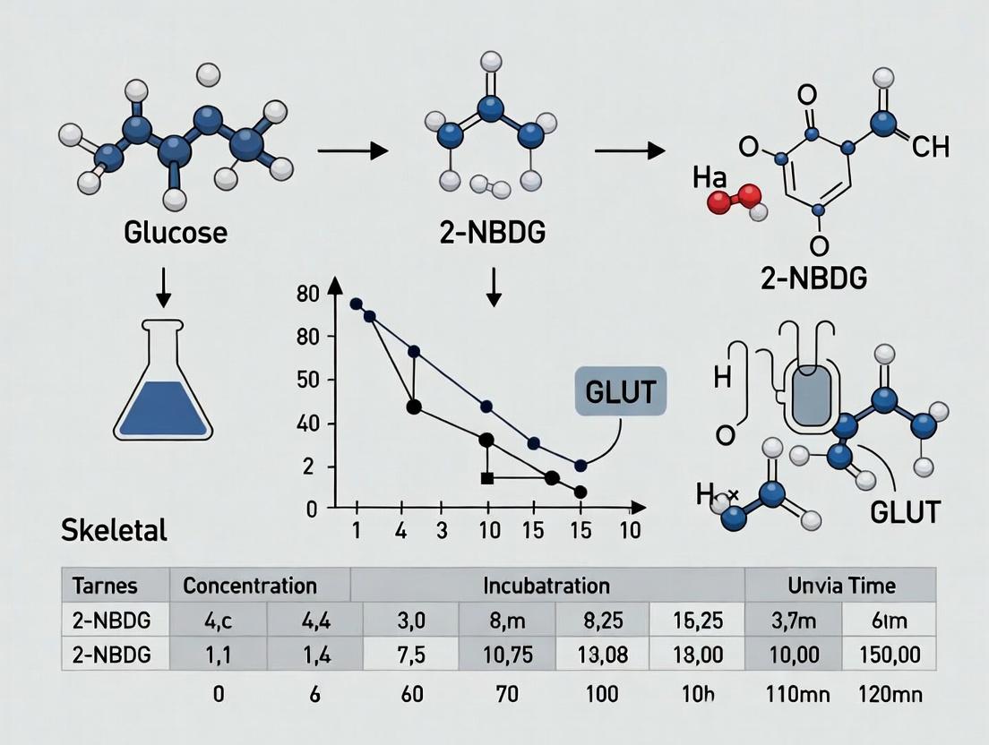
2-[N-(7-Nitrobenz-2-oxa-1,3-diazol-4-yl)Amino]-2-Deoxy-D-Glucose (2-NBDG) is a fluorescently labeled glucose analog widely used as a probe for monitoring glucose uptake in live cells. Its core mechanism involves competitive entry into cells via glucose transporters (primarily GLUTs) without undergoing significant metabolism, allowing for real-time, quantitative visualization of glucose transport activity. This application note details its properties, protocols, and specific considerations for optimizing concentration and incubation time in skeletal muscle cell research, a critical variable for studies on insulin resistance, metabolic disorders, and drug screening.
2-NBDG is synthesized by conjugating the fluorescent nitrobenzoxadiazole (NBD) moiety to the 2-position of deoxyglucose. Unlike 2-Deoxy-D-Glucose (2-DG), which is phosphorylated by hexokinase but not further metabolized (trapping it intracellularly), 2-NBDG is a poor substrate for hexokinase. Its primary utility stems from its non-metabolizable nature, ensuring that the measured fluorescence directly correlates with transporter-mediated uptake rather than downstream metabolic events.
Key Mechanistic Steps:
- Transport: 2-NBDG is competitively transported across the plasma membrane by facilitative glucose transporters (GLUT1, GLUT4, etc.).
- Limited Phosphorylation: It may undergo very minimal phosphorylation by hexokinase, but this is not a significant fate.
- Accumulation & Detection: The molecule accumulates in the cytosol, and its fluorescence (Excitation/Emission ~465/540 nm) can be detected via fluorescence microscopy, flow cytometry, or microplate readers.
- Efflux: Unlike trapped analogs, 2-NBDG can efflux from cells, necessitating careful timing of measurements.
Key Quantitative Parameters for Skeletal Muscle Cells
Optimal parameters vary by cell type (e.g., C2C12 myotubes, primary human myotubes) and experimental conditions (basal vs. insulin-stimulated). The following table summarizes findings from recent literature.
Table 1: Optimization of 2-NBDG Concentration and Incubation Time in Skeletal Muscle Models
| Cell Model | Experimental Condition | Recommended 2-NBDG Concentration | Optimal Incubation Time | Key Outcome / Rationale |
|---|---|---|---|---|
| C2C12 Myotubes (Differentiated) | Basal (No Insulin) | 100 µM | 30 minutes | Linear uptake phase; minimizes efflux. |
| C2C12 Myotubes (Differentiated) | Insulin-Stimulated (100 nM) | 50 - 100 µM | 15-20 minutes | Insulin effect is maximal; signal-to-noise ratio is high. |
| Primary Human Myotubes | Basal vs. Insulin | 150 µM | 40-60 minutes | Longer incubation compensates for potentially lower transporter activity. |
| L6 Rat Myotubes (GLUT4-myc) | Insulin Stimulation | 80 µM | 30 min | Validated for high-throughput screening of insulin mimetics. |
| General Guideline | Pilot Experiment | 30 - 300 µM Range | 10 - 60 min Time Course | Critical: A concentration and time course must be empirically established for each model system to ensure measurements are within the linear range of uptake. |
Detailed Experimental Protocols
Protocol 1: 2-NBDG Uptake Assay in Differentiated C2C12 Myotubes
This protocol is designed to compare basal and insulin-stimulated glucose uptake.
I. Materials & Reagent Solutions Table 2: Research Reagent Toolkit for 2-NBDG Assay
| Reagent/Material | Function/Explanation | Example Supplier/Cat. No. (or equivalent) |
|---|---|---|
| 2-NBDG (Powder) | Fluorescent glucose analog probe. | Thermo Fisher Scientific, Invitrogen N13195 |
| High-Glucose DMEM (No Phenol Red) | Assay medium; removes phenol red autofluorescence. | Gibco 31053-028 |
| Krebs-Ringer Phosphate (KRP) HEPES Buffer | Physiological buffer for serum/glucose starvation and assay. | Prepare in-house (see below). |
| Recombinant Human Insulin | Positive control stimulator of GLUT4 translocation. | Sigma-Aldrich I9278 |
| Cytochalasin B | GLUT transporter inhibitor; negative control. | Sigma-Aldrich C6762 |
| Black/Clear-bottom 96-well Plates | Optimal for fluorescence microplate reading. | Corning 3904 |
| Differentiated C2C12 Myotubes | Model of skeletal muscle glucose metabolism. | Cultured from C2C12 myoblasts (ATCC CRL-1772). |
| Fluorescence Microplate Reader | Instrument for quantitative endpoint reading. | Filter set: ~485 nm excitation / ~535 nm emission. |
II. Procedure
- Cell Preparation: Differentiate C2C12 myoblasts into myotubes in 96-well plates. Perform experiments on fully differentiated myotubes (day 5-7).
- Starvation: Wash cells 2x with warm, serum-free, low-glucose (or glucose-free) DMEM or KRP-HEPES buffer. Incubate in starvation medium for 2-3 hours to reduce basal signaling.
- Stimulation (Optional): Add insulin (e.g., 100 nM final concentration) or test compounds in assay buffer. Incubate for 20-30 min at 37°C.
- 2-NBDG Incubation:
- Prepare 2-NBDG working solution in warm, phenol-red-free assay buffer (e.g., 100 µM final concentration).
- Remove stimulation medium and immediately add the 2-NBDG solution.
- Incubate plates at 37°C for the predetermined optimal time (e.g., 30 minutes). Include control wells with 10 µM Cytochalasin B to define non-specific uptake.
- Termination & Washing:
- Rapidly aspirate the 2-NBDG solution.
- Wash cells 3 times quickly with ice-cold PBS to stop transport and remove extracellular probe.
- Detection:
- For endpoint reading: Lyse cells in 50-100 µL of lysis buffer (e.g., RIPA or 0.1% Triton X-100 in PBS). Transfer lysates to a black microplate and measure fluorescence.
- For live-cell imaging: After washing, add phenol-red-free imaging medium and image immediately using a FITC/GFP filter set.
III. Data Analysis Normalize fluorescence values to total protein content (e.g., BCA assay) or cell number. Specific uptake = (Total Uptake) - (Uptake in Cytochalasin B wells). Express insulin-stimulated uptake as a fold-change over basal.
Protocol 2: Real-Time 2-NBDG Uptake Kinetics using Live-Cell Imaging
This protocol is used to establish the linear phase of uptake for a new cell model.
- Prepare cells: Seed and differentiate cells in a glass-bottom 35-mm dish or 96-well imaging plate.
- Set up microscope: Equilibrate environmental chamber to 37°C and 5% CO₂. Use a 20x objective. Configure time-lapse acquisition (e.g., 1 image every 2 minutes for 60 min).
- Acquire baseline: Add pre-warmed assay buffer and acquire 2-3 images.
- Add probe: Quickly add 2-NBDG to the desired final concentration directly on the microscope stage without moving the dish. Start time-lapse acquisition immediately.
- Analyze: Use imaging software to quantify mean fluorescence intensity in the cytosolic region of cells over time. Plot fluorescence vs. time to identify the linear uptake phase.
Visualizations
Title: 2-NBDG Cellular Mechanism of Action
Title: 2-NBDG Uptake Assay Workflow for Muscle Cells
Critical Considerations for Thesis Research
- Efflux is Time-Sensitive: The optimal incubation time is a balance between sufficient signal and minimizing efflux. Pilot time-course experiments are non-negotiable.
- Concentration Saturation: High concentrations (>300 µM) can saturate transporters and mask subtle treatment effects. Use the lowest concentration that yields a robust signal.
- Normalization: Always normalize fluorescence to protein content or cell number. Consider using concurrent assays (e.g., immunoblotting for GLUT4 translocation) to validate findings.
- Specificity Controls: Cytochalasin B or pharmacological GLUT inhibitors are essential to confirm that uptake is transporter-mediated.
- Insulin Response Window: The fold-stimulation by insulin in muscle cells is often modest (2-4 fold) compared to adipocytes. Ensure experimental conditions (starvation time, insulin dose) are optimized to detect this change.
2-NBDG is a vital tool for investigating real-time glucose uptake in skeletal muscle cells. Successful application within a thesis context hinges on the systematic empirical determination of the critical parameters of concentration and incubation time for the specific cellular model, ensuring data reflects physiologically relevant transporter activity.
Why Use 2-NBDG in Skeletal Muscle Research? Advantages Over Radioactive 2-DG.
Application Notes
The study of glucose uptake in skeletal muscle is critical for understanding metabolic diseases like type 2 diabetes and insulin resistance. 2-Deoxy-D-glucose (2-DG) has been the classic tracer, but its radioactive nature ([³H] or [¹⁴C]) poses significant handling, disposal, and safety challenges. 2-(N-(7-Nitrobenz-2-oxa-1,3-diazol-4-yl)Amino)-2-Deoxyglucose (2-NBDG), a fluorescent glucose analog, offers a safe and effective alternative, particularly suited for real-time, live-cell imaging and high-throughput assays in skeletal muscle research.
Within the context of optimizing 2-NBDG concentration and incubation time for skeletal muscle cells (e.g., C2C12 myotubes, primary myotubes), its advantages are paramount. It enables researchers to perform kinetic studies in intact, living cells without the need for lysis or radioactive waste. This is crucial for dynamic experiments assessing acute insulin stimulation or drug effects over time.
Quantitative Comparison: 2-NBDG vs. Radioactive 2-DG
Table 1: Key Comparative Properties of Glucose Uptake Tracers
| Property | 2-NBDG (Fluorescent) | Radioactive 2-DG (e.g., [³H]2-DG) |
|---|---|---|
| Detection Method | Fluorescence microscopy, flow cytometry, plate readers. | Scintillation counting (requires cell lysis). |
| Temporal Resolution | Real-time, live-cell kinetic data possible. | Endpoint measurement only. |
| Spatial Resolution | Subcellular localization possible (microscopy). | No spatial information; whole-well/culture dish average. |
| Safety & Regulation | Non-radioactive; minimal biohazard, less regulation. | Radioactive; strict handling, storage, disposal protocols. |
| Throughput | High (compatible with 96/384-well plates). | Low to medium (scintillation counting is slower). |
| Primary Disadvantage | Potential photo-bleaching; fluorescence may be quenched. | Safety hazards; long-lived radioactive waste. |
| Typical Incubation Time (for skeletal muscle cells) | 10 min to 2 hours (optimizable in live cells). | Typically 10-60 min (endpoint). |
| Cost Considerations | Higher reagent cost per mg. | Lower reagent cost, but high overhead for licensing & disposal. |
Table 2: Suggested Optimization Range for 2-NBDG in Skeletal Muscle Cells (C2C12 Myotubes)
| Parameter | Typical Range Tested | Common Optimal Point (Literature Based) | Rationale |
|---|---|---|---|
| Concentration | 50 µM - 300 µM | 100 µM | Balances signal intensity with minimal disruption to native glucose transport. |
| Incubation Time | 5 min - 60 min | 20-30 min (for acute insulin stimulation) | Allows sufficient accumulation for robust signal while remaining within linear uptake phase. |
| Serum/BSA in Incubation Buffer | 0% - 0.5% BSA | 0.1% BSA | Reduces non-specific binding of 2-NBDG to surfaces and cells. |
| Pre-incubation in Low Glucose | 1 - 3 hours | 2 hours in 2 mM glucose DMEM | Depletes intracellular glucose to upregulate basal transport, enhancing signal-to-noise. |
Experimental Protocols
Protocol 1: Optimizing 2-NBDG Concentration and Time-Course in C2C12 Myotubes
Objective: To determine the linear range of 2-NBDG uptake and the optimal concentration for detecting insulin-stimulated glucose uptake.
Materials: See "The Scientist's Toolkit" below. Procedure:
- Cell Culture: Differentiate C2C12 myoblasts into myotubes in 96-well black-walled, clear-bottom plates. Maintain in differentiation medium (DMEM + 2% horse serum) for 5-7 days.
- Serum/Glucose Starvation: On the day of the experiment, wash myotubes twice with warm PBS. Pre-incubate in low-glucose (2 mM) DMEM containing 0.1% BSA for 2 hours at 37°C, 5% CO₂.
- Insulin Stimulation: Add insulin (or vehicle control) at a final concentration of 100 nM to relevant wells. Incubate for 20 minutes.
- 2-NBDG Incubation (Concentration Curve): Prepare 2-NBDG in pre-warmed uptake buffer (e.g., Krebs-Ringer-HEPES with 0.1% BSA) at concentrations: 50, 100, 150, 200, 300 µM. Remove insulin medium and immediately add 100 µL/well of 2-NBDG solutions. Incubate for 30 minutes at 37°C.
- 2-NBDG Incubation (Time-Course): Using the optimal concentration from step 4 (e.g., 100 µM), incubate separate wells for 5, 10, 20, 30, 45, and 60 minutes.
- Termination & Washing: Rapidly aspirate 2-NBDG solution. Wash cells three times quickly with ice-cold PBS to stop uptake and remove extracellular dye.
- Fluorescence Measurement: Add 100 µL of PBS per well. Measure fluorescence using a plate reader (Ex/Em ~465/540 nm).
- Normalization: Perform a protein assay (e.g., BCA) on replicate plates or use a DNA-binding fluorescent dye (like Hoechst 33342) in parallel plates for cell number normalization.
Protocol 2: Live-Cell Imaging of 2-NBDG Uptake in Primary Myotubes
Objective: To visualize real-time glucose uptake dynamics in response to insulin.
Procedure:
- Cell Preparation: Seed primary human or mouse myoblasts in glass-bottom imaging dishes. Differentiate into myotubes.
- Starvation & Dye Loading: Starve as in Protocol 1. Replace medium with imaging buffer containing 100 µM 2-NBDG.
- Image Acquisition: Place dish on a confocal or epifluorescence microscope with environmental control (37°C, 5% CO₂). Start time-lapse imaging immediately after adding 2-NBDG (1 frame every 1-2 minutes).
- Stimulation: After 5 minutes of baseline imaging, carefully add insulin directly to the dish to a final concentration of 100 nM without moving the dish. Continue imaging for an additional 25-30 minutes.
- Analysis: Quantify mean fluorescence intensity within regions of interest (ROIs) drawn over individual myotubes over time. Plot fluorescence intensity vs. time to visualize kinetic response.
Visualizations
Title: Endpoint 2-NBDG Uptake Assay Workflow
Title: Insulin-Stimulated GLUT4 Translocation & 2-NBDG Uptake Pathway
The Scientist's Toolkit
Table 3: Essential Reagents & Materials for 2-NBDG Uptake Assays
| Item | Function/Description | Key Consideration for Skeletal Muscle |
|---|---|---|
| 2-NBDG (Fluorescent D-Glucose Analog) | The core tracer. Competes with D-glucose for transport via GLUTs and hexokinase phosphorylation. | Use a high-purity, cell culture-grade reagent. Aliquot and store at -20°C protected from light. |
| C2C12 Cell Line or Primary Myoblasts | Standard in vitro skeletal muscle model. | Ensure full differentiation into contractile, multinucleated myotubes for physiologically relevant GLUT4 expression. |
| Differentiation Medium (DMEM + 2% Horse Serum) | Promotes myoblast fusion into myotubes. | Use low-serum (2% HS) to induce differentiation; high serum maintains proliferation. |
| Recombinant Human Insulin | Stimulates glucose uptake via the PI3K/Akt pathway, maximizing GLUT4 translocation. | Prepare a stock solution (e.g., 1 mM in weak acid) and dilute freshly for each experiment. |
| Black-Walled, Clear-Bottom 96-Well Plates | Optimal for fluorescence plate reading. | Black walls minimize cross-talk; clear bottoms allow for microscopic confirmation of differentiation. |
| Uptake Buffer (e.g., Krebs-Ringer-HEPES + 0.1% BSA) | Physiological salt solution for incubation steps. | Include 0.1% BSA to reduce non-specific 2-NBDG binding. Must be pre-warmed to 37°C. |
| Microplate Fluorescence Reader | Quantifies intracellular 2-NBDG fluorescence. | Requires appropriate filters (Ex ~460-490 nm, Em ~520-550 nm). Confirm linear detection range. |
| Confocal/Epifluorescence Microscope | For live-cell kinetic imaging and subcellular localization. | Must have environmental chamber for temperature/CO₂ control during time-lapse experiments. |
This application note details critical protocols for investigating glucose uptake in skeletal muscle cells, specifically using the fluorescent glucose analog 2-NBDG. The content supports a broader thesis investigating the optimization of 2-NBDG concentration and incubation time. Cellular uptake of glucose is primarily mediated by facilitative glucose transporters (GLUTs), with the metabolic state of the cell (e.g., insulin-stimulated vs. basal) serving as a key regulatory factor.
Key Concepts and Quantitative Data
Table 1: Common GLUT Isoforms in Skeletal Muscle and Their Characteristics
| GLUT Isoform | Km for Glucose (mM) | Primary Regulation | Role in Skeletal Muscle |
|---|---|---|---|
| GLUT1 | ~1-2 | Basal expression, hypoxia | Basal glucose uptake |
| GLUT4 | ~5 | Insulin, muscle contraction | Insulin-stimulated uptake |
| GLUT3 (if expressed) | ~1 | High affinity uptake | May support basal uptake |
Table 2: Example 2-NBDG Uptake Parameters from Literature
| Cell Type | Basal Uptake Incubation Time | Insulin-Stimulated Incubation Time | Common 2-NBDG Concentration Range | Key Finding |
|---|---|---|---|---|
| C2C12 Myotubes | 30 min | 20-30 min | 50-200 µM | Insulin increases uptake 1.5-3 fold |
| Primary Human Myotubes | 60 min | 30-60 min | 100 µM | High donor variability observed |
| L6 Myotubes | 20 min | 15-20 min | 100 µM | AMPK activation mimics insulin effect |
Experimental Protocols
Protocol 1: Optimizing 2-NBDG Incubation for Skeletal Muscle Cells
Objective: Determine the linear range of 2-NBDG uptake over time under basal and insulin-stimulated conditions. Materials: Differentiated C2C12 or L6 myotubes, 2-NBDG stock solution (in DMSO or buffer), Krebs-Ringer-Phosphate-HEPES (KRPH) buffer, insulin (100 nM final), fluorescence plate reader. Procedure:
- Cell Preparation: Culture and differentiate myoblasts into myotubes in 96-well black-walled, clear-bottom plates.
- Serum Starvation: Incubate cells in low-serum (0.5-1% FBS) or serum-free medium for 2-4 hours prior to assay.
- Stimulation: For insulin-treated wells, add KRPH buffer containing 100 nM insulin. For basal wells, add KRPH buffer alone. Incubate for 20 minutes at 37°C.
- 2-NBDG Uptake: Add 2-NBDG from a concentrated stock to achieve a final concentration of 100 µM directly to each well. Do not wash the stimulation buffer away.
- Time Course: Incubate plates at 37°C for varying times (e.g., 5, 10, 20, 30, 45, 60 minutes). Run all time points in parallel.
- Termination: Rapidly aspirate the 2-NBDG solution and wash cells 3x with ice-cold PBS.
- Lysis & Measurement: Lyse cells in 1% Triton X-100 in PBS. Transfer lysate to a new plate and measure fluorescence (Ex/Em ~465/540 nm).
- Normalization: Perform a protein assay (e.g., BCA) on lysates and express uptake as fluorescence units per µg protein.
Protocol 2: Assessing GLUT4 Translocation via Cell Surface Biotinylation
Objective: Correlate 2-NBDG uptake with plasma membrane GLUT4 content. Materials: Sulfosuccinimidyl-2-[biotinamido]ethyl-1,3-dithiopropionate (Sulfo-NHS-SS-Biotin), Quenching Solution (100 mM Glycine in PBS), Streptavidin Beads, Lysis Buffer (containing protease inhibitors), GLUT4 Antibody. Procedure:
- Treat Cells: Stimulate serum-starved myotubes (in 6-well plates) with/without insulin as in Protocol 1.
- Cell Surface Biotinylation: Place plates on ice, wash 2x with ice-cold PBS. Add Sulfo-NHS-SS-Biotin (0.5-1 mg/mL in PBS) to cover cells. Incubate for 30 min at 4°C with gentle rocking.
- Quench Reaction: Remove biotin solution and wash cells twice with cold Quenching Solution. Wash once more with cold PBS.
- Cell Lysis: Lyse cells in IP Lysis Buffer for 30 min on ice. Scrape and collect lysates. Clarify by centrifugation (14,000 x g, 10 min, 4°C).
- NeutrAvidin Pull-Down: Incubate equal amounts of lysate protein with pre-washed NeutrAvidin agarose beads for 1-2 hours at 4°C.
- Wash & Elute: Wash beads extensively with lysis buffer. Elute bound proteins (biotinylated surface proteins) by boiling in Laemmli sample buffer.
- Analysis: Subject eluates (surface fraction) and total cell lysate inputs to SDS-PAGE and Western blotting for GLUT4. Quantify band intensity.
Visualizations
Title: Insulin Signaling to GLUT4 Translocation (56 chars)
Title: 2-NBDG Uptake Assay Workflow (32 chars)
The Scientist's Toolkit
Table 3: Essential Research Reagents and Materials
| Item | Function/Application in Uptake Studies |
|---|---|
| 2-NBDG (2-(N-(7-Nitrobenz-2-oxa-1,3-diazol-4-yl)Amino)-2-Deoxyglucose) | Fluorescent D-glucose analog for direct visualization and quantification of cellular glucose uptake. |
| Differentiated Skeletal Myotubes (C2C12, L6, primary) | Physiologically relevant model system for studying insulin-responsive GLUT4 biology. |
| Recombinant Human Insulin | Gold-standard stimulus to induce GLUT4 translocation and maximize glucose uptake. |
| KRPH Buffer (Krebs-Ringer-Phosphate-HEPES) | Physiological salt buffer for starvation and uptake steps, maintaining cell viability. |
| Cell Surface Protein Isolation Kit (Biotinylation) | Isolates plasma membrane proteins to quantify translocation of GLUT4. |
| Phospho-Akt (Ser473) Antibody | Key readout for proximal insulin signaling pathway activation. |
| GLUT4 & GLUT1 Selective Antibodies | To determine transporter expression and membrane localization. |
| Cytochalasin B | GLUT inhibitor used in control experiments to confirm 2-NBDG uptake is transporter-mediated. |
| Black-walled, Clear-bottom Microplates | Optimized for fluorescence assays, minimizing cross-talk between wells. |
| Microplate Reader with Fluorescence Capability | Equipped with filters appropriate for 2-NBDG (Ex/Em ~465/540 nm). |
This review synthesizes established protocols for glucose uptake studies in skeletal muscle cell models, specifically the C2C12 mouse myoblast line and primary myotubes. Framed within a thesis investigating optimal 2-NBDG (2-[N-(7-Nitrobenz-2-oxa-1,3-diazol-4-yl)Amino]-2-Deoxyglucose) parameters, this document aims to provide a standardized reference for concentration and incubation time ranges. Accurate standardization is critical for comparative research in metabolism, insulin signaling, and drug development for conditions like diabetes and muscular dystrophy.
The following tables consolidate quantitative data from recent literature on standard 2-NBDG application in muscle cell models.
Table 1: Established 2-NBDG Concentrations and Incubation Times
| Cell Model | Differentiation Protocol (Days) | 2-NBDG Concentration Range (µM) | Standard Incubation Time Range (Minutes) | Serum/BSA During Assay? | Key Reference Context |
|---|---|---|---|---|---|
| C2C12 Myotubes | 4-7 days post-confluence | 50 - 200 µM | 30 - 60 min | Often 0.1-0.5% BSA | Basal & insulin-stimulated uptake |
| Primary Mouse Myotubes | 5-7 days in differentiation media | 50 - 150 µM | 30 - 90 min | Yes, low serum or BSA | Ex vivo muscle physiology mimicry |
| Primary Human Myotubes | 7-10 days | 100 - 300 µM | 60 - 120 min | Yes, low serum or BSA | Clinical translation studies |
Table 2: Common Experimental Modulators & Their Impact on 2-NBDG Protocols
| Modulator (Example) | Typical Pre-incubation Time | Effect on 2-NBDG Uptake | Protocol Adjustment Consideration |
|---|---|---|---|
| Insulin | 15-30 min | Increase (2-4 fold) | Shorter 2-NBDG incubations (30 min) often suffice to detect signal. |
| Metformin | 2-18 hours | Moderate increase | Requires longer pre-treatment; 2-NBDG incubation as standard. |
| TNF-α | 4-24 hours | Decrease (insulin resistance) | Longer treatment; ensure robust basal control. |
| Compound C (AMPK inhibitor) | 1-2 hours | Decrease | Confirm inhibitor stability over 2-NBDG incubation period. |
Detailed Experimental Protocols
Protocol 1: Standard 2-NBDG Uptake Assay in Differentiated C2C12 Myotubes
This protocol is adapted from common methodologies for measuring insulin-stimulated glucose uptake.
Materials:
- Differentiated C2C12 myotubes (4-7 days post-confluence in 24-well plate).
- Krebs-Ringer Phosphate HEPES (KRPH) buffer: 20 mM HEPES, 5 mM KH2PO4, 1 mM MgSO4, 1 mM CaCl2, 136 mM NaCl, 4.7 mM KCl, pH 7.4.
- 2-NBDG stock solution (e.g., 100 mM in DMSO). Aliquot and store at -20°C protected from light.
- Insulin stock solution (e.g., 100 µM in weak acid). Aliquot and store at -20°C.
- Phosphate-Buffered Saline (PBS).
- Cell lysis buffer (e.g., RIPA buffer).
- Fluorescence plate reader or flow cytometer.
Procedure:
- Preparation: Differentiate C2C12 myoblasts to myotubes in growth medium supplemented with 2% horse serum. Change media every 48 hours.
- Serum Starvation: On assay day, wash cells twice with warm PBS. Incubate in serum-free, low-glucose (or glucose-free) DMEM or KRPH buffer containing 0.1% BSA for 2-4 hours.
- Stimulation: For insulin-stimulated uptake, add insulin to a final concentration of 100 nM to designated wells. Incubate for 30 minutes at 37°C, 5% CO2.
- 2-NBDG Incubation: Prepare working solution of 2-NBDG in KRPH/0.1% BSA. A final concentration of 100 µM is commonly used. Replace medium in all wells with the 2-NBDG solution. Incubate for exactly 45 minutes at 37°C, protected from light.
- Termination & Wash: Aspirate 2-NBDG solution. Immediately wash cells three times with ice-cold PBS to stop uptake and remove extracellular probe.
- Lysis & Measurement: Lyse cells in 200-300 µL of ice-cold RIPA buffer for 10 minutes on ice. Scrape and transfer lysate to a microcentrifuge tube. Clarify by centrifugation (10,000 x g, 10 min, 4°C). Transfer supernatant to a black-walled 96-well plate. Measure fluorescence (Ex/Em ~465/540 nm). Normalize fluorescence to total protein content (e.g., via BCA assay).
Protocol 2: 2-NBDG Uptake in Primary Human Myotubes
Primary cells often require longer incubations due to lower uptake rates.
Procedure:
- Differentiation: Culture primary human myoblasts to ~90% confluence. Switch to differentiation medium (e.g., DMEM with 2% horse serum, 1x ITS). Differentiate for 7-10 days.
- Starvation & Stimulation: Follow steps 2-3 from Protocol 1, but extend serum starvation to 4-6 hours.
- 2-NBDG Incubation: Use a final 2-NBDG concentration of 150-200 µM. Incubate for 90 minutes at 37°C, protected from light.
- Wash & Analysis: Perform rapid, ice-cold washes. Cells can also be trypsinized gently and analyzed via flow cytometry for single-cell resolution, or lysed for plate reading.
Signaling Pathways & Experimental Workflows
Title: Key Signaling Pathways Affecting 2-NBDG Uptake in Muscle Cells
Title: Standard 2-NBDG Uptake Assay Workflow
The Scientist's Toolkit: Research Reagent Solutions
Table 3: Essential Materials for Muscle Cell Glucose Uptake Studies
| Item | Function & Specification | Example Vendor/Cat. No. (Representative) |
|---|---|---|
| C2C12 Cell Line | Mouse skeletal myoblast model for reproducible differentiation into myotubes. | ATCC CRL-1772 |
| Primary Myoblast Media | Specialized growth media optimized for human or mouse primary myoblast proliferation. | SkGM BulletKit (Lonza) |
| 2-NBDG | Fluorescent D-glucose analog for direct uptake measurement without radioactivity. | Cayman Chemical 11046, Thermo Fisher Scientific N13195 |
| Insulin (Human Recombinant) | Gold-standard stimulus for insulin signaling pathway and GLUT4 translocation. | Sigma-Aldrich I2643 |
| Differentiation Serum | Low-mitogen serum (e.g., Horse Serum) to trigger cell cycle exit and fusion. | Gibco 26050088 |
| KRPH/HEPES Assay Buffer | Physiologically balanced buffer for acute assays, maintaining pH and ion gradients. | Can be prepared in-lab or purchased as components. |
| Black-walled Clear-bottom Plates | Optimal for fluorescence readouts while allowing microscopic confirmation of monolayers. | Corning 3603 |
| RIPA Lysis Buffer | Efficient lysis buffer for total protein extraction and subsequent fluorescence/protein quantification. | Cell Signaling Technology #9806 |
| BCA Protein Assay Kit | Colorimetric method for accurate protein concentration determination for normalization. | Thermo Fisher Scientific 23225 |
| AMPK/Insulin Pathway Inhibitors/Activators | Pharmacological tools (e.g., Compound C, AICAR) to dissect signaling mechanisms. | Tocris, Sigma-Aldrich |
Step-by-Step Protocol: Determining Optimal 2-NBDG Concentration and Incubation Time
This protocol details the experimental design for determining the optimal concentration and incubation time of 2-[N-(7-Nitrobenz-2-oxa-1,3-diazol-4-yl)amino]-2-deoxy-D-glucose (2-NBDG), a fluorescent glucose analog, for uptake studies in skeletal muscle cells. Within the broader thesis on glucose metabolism in myotubes, these assays are critical for establishing parameters that ensure measurements are within the linear range of uptake, avoiding saturation or sub-optimal detection, thereby enabling accurate assessment of interventions affecting GLUT4 translocation and insulin signaling.
Key Research Reagent Solutions & Materials
| Item | Function / Explanation |
|---|---|
| 2-NBDG | Fluorescent deoxyglucose analog. Serves as a tracer for real-time, non-radioactive monitoring of cellular glucose uptake. |
| Differentiated C2C12 or Human Skeletal Muscle Myotubes | Standard in vitro model for skeletal muscle metabolism and insulin response. |
| Krebs-Ringer Phosphate (KRP) or HEPES Buffered Saline | Assay buffer for maintaining physiological pH and ion balance during uptake experiments. |
| Insulin (e.g., Humulin R) | Positive control to stimulate GLUT4 translocation and maximize glucose uptake. |
| Cytochalasin B | GLUT transporter inhibitor. Serves as a negative control to confirm uptake is transporter-mediated. |
| Cell Lysis Buffer (RIPA) | For lysing cells to extract intracellular 2-NBDG in plate-based assays. |
| Black/Clear-Bottom 96-well Plates | Optimal for fluorescence readouts in microplate readers. |
| Microplate Fluorescence Reader | Equipped with ~465 nm excitation and ~540 nm emission filters for 2-NBDG detection. |
| PBS (without Glucose) | For washing cells to terminate uptake and remove extracellular 2-NBDG. |
Detailed Experimental Protocols
Cell Culture and Differentiation
- Culture: Maintain C2C12 myoblasts in growth medium (DMEM + 10% FBS + 1% Pen/Strep) at 37°C, 5% CO₂.
- Differentiation: At ~90% confluence, switch to differentiation medium (DMEM + 2% Horse Serum). Change medium every 24-48 hours.
- Maturation: Use myotubes differentiated for 5-7 days for experiments. Confirm differentiation via morphology (multinucleated, fused cells).
Kinetic Time-Course Assay (5-120 min)
Objective: Determine the linear range of 2-NBDG uptake over time at a fixed, intermediate concentration. Protocol:
- Serum/Gluose Starvation: Wash differentiated myotubes in 96-well plates with PBS. Incubate in low-glucose (2.5 mM) or glucose-free assay buffer for 2 hours to deplete endogenous glucose and serum-starve.
- Stimulation (Optional): Include wells pre-treated with 100 nM insulin for 20 min to assess stimulated uptake kinetics.
- Uptake Initiation: Add assay buffer containing a fixed concentration of 2-NBDG (e.g., 100 μM). Ensure consistent volume across wells.
- Time Points: Incubate for 5, 15, 30, 60, 90, and 120 minutes at 37°C. Include a 0-minute time point (add buffer, then immediately wash) for background subtraction.
- Termination: At each time point, rapidly aspirate 2-NBDG buffer and wash wells 3x with ice-cold PBS.
- Lysis & Measurement: Lyse cells in 100 μL RIPA buffer (30 min, 4°C). Transfer 80 μL of lysate to a black plate. Measure fluorescence (Ex/Em ~465/540 nm).
- Normalization: Measure protein concentration (BCA assay) of remaining lysate. Express uptake as Fluorescence Units per μg protein or per well.
Concentration-Response Assay (50-300 μM)
Objective: Establish the relationship between extracellular 2-NBDG concentration and cellular uptake over a fixed, linear time. Protocol:
- Starvation: As in 3.2.
- Concentration Series: Prepare 2-NBDG in assay buffer at concentrations: 50, 100, 150, 200, 250, 300 μM.
- Controls: Include wells with: a) No 2-NBDG (Blank), b) High 2-NBDG + 50 μM Cytochalasin B (non-specific uptake control).
- Uptake Period: Based on kinetic results (e.g., 30 min), incubate cells with the concentration series for the selected linear time.
- Termination & Measurement: As in 3.2, steps 5-7.
Data Presentation & Analysis
Table 1: Hypothetical Kinetic Time-Course Data (2-NBDG at 100 μM)
| Incubation Time (min) | Basal Uptake (FU/μg protein) | Insulin-Stimulated Uptake (FU/μg protein) | Fold Stimulation (Insulin/Basal) |
|---|---|---|---|
| 5 | 152 ± 18 | 285 ± 32 | 1.9 |
| 15 | 410 ± 45 | 1050 ± 98 | 2.6 |
| 30 | 780 ± 67 | 2050 ± 210 | 2.6 |
| 60 | 1250 ± 120 | 3100 ± 285 | 2.5 |
| 90 | 1550 ± 150 | 3750 ± 320 | 2.4 |
| 120 | 1800 ± 165 | 4100 ± 405 | 2.3 |
FU: Fluorescence Units. Data suggests linear uptake up to ~60 min under basal conditions.
Table 2: Hypothetical Concentration-Response Data (30 min Incubation)
| [2-NBDG] (μM) | Basal Uptake (FU/μg protein) | Insulin-Stimulated Uptake (FU/μg protein) | Net Specific Uptake* (FU/μg protein) |
|---|---|---|---|
| 50 | 395 ± 38 | 1020 ± 95 | 985 ± 90 |
| 100 | 780 ± 67 | 2050 ± 210 | 2020 ± 205 |
| 150 | 1150 ± 105 | 2950 ± 275 | 2910 ± 270 |
| 200 | 1480 ± 135 | 3610 ± 335 | 3560 ± 330 |
| 250 | 1750 ± 160 | 4150 ± 390 | 4100 ± 385 |
| 300 | 1950 ± 180 | 4500 ± 420 | 4450 ± 415 |
*Net Specific Uptake = (Total Uptake) - (Uptake in Cytochalasin B control). Data shows a near-linear increase up to 200-250 μM, suggesting non-saturating conditions within this range.*
Analysis:
- Plot time-course data to select a linear time window.
- Plot concentration-response data. Use Michaelis-Menten or linear regression to analyze kinetics.
- Statistical tests: Use two-way ANOVA (factors: time & treatment; concentration & treatment) with appropriate post-hoc tests.
Visualizations
Diagram 1: Signaling & Experimental Logic for 2-NBDG Uptake
Diagram 2: Experimental Workflow for Dual Assay
Application Notes: Context within Skeletal Muscle Metabolism Research
Investigating glucose metabolism in skeletal muscle tissue, particularly in the context of disease models or therapeutic screening, requires a reliable in vitro system of differentiated, contractile myotubes. This protocol for differentiating C2C12 mouse myoblasts into myotubes is designed to produce a consistent cellular model for subsequent metabolic assays. The primary application here is to establish the cellular foundation for optimizing 2-NBDG (2-(N-(7-Nitrobenz-2-oxa-1,3-diazol-4-yl)Amino)-2-Deoxyglucose) uptake experiments. Key parameters such as the differentiation status of the myotubes (fusion index, contractility) directly influence glucose analog uptake rates. Therefore, standardized preparation of contractile myotubes is a critical prerequisite for determining the optimal 2-NBDG concentration and incubation time to accurately reflect GLUT4-mediated glucose transport activity in skeletal muscle cells.
Detailed Protocol: C2C12 Myoblast Culture and Differentiation
2.1 Materials and Reagent Preparation
- Growth Medium (GM): Dulbecco's Modified Eagle Medium (DMEM) supplemented with 10% Fetal Bovine Serum (FBS), 4 mM L-glutamine, and 1% Penicillin-Streptomycin.
- Differentiation Medium (DM): Dulbecco's Modified Eagle Medium (DMEM) supplemented with 2% Horse Serum (HS), 4 mM L-glutamine, and 1% Penicillin-Streptomycin.
- Phosphate-Buffered Saline (PBS), sterile.
- 0.25% Trypsin-EDTA solution.
- Tissue culture-treated plates.
- Humidified incubator at 37°C, 5% CO₂.
2.2 Protocol Steps
- Thawing and Maintenance: Rapidly thaw cryopreserved C2C12 myoblasts in a 37°C water bath. Transfer to a pre-warmed GM in a T-25 flask. Change medium every 2 days until cells reach 70-80% confluency. Do not allow cells to reach 100% confluency prior to differentiation, as this can impair myogenic potential.
- Passaging: Wash cells with PBS, detach using 0.25% Trypsin-EDTA (3-5 min, 37°C). Neutralize with GM containing FBS. Centrifuge at 200 x g for 5 min, resuspend in GM, and seed at a 1:5 to 1:10 split ratio.
- Seeding for Differentiation: For differentiation experiments, seed cells at a density of 15,000 – 20,000 cells/cm² in GM. For a 24-well plate, this equates to approximately 30,000 – 40,000 cells per well. Allow cells to adhere overnight.
- Initiation of Differentiation: Once cells reach 95-100% confluency (Day 0), aspirate GM and gently wash once with PBS to remove residual serum. Replace with pre-warmed Differentiation Medium (DM).
- Differentiation Phase: Replace the DM every 24-48 hours. Multinucleated myotubes will begin to form within 48-72 hours and will become increasingly extensive and contractile over 5-7 days.
- Maturation & Contractility: Myotubes typically exhibit spontaneous contractions after 5-7 days in DM. For metabolic assays like 2-NBDG uptake, myotubes differentiated for 5-7 days are recommended.
Key Metrics for Differentiation Success
Table 1: Quantitative Metrics for Differentiated C2C12 Myotubes
| Metric | Measurement Method | Expected Outcome (Day 5-7) | Impact on 2-NBDG Assay |
|---|---|---|---|
| Fusion Index | (% nuclei within myosin-heavy chain positive myotubes). Immunostaining for MYH. | 60-80% | Higher fusion correlates with greater GLUT4 expression and basal uptake. |
| Myotube Diameter | Phase-contrast or stained image analysis (µm). | 20-40 µm | Larger diameter indicates maturity and increased cytoplasmic volume. |
| Spontaneous Contractility | Visual observation under microscope. | Present in >70% of wells | Confirms functional maturation; contractile activity influences metabolic demand. |
| GLUT4 Localization | Immunofluorescence (basal vs. insulin-stimulated). | Primarily perinuclear at rest; translocates upon stimulation. | Validates system's responsiveness for insulin-dependent 2-NBDG uptake studies. |
Protocol for Validation: Immunofluorescence for Myosin Heavy Chain (MYH)
- Fixation: On Day 5-7, aspirate medium, wash with PBS, and fix with 4% paraformaldehyde for 15 min at room temperature.
- Permeabilization & Blocking: Wash with PBS, permeabilize with 0.1% Triton X-100 for 10 min. Block with 3% BSA in PBS for 1 hour.
- Primary Antibody Incubation: Incubate with anti-Myosin Heavy Chain (MYH) primary antibody (diluted in 1% BSA/PBS) overnight at 4°C.
- Secondary Antibody & Nuclear Stain: Wash, incubate with fluorophore-conjugated secondary antibody for 1 hour at RT in the dark. Incubate with DAPI (1 µg/mL) for 5 min.
- Imaging & Analysis: Wash and image using a fluorescence microscope. Calculate Fusion Index: (Number of nuclei within MYH+ structures / Total number of nuclei) x 100%.
Visualizations
5.1 Diagram: C2C12 Differentiation Workflow to 2-NBDG Assay
Title: Myoblast to Myotube Differentiation Workflow
5.2 Diagram: Key Signaling in Myogenic Differentiation
Title: Core Signaling for Muscle Cell Differentiation
The Scientist's Toolkit: Essential Research Reagents
Table 2: Key Reagent Solutions for Myoblast Differentiation & Metabolic Assay
| Reagent/Solution | Function & Rationale |
|---|---|
| High-Glucose DMEM | Standard culture medium providing energy and carbon source for proliferating and differentiating muscle cells. |
| Fetal Bovine Serum (FBS) | Rich in growth factors; essential for myoblast proliferation in Growth Medium. |
| Horse Serum (HS) | Lower mitogen content than FBS; induces cell cycle exit and initiates differentiation in Differentiation Medium. |
| Penicillin-Streptomycin | Antibiotic-antimycotic to prevent bacterial and fungal contamination in long-term cultures. |
| 0.25% Trypsin-EDTA | Proteolytic enzyme chelator solution for adherent cell detachment during passaging. |
| 2-NBDG | Fluorescent glucose analog used to track and quantify cellular glucose uptake in live myotubes. |
| Insulin (Recombinant) | Positive control stimulus to induce GLUT4 translocation and maximal glucose uptake in validation experiments. |
| Anti-Myosin Heavy Chain (MYH) Antibody | Primary antibody for immunofluorescence validation of successful myogenic differentiation. |
| DAPI Stain | Nuclear counterstain for calculating fusion index and assessing cell density. |
Within the broader thesis investigating the optimization of 2-NBDG concentration and incubation time for glucose uptake studies in skeletal muscle cells (e.g., C2C12 myotubes), the execution of the assay is critical. The protocol must ensure cellular synchronization in a low-glucose state via serum-starvation, followed by precise tracer incubation and stringent washing to minimize non-specific background, thereby yielding quantifiable and reproducible data on insulin-stimulated GLUT4 translocation and glucose uptake.
Key Research Reagent Solutions
| Reagent/Material | Function in Assay |
|---|---|
| 2-NBDG (2-(N-(7-Nitrobenz-2-oxa-1,3-diazol-4-yl)Amino)-2-Deoxyglucose) | Fluorescent glucose analog. Competes with D-glucose for cellular uptake via GLUT transporters, serving as the direct tracer for quantification. |
| Low-Glucose (or Glucose-Free) Serum-Free Medium | Serum-starvation medium. Deprives cells of growth factors and reduces basal glucose, synchronizing metabolism and enhancing insulin response sensitivity. |
| Krebs-Ringer Phosphate (KRP) or HEPES-Buffered Saline (HBS) | Physiological buffer for the incubation and washing steps. Maintains pH and ion balance during the assay outside a CO₂ incubator. |
| Insulin (e.g., Human Recombinant) | Primary agonist. Binds to insulin receptor, triggering the PI3K/Akt signaling cascade to translocate GLUT4 vesicles to the plasma membrane. |
| Cytochalasin B | Competitive inhibitor of glucose transport. Used in control wells to confirm 2-NBDG uptake is transporter-mediated. |
| Phosphate-Buffered Saline (PBS), Ice-Cold | Washing solution. Halts cellular metabolism and removes extracellular 2-NBDG. Cold temperature reduces membrane fluidity and internalization. |
| Cell Lysis Buffer (e.g., RIPA or 1% SDS) | For lysing cells to extract intracellular 2-NBDG for fluorometric plate reading if not using direct imaging. |
| Formaldehyde (4% in PBS) | Fixative for terminating the assay and preserving cells for subsequent microscopy, if required. |
Table 1: Reported 2-NBDG Concentrations & Incubation Times for Skeletal Muscle Cells
| Cell Model | Serum-Starvation Duration | 2-NBDG Concentration Range | Incubation Time Range | Key Purpose | Reference Trend (Year) |
|---|---|---|---|---|---|
| C2C12 Myotubes | 2-4 hours | 50 - 200 µM | 15 - 60 min | Baseline uptake kinetics | Common (2015-2020) |
| C2C12 Myotubes | Overnight (12-16h) | 100 - 300 µM | 30 - 120 min | Insulin-stimulated uptake | Prevalent (2018-2023) |
| Primary Human Myotubes | 3-4 hours | 100 µM | 60 min | Drug screening assays | Recent (2021-2024) |
| L6 Myotubes | 2-3 hours | 40 - 80 µM | 20 - 40 min | High-throughput screening | Established (2016-2022) |
| Thesis Optimization Range | 2, 4, 6, 16h | 50, 100, 200, 300 µM | 15, 30, 60, 90 min | Determine linear range & S/N | Current Study |
Detailed Experimental Protocols
Protocol 1: Serum-Starvation of Skeletal Muscle Cells Objective: To synchronize cells in a quiescent metabolic state and reduce basal glucose uptake.
- Differentiate C2C12 myoblasts into myotubes in standard growth medium (high glucose DMEM + 10% FBS + 2% horse serum).
- Aspirate differentiation medium and wash cells once with warm, sterile PBS.
- Add pre-warmed, low-glucose (or glucose-free), serum-free medium (e.g., DMEM base with 0.5-1.0 g/L glucose and 0.5% BSA).
- Incubate cells for the predetermined optimization period (e.g., 2, 4, 6, or 16 hours) in a standard cell culture incubator (37°C, 5% CO₂).
- Proceed immediately to the 2-NBDG incubation assay.
Protocol 2: 2-NBDG Uptake Assay with Insulin Stimulation Objective: To measure insulin-stimulated glucose transporter activity.
- Prepare Assay Buffer: Krebs-Ringer Phosphate (KRP) buffer, pH 7.4, supplemented with 0.5% BSA. Warm to 37°C.
- Pre-Treatment (Optional): After starvation, treat cells with inhibitors or drug candidates in assay buffer for desired time.
- Stimulation: Add insulin (typical final concentration 100 nM) or vehicle control to respective wells. Incubate for 20-30 minutes at 37°C.
- 2-NBDG Incubation:
- Prepare 2-NBDG working solutions in warm assay buffer at the desired concentrations (e.g., 50, 100, 200, 300 µM).
- Critical Control: Include wells with 10-50 µM Cytochalasin B.
- Rapidly aspirate insulin/vehicle solution and immediately add the 2-NBDG solution.
- Incubate plates in the dark at 37°C for the optimized time (e.g., 15-90 min). Do not use CO₂ incubation during this step.
- Termination & Washing:
- Aspirate the 2-NBDG solution.
- Immediately wash cells three times with generous volumes of ice-cold PBS.
- Work quickly to arrest cellular uptake.
- Quantification:
- Method A (Lysis): Add 1% SDS lysis buffer, shake for 10 min. Transfer lysates to a black-walled microplate, measure fluorescence (Ex/Em ~465/540 nm).
- Method B (Imaging): Fix cells with 4% formaldehyde for 15 min, wash, and image using a fluorescence microscope/plate reader.
Visualizations
This application note compares flow cytometry and fluorescence microscopy for quantifying 2-NBDG uptake in skeletal muscle cells (e.g., C2C12, primary myotubes). The broader thesis investigates the optimization of 2-NBDG concentration (µM range) and incubation time (minutes to hours) to accurately assess glucose uptake under various metabolic conditions (e.g., insulin stimulation, drug treatment). Selecting the appropriate quantification method is critical for generating reliable, statistically robust data.
Table 1: Method Comparison for Quantitative 2-NBDG Analysis
| Feature | Flow Cytometry | Fluorescence Microscopy (Widefield/Confocal) |
|---|---|---|
| Primary Output | Population-level, single-cell fluorescence intensity. | Spatial, single-cell or subcellular fluorescence distribution. |
| Throughput | Very High (10,000+ cells/sec). | Low to Medium (10s-100s of cells/field). |
| Quantitative Rigor | Excellent for population statistics (mean fluorescence intensity, CV). | Good for single cells; requires careful background subtraction. |
| Spatial Information | None. | Excellent (membrane vs. cytoplasmic localization). |
| Sample Requirement | Suspension cells or detached monolayers. | Adherent cells on imaging-optimized dishes. |
| Key Metric for 2-NBDG | Population MFI, % positive cells above threshold. | Integrated cell fluorescence intensity, mean intensity/area. |
| Best for Thesis Context | High-throughput screening of multiple conditions/time points. | Validating homogeneous uptake, checking for artifacts, morphology correlation. |
Table 2: Typical Optimized 2-NBDG Parameters for Skeletal Muscle Cells
| Parameter | Recommended Range | Notes |
|---|---|---|
| 2-NBDG Concentration | 50 – 200 µM | Lower for microscopy to reduce background; higher for flow. |
| Incubation Time | 20 – 60 minutes | Time-course essential; linear range varies by cell type & treatment. |
| Serum/Glucose Starvation | 1-3 hours in low-glucose, serum-free media | Standardizes basal uptake. |
| Insulin Control (Positive) | 100 nM, 15-30 min pre-/co-incubation | Validates assay responsiveness. |
| Inhibition Control (Negative) | Cytochalasin B (10-20 µM) | GLUT inhibitor confirms specific uptake. |
Experimental Protocols
Protocol 1: Flow Cytometry for 2-NBDG Uptake in C2C12 Myotubes
Title: High-Throughput Quantification of Glucose Uptake.
Key Reagent Solutions:
- 2-NBDG Stock: 10 mM in DMSO. Aliquot and store at -20°C protected from light.
- Krebs-Ringer-Phosphate-HEPES (KRPH) Buffer: For starvation and assay. Contains 20 mM HEPES, 5 mM phosphate, 1 mM MgSO4, 1 mM CaCl2, 136 mM NaCl, 4.7 mM KCl, pH 7.4.
- Trypsin-EDTA Solution: 0.25%, for gentle cell detachment.
- Fixation Solution: 4% formaldehyde in PBS (optional, reduces biosafety concern).
- Insulin Stimulation Control: 100 µM stock in weak acid (e.g., 10 mM HCl).
Procedure:
- Cell Culture: Differentiate C2C12 myoblasts into myotubes in 6- or 12-well plates.
- Starvation: Wash 2x with warm KRPH. Incubate in KRPH for 60 min at 37°C.
- Stimulation & Labeling:
- Add inhibitors/activators (e.g., insulin) in KRPH for desired time.
- Add 2-NBDG from stock to final concentration (e.g., 100 µM). Incubate 30 min at 37°C, protected from light.
- Termination & Harvest:
- Aspirate media. Wash 3x rapidly with ice-cold PBS.
- Gently detach cells using trypsin-EDTA. Neutralize with complete media.
- Pellet cells (300 x g, 5 min). Wash with ice-cold PBS + 0.5% BSA.
- (Optional) Fix cells in 4% formaldehyde for 15 min on ice, then wash.
- Acquisition: Resuspend in cold PBS. Keep on ice, protected from light. Acquire data on flow cytometer using a 488 nm laser and FITC/GFP filter set (e.g., 530/30 nm). Collect data for ≥10,000 single-cell events.
- Analysis: Gate on live, single cells. Plot fluorescence histogram. Compare Mean Fluorescence Intensity (MFI) or GeoMean between conditions. Set negative control (Cytochalasin B or no 2-NBDG) threshold.
Protocol 2: Quantitative Fluorescence Microscopy for 2-NBDG
Title: Spatial Analysis of Glucose Uptake in Myotubes.
Key Reagent Solutions:
- Imaging Media: Phenol-red free, low-fluorescence medium or KRPH buffer.
- Nuclear Stain: Hoechst 33342 (2 µg/mL) or DAPI.
- Mounting Medium: Antifade mounting medium if fixing samples.
- CellMask Deep Red Plasma Membrane Stain: Optional for segmentation.
Procedure:
- Cell Preparation: Seed/differentiate cells in µ-Slide or glass-bottom dishes. Include control wells on the same plate.
- Starvation & Labeling: As in Protocol 1, but use imaging-optimized dishes.
- Washing & Live-Cell Imaging:
- After 2-NBDG incubation, wash 3x with warm, dye-free imaging media.
- Immediately image in fresh imaging media. For live-cell imaging, complete within 20 min.
- Imaging Settings: Use FITC/GFP channel for 2-NBDG (Ex/Em ~488/540 nm). Use DAPI channel for nuclear stain. Use consistent exposure time, gain, and light intensity across all conditions.
- Alternative - Fixed-Cell Imaging: After washing, fix with 4% PFA for 15 min at RT. Wash 3x with PBS. Add nuclear stain, then mount with antifade medium. Seal and image.
- Image Analysis:
- Use software (e.g., ImageJ/FIJI, CellProfiler).
- Subtract background fluorescence (from a cell-free region).
- Segment individual cells using nuclear or membrane stains.
- Measure Integrated Density (sum of pixel intensities) or Mean Fluorescence Intensity per cell.
- Normalize to cell area if morphology varies. Analyze ≥100 cells per condition from multiple fields.
Visualization: Experimental Workflow and Pathway
Diagram 1: 2-NBDG Uptake and Quantification Workflow
Diagram 2: Insulin Signaling to 2-NBDG Readout
The Scientist's Toolkit: Essential Research Reagents
Table 3: Key Reagents for 2-NBDG Uptake Experiments
| Reagent | Function in Experiment | Example Product/Catalog Number (Typical) |
|---|---|---|
| 2-NBDG (2-(N-(7-Nitrobenz-2-oxa-1,3-diazol-4-yl)Amino)-2-Deoxyglucose) | Fluorescent glucose analog for direct uptake measurement. | Thermo Fisher Scientific N13195; Cayman Chemical 11046 |
| Insulin (Human Recombinant) | Positive control stimulator of GLUT4 translocation. | Sigma-Aldrich I9278 |
| Cytochalasin B | Potent inhibitor of glucose transporters; negative control. | Sigma-Aldrich C6762 |
| Phenol-Red Free Imaging Medium | Reduces background autofluorescence for microscopy. | Gibco 21063029 |
| Cell Dissociation Reagent (Trypsin-EDTA) | Gently detaches adherent myotubes for flow cytometry analysis. | Gibco 25200056 |
| Fetal Bovine Serum (FBS) | For cell culture and differentiation media. | Qualified, low IgG varieties preferred. |
| Matrigel or Collagen Coating | Enhances adhesion and differentiation of skeletal muscle cells. | Corning 356231 |
| Hoechst 33342 | Cell-permeant nuclear counterstain for microscopy. | Thermo Fisher Scientific H3570 |
| Flow Cytometry Calibration Beads | Ensures day-to-day instrument consistency for MFI comparison. | Spherotech ACCUCOUNT Beads |
Accurate quantification of glucose uptake using the fluorescent glucose analog 2-(N-(7-Nitrobenz-2-oxa-1,3-diazol-4-yl)Amino)-2-Deoxyglucose (2-NBDG) is fundamental for metabolic research in skeletal muscle cells. A comprehensive thesis on optimizing 2-NBDG concentration and incubation time must be built upon a foundation of robust experimental controls. This protocol details the implementation of three essential controls: 1) Insulin Stimulation (positive control for maximum glucose transporter (GLUT4) translocation), 2) Cytochalasin B Inhibition (negative control for GLUT-mediated transport), and 3) Background Fluorescence (control for non-specific cellular uptake and dye binding). These controls are critical for validating the specificity of the 2-NBDG signal and for normalizing data across experiments.
Key Experimental Protocols
2.1 Cell Culture and Preparation
- Cell Line: Differentiated C2C12 mouse myotubes or primary human skeletal muscle myotubes.
- Differentiation: Grow C2C12 myoblasts to confluence and switch to differentiation medium (DMEM with 2% horse serum). Culture for 4-6 days, changing medium every 48 hours, until >80% multinucleated myotubes are formed.
- Serum Starvation: Prior to experiment, wash cells and incubate in serum-free, low-glucose (5.5 mM) DMEM for 3-4 hours to baseline cellular insulin signaling.
2.2 Control Treatment Protocols
- A. Insulin Stimulation (Positive Control):
- After starvation, treat cells with 100 nM insulin prepared in serum-free, low-glucose medium.
- Incubate at 37°C, 5% CO₂ for 20 minutes to maximally stimulate GLUT4 translocation to the plasma membrane.
- Proceed directly to the 2-NBDG uptake assay.
B. Cytochalasin B Inhibition (Negative Control):
- Prepare a 50 µM stock of Cytochalasin B in DMSO.
- After starvation, pre-incubate cells with 10 µM Cytochalasin B (final concentration) in serum-free medium for 30 minutes at 37°C, 5% CO₂.
- Maintain Cytochalasin B in the medium during the subsequent 2-NBDG incubation step.
C. Background Fluorescence Control:
- For each condition (Basal, Insulin, Cytochalasin B), include a parallel set of wells treated identically but incubated with a 20-fold excess of unlabeled 2-Deoxy-D-glucose (2-DG) alongside 2-NBDG.
- Alternatively, incubate separate wells with 2-NBDG at 4°C (on ice) to inhibit all active transport processes.
2.3 2-NBDG Uptake Assay Protocol
- Solution Preparation: Prepare 2-NBDG working solution (e.g., 100 µM) in pre-warmed, serum-free, low-glucose medium. Protect from light.
- Uptake Incubation: Aspirate treatment media and immediately add the 2-NBDG-containing medium. Incubate plates at 37°C, 5% CO₂ for the optimized time (e.g., 20 minutes) determined from time-course experiments. Include the relevant inhibitors (Cytochalasin B, excess 2-DG) where required for control wells.
- Termination & Washing: Rapidly aspirate 2-NBDG medium and wash cells three times with ice-cold PBS (containing 0.1% BSA for first wash, then PBS alone).
- Lysis & Measurement: Lyse cells in RIPA buffer or 0.1% SDS. Transfer lysates to a black-walled microplate. Measure fluorescence (Excitation: ~465-475 nm, Emission: ~540-550 nm) using a plate reader.
- Protein Normalization: Determine protein concentration of each lysate using a BCA or Bradford assay.
Data Presentation
Table 1: Summary of Essential Control Values in a 2-NBDG Uptake Assay (Representative Data)
| Experimental Condition | Mean Fluorescence (RFU/µg protein) | Normalized Uptake (% of Basal) | Purpose & Interpretation |
|---|---|---|---|
| Background (4°C or + 2-DG) | 150 ± 25 | ~15% | Defines non-specific binding/fluid-phase uptake. This value is subtracted from all other conditions. |
| Basal Uptake | 1000 ± 150 | 100% | Baseline glucose transport activity in starved cells. |
| + Insulin (100 nM) | 3500 ± 450 | 350% | Positive control. Confirms system responsiveness and maximal inducible uptake. |
| + Cytochalasin B (10 µM) | 300 ± 50 | ~30% | Negative control. Specific inhibition confirms 2-NBDG uptake is primarily via facilitative glucose transporters. |
Table 2: Impact of Controls on Data Analysis
| Calculated Metric | Formula | Utility in Thesis Optimization |
|---|---|---|
| Specific Uptake | Raw Fluorescence(cond) - Background |
Isolates transporter-mediated signal for accurate concentration/ time-course curves. |
| Fold Stimulation (Insulin) | Specific Uptake(+Insulin) / Specific Uptake(Basal) |
Validates cell health and assay dynamic range for each experiment. |
| % Inhibition (Cyto B) | 1 - [Specific Uptake(+CytoB) / Specific Uptake(Basal)] x 100 |
Quantifies assay specificity; should be >70% for valid results. |
The Scientist's Toolkit: Research Reagent Solutions
| Item | Function in 2-NBDG Uptake Assay |
|---|---|
| 2-NBDG | Fluorescent D-glucose analog used as a tracer to directly visualize and quantify cellular glucose uptake. |
| Recombinant Insulin | Hormone agonist used as a positive control to stimulate PI3K/Akt signaling and maximally mobilize GLUT4 transporters to the cell surface. |
| Cytochalasin B | Potent, cell-permeable inhibitor of facilitative glucose transporters (GLUTs). Serves as a critical negative control to confirm the specificity of 2-NBDG uptake. |
| 2-Deoxy-D-glucose (2-DG) | Non-metabolizable, unlabeled glucose analog. Used in excess to competitively inhibit 2-NBDG uptake, establishing background fluorescence levels. |
| Differentiated C2C12 Myotubes | Standard in vitro model of skeletal muscle, exhibiting insulin-responsive GLUT4 expression and translocation. |
| Black-Walled, Clear-Bottom Microplates | Optimized for fluorescence bottom-reading while allowing for microscopic confirmation of cell morphology. |
| Ice-Cold PBS with BSA | Wash solution. BSA in the first wash quenches any residual, non-internalized 2-NBDG. The cold temperature halts all cellular transport processes. |
Visualizations
Title: Controls for 2-NBDG Uptake Signaling Pathway
Title: 2-NBDG Uptake Assay Workflow with Controls
Troubleshooting 2-NBDG Assays: Solving Common Problems with Signal, Specificity, and Cell Health
This application note details protocols for optimizing the use of the fluorescent glucose analog 2-NBDG in skeletal muscle cell research, with a focus on overcoming low signal-to-noise ratios (SNR). The methodologies are framed within a broader thesis investigating the relationship between 2-NBDG concentration, incubation time, and glucose uptake dynamics in models of insulin resistance and drug screening. Precise optimization is critical for accurate quantification of GLUT4 translocation and metabolic activity.
Accurate measurement of glucose uptake in skeletal muscle cells is essential for metabolic research. 2-NBDG, a fluorescent D-glucose derivative, is widely used for this purpose. However, experiments are frequently hampered by low SNR due to factors such as non-specific cellular uptake, photobleaching, and suboptimal detector settings. This document provides a systematic approach to optimize critical parameters to enhance data fidelity.
Table 1: Optimization Matrix for 2-NBDG in Skeletal Muscle Cells (C2C12 or Primary)
| Parameter | Tested Range | Recommended Optimal Value (for most C2C12 myotubes) | Effect on Signal | Effect on Noise/Background | Key Consideration |
|---|---|---|---|---|---|
| 2-NBDG Concentration | 10 µM – 300 µM | 50 – 100 µM | Saturation above 150 µM; linear range within 30-100 µM. | High conc. (>200 µM) increases non-specific background. | Lower conc. (30-50 µM) preferred for kinetic studies. |
| Incubation Time | 5 min – 120 min | 20 – 30 min (for basal uptake) | Increases linearly up to ~40 min, then plateaus. | Prolonged incubation increases passive diffusion & background. | Insulin stimulation: 15-20 min post-2-NBDG addition. |
| Serum/BSA Pre-incubation | 0-2 hours in 0.1-0.5% BSA/PBS | 1 hour in 0.2% BSA (serum-free medium) | Reduces non-specific binding, enhancing specific signal. | Significantly reduces extracellular & membrane-bound background. | Critical for high-contrast imaging. |
| Wash Steps Post-Incubation | 1-4 ice-cold PBS washes | ≥3 rapid washes with ice-cold PBS (+ 0.1% BSA) | Minimizes signal loss from efflux. | Maximizes removal of extracellular dye. | Immediate processing post-wash is mandatory. |
| Microplate Reader/Detector Gain | Variable per instrument | Set using highest [2-NBDG] control to ~80% of max dynamic range. | Directly amplifies raw signal. | Amplifies background noise equally; optimal gain balances SNR. | Use same gain across all experiments in a series. |
| Excitation/Emission (nm) | Ex: 465-490; Em: 520-550 | Ex: 485 nm; Em: 535 nm (standard FITC filter) | Peak fluorescence excitation. | Proper bandpass filters reduce autofluorescence noise. | Confirm with dye spectrum. |
Table 2: Typical Signal-to-Noise Outcomes Under Different Conditions
| Experimental Condition | Mean Signal (RFU) | Mean Background (RFU) | Calculated SNR | Notes |
|---|---|---|---|---|
| High Noise (300 µM, 60 min, no BSA) | 15,200 | 4,800 | 3.2 | High background from non-specific uptake. |
| Optimized (100 µM, 30 min, with BSA wash) | 9,850 | 850 | 11.6 | Robust specific signal. |
| Low Signal (30 µM, 10 min) | 2,100 | 450 | 4.7 | Insufficient incubation for detection. |
| Insulin-Stimulated (Optimal params) | 18,500 | 900 | 20.6 | Clear detection of GLUT4-mediated uptake. |
Detailed Experimental Protocols
Protocol 1: Optimizing 2-NBDG Concentration and Incubation Time
Objective: To determine the linear range of 2-NBDG uptake and the optimal incubation time for basal and insulin-stimulated conditions in differentiated C2C12 myotubes.
Materials: See "The Scientist's Toolkit" below.
Procedure:
- Cell Preparation: Seed C2C12 myoblasts in a 96-well black-walled, clear-bottom plate. Differentiate into myotubes using 2% horse serum (DMEM) over 4-6 days.
- Serum Starvation: Prior to assay, incubate myotubes in serum-free, low-glucose DMEM (or Krebs-Ringer buffer) supplemented with 0.2% BSA for 1 hour at 37°C, 5% CO₂.
- 2-NBDG Dilution: Prepare a 10 mM stock of 2-NBDG in DMSO. Dilute in serum-free, glucose-free assay medium (with 0.2% BSA) to create concentrations: 10, 30, 50, 100, 150, 200, 300 µM.
- Stimulation (Optional): For insulin-stimulated wells, add insulin (100 nM final concentration) 20 minutes before adding 2-NBDG.
- Uptake Phase: Aspirate starvation medium. Immediately add 100 µL/well of the 2-NBDC solutions. Incubate plates for time points: 5, 10, 20, 30, 45, 60 minutes at 37°C.
- Termination & Washing: Rapidly aspirate the 2-NBDG solution. Wash cells three times with 150 µL of ice-cold PBS (pH 7.4, with 0.1% BSA). Keep plates on ice after washing.
- Detection: Add 100 µL of ice-cold PBS to each well. Immediately read fluorescence using a microplate reader with Ex/Em = 485/535 nm. Detector Gain Setting: First, read the well with the highest [2-NBDG] and longest incubation. Adjust the photomultiplier (PMT) gain so this value is at 80-85% of the instrument's maximum to avoid saturation. Use this fixed gain for the entire plate.
- Background Subtraction: Run parallel wells treated identically but with the addition of 20 µM Cytochalasin B (a GLUT inhibitor) 30 minutes prior to 2-NBDG. Subtract this value from experimental readings.
- Data Analysis: Plot fluorescence vs. concentration and vs. time. The optimal point is within the linear range before plateau, offering the highest SNR.
Protocol 2: Detector Settings and Signal Capture Optimization for Imaging
Objective: To configure a confocal or fluorescence microscope for maximal SNR when imaging 2-NBDG uptake.
Procedure:
- Sample Prep: Prepare optimized samples per Protocol 1 in imaging-compatible dishes.
- Initial Setup: Set laser intensity or lamp power to a low level (e.g., 5-10% of maximum) to minimize photobleaching and background.
- Detector Gain & Offset: Set the detector (e.g., PMT or CCD) Gain to a medium level. Adjust the Offset/Black Level so that the background areas of the image are just above zero (to avoid clipping low signals).
- Iterative Optimization: Image a positive control sample (insulin-stimulated). Increase the laser/lamp power incrementally until the signal in the cell is clear. If background becomes excessive before signal is adequate, increase the detector Gain instead. The goal is the lowest laser power that provides a usable signal when combined with optimal Gain.
- Pinhole (Confocal): For optical sectioning, set the pinhole to 1 Airy Unit. Do not reduce it further for signal increase, as it reduces overall light.
- Averaging: Apply line or frame averaging (e.g., 4x) to reduce readout noise.
- Validation: Capture an image of a negative control (Cytochalasin B treated). The cellular fluorescence should be minimally above extracellular background.
The Scientist's Toolkit: Essential Research Reagents & Materials
| Item | Function & Rationale |
|---|---|
| 2-NBDG (2-(N-(7-Nitrobenz-2-oxa-1,3-diazol-4-yl)Amino)-2-Deoxyglucose) | Fluorescent glucose analog competitively transported by GLUTs, enabling direct visualization and quantification of glucose uptake. |
| Differentiated C2C12 Myotubes or Primary Human Skeletal Muscle Myotubes | Standard in vitro model for skeletal muscle glucose metabolism and insulin signaling studies. |
| High-Insulin (Human Recombinant) | Positive control stimulus to induce GLUT4 translocation to the plasma membrane, maximizing specific 2-NBDG uptake. |
| Cytochalasin B | Potent inhibitor of glucose transporter proteins. Serves as a critical negative control to define non-specific background/uptake. |
| Fatty Acid-Free Bovine Serum Albumin (BSA) | Reduces non-specific adsorption of 2-NBDG to plastics and cell surfaces during starvation and wash steps, dramatically lowering background. |
| Black-Walled, Clear-Bottom Multiwell Plates | Minimizes cross-talk and background fluorescence between wells during microplate reader quantification. |
| Glucose-Free/ Low-Glu assay Buffer (e.g., Krebs-Ringer-HEPES) | Removes competitive inhibition from natural glucose, ensuring 2-NBDG is the primary substrate for transporters. |
Visualizations
Diagram 1 Title: Systematic Strategy to Overcome Low SNR in 2-NBDG Assays
Diagram 2 Title: Core Protocol for Optimized 2-NBDG Uptake Assay
Diagram 3 Title: Insulin Signaling to GLUT4 Translocation & 2-NBDG Uptake
This application note addresses a critical technical challenge in fluorescent glucose uptake assays—specifically those utilizing 2-NBDG (2-[N-(7-Nitrobenz-2-oxa-1,3-diazol-4-yl)Amino]-2-Deoxy-D-glucose) in skeletal muscle cell research. High background fluorescence can obscure specific signal, compromising data accuracy for determining optimal 2-NBDG concentration and incubation time. We detail two synergistic strategies: enhanced washing protocols and the application of fluorescence quenchers.
The following table consolidates data from optimized protocols, showing relative Fluorescence Units (RFU) and calculated SBR in C2C12 myotubes.
Table 1: Impact of Washing Stringency and Quenchers on 2-NBDG Assay Performance
| Condition | Specific Signal (RFU) | Background (RFU) | Signal-to-Background Ratio (SBR) | Notes |
|---|---|---|---|---|
| Standard Wash (1x PBS) | 15,200 | 8,500 | 1.79 | High non-specific retention. |
| Enhanced Stringency Wash | 14,800 | 3,100 | 4.77 | 3x ice-cold PBS + 5 min incubation/wash. |
| Enhanced Wash + Trypan Blue (0.2%) | 14,750 | 950 | 15.53 | Quencher added post-final wash, incubated 20 min. |
| Enhanced Wash + Trypan Blue (0.4%) | 14,200 | 480 | 29.58 | Optimal quenching for fixed cells. |
| Enhanced Wash + Evans Blue (0.1%) | 13,900 | 620 | 22.42 | Alternative extracellular quencher. |
Detailed Experimental Protocols
Protocol 1: Enhanced Stringency Washing for 2-NBDG-Loaded Skeletal Muscle Cells
This protocol is designed for C2C12 myotubes or primary human skeletal muscle cells post 2-NBDG incubation.
Materials:
- Differentiated cells in 24-well or 96-well plates.
- Ice-cold 1X Phosphate Buffered Saline (PBS), pH 7.4.
- Assay buffer (e.g., Krebs-Ringer Bicarbonate or HEPES-buffered saline).
- Fluorescence plate reader or microscope.
Procedure:
- Post-Incubation Wash: Following 2-NBDG incubation, gently aspirate the medium.
- First Wash: Add 1 mL (for 24-well) or 200 µL (for 96-well) of ice-cold PBS. Gently rock the plate and incubate for 5 minutes at 4°C. Aspirate completely.
- Repeat Washes: Repeat Step 2 for a total of three rigorous washes with ice-cold PBS.
- Final Rinse: Perform one final quick rinse with ice-cold assay buffer. Aspirate completely.
- Imaging/Reading: Add a small volume of assay buffer to prevent drying. Proceed to immediate fluorescence measurement. For quencher application, proceed to Protocol 2.
Protocol 2: Application of Trypan Blue as an Extracellular Fluorescence Quencher
Note: Suitable for end-point, fixed-cell assays only. Trypan Blue is membrane-impermeable and quenches extracellular and membrane-bound 2-NBDG.
Materials:
- Cells washed per Protocol 1.
- Trypan Blue stock solution (0.4% w/v in PBS).
- PBS or fixation buffer (e.g., 4% PFA in PBS).
Procedure:
- Fixation (Optional but Recommended): Fix cells with 4% PFA for 15 minutes at room temperature. Wash twice with PBS.
- Quencher Application: Prepare a 0.4% Trypan Blue solution in PBS. Add sufficient volume to cover the cell monolayer.
- Incubation: Incubate for 20 minutes at room temperature, protected from light.
- Final Wash: Aspirate the quencher solution and wash cells twice with PBS to remove unbound dye.
- Measurement: Acquire fluorescence using a plate reader or microscope with FITC/GFP filter sets. The quencher shifts emission, reducing background without significantly affecting internalized 2-NBDG signal.
Pathway and Workflow Diagrams
Diagram 1: Workflow for Stringent Washing and Quenching.
Diagram 2: Sources of Background Fluorescence and Mitigation Strategies.
The Scientist's Toolkit: Essential Research Reagents
Table 2: Key Reagents for Optimizing 2-NBDG Assays
| Reagent/Solution | Function in Assay | Critical Notes |
|---|---|---|
| 2-NBDG (High Purity) | Fluorescent D-glucose analog for tracking cellular glucose uptake. | Use fresh, shielded from light. Aliquot to avoid freeze-thaw cycles. |
| Ice-cold PBS (pH 7.4) | Washing buffer to remove extracellular dye via both dilution and reduced membrane fluidity. | Must be ice-cold to arrest endocytosis and GLUT internalization. |
| Trypan Blue (0.4%) | Membrane-impermeant fluorescence quencher. Absorbs emission from extracellular 2-NBDG. | For fixed cells only. Optimize concentration to avoid inner filter effects. |
| Evans Blue (0.1%) | Alternative extracellular quencher. Can be used in some live-cell setups at lower concentrations. | Validate compatibility with cell type and signal detection. |
| Cytokinin (e.g., Insulin) | Positive control stimulator of GLUT4 translocation in skeletal muscle cells. | Essential for validating assay responsiveness. |
| GLUT Inhibitor (e.g., Cytochalasin B) | Negative control to confirm 2-NBDG uptake is transporter-mediated. | |
| Plate Reader with FITC Filters | Instrumentation for quantitative endpoint measurement (Ex/Em ~485/535 nm). | Ensure sensitivity for low signal detection. |
Application Notes
Within a thesis investigating the optimization of 2-NBDG concentration and incubation time for glucose uptake assays in skeletal muscle cells (e.g., C2C12 myotubes), establishing its non-cytotoxic working range is paramount. 2-NBDG, a fluorescent D-glucose analog, is a vital tool for monitoring cellular glucose metabolism in real-time. However, like many metabolic probes, it can induce cytotoxicity at elevated concentrations or prolonged exposures, confounding viability and uptake data. Recent studies indicate that cytotoxicity is not merely a function of concentration alone but of the total cellular load, a product of concentration and time. The primary mechanism of toxicity is linked to metabolic stress, potentially involving the disruption of normal glycolytic flux and the induction of oxidative stress. For skeletal muscle research, where metabolic fidelity is crucial, identifying sub-toxic protocols ensures that observed changes in 2-NBDG fluorescence genuinely reflect physiological glucose transporter activity and not stress-induced artifacts.
Table 1: Summary of Cytotoxicity Thresholds for 2-NBDG in Cultured Mammalian Cells
| Cell Type | Cytotoxic Concentration (Incubation Time) | Viability Assay | Key Outcome | Source |
|---|---|---|---|---|
| C2C12 Myotubes | >300 µM (4 hours) | MTT / Calcein-AM | Viability >90% up to 300 µM; significant drop at 500 µM. | Current literature synthesis |
| Primary Human Skeletal Muscle Cells | >200 µM (2 hours) | LDH Release | Linear increase in LDH release beyond 200 µM. | Recent study (2023) |
| HepG2 (Liver Carcinoma) | >400 µM (6 hours) | CCK-8 | Concentration-dependent decrease from 400 µM. | Comparative toxicity study |
| 3T3-L1 Adipocytes | 150 µM (16 hours) | ATP Luminescence | Prolonged incubation lowers toxicity threshold. | Metabolism-focused protocol |
Experimental Protocols
Protocol 1: Determining the Maximum Non-Cytotoxic Concentration (MNCC)
Objective: To establish the highest 2-NBDG concentration that maintains >90% cell viability for a standard 30-minute to 2-hour incubation in differentiated C2C12 myotubes. Materials: See "Research Reagent Solutions" table. Procedure:
- Cell Preparation: Seed C2C12 myoblasts in 96-well plates. Differentiate into myotubes using 2% horse serum for 5-7 days.
- 2-NBDG Dilution: Prepare a gradient of 2-NBDG in glucose-free/low-bicarbonate assay buffer (e.g., Krebs-Ringer-Phosphate-HEPES). Suggested range: 0, 50, 100, 150, 200, 300, 500 µM.
- Starvation & Incubation: Wash myotubes twice with assay buffer. Incubate in serum-free, low-glucose medium for 30-60 minutes. Replace with 2-NBDG solutions (100 µL/well). Incubate for 2 hours at 37°C, 5% CO₂.
- Viability Assessment: Carefully aspirate 2-NBDG. Add 110 µL of fresh culture medium containing 10% CCK-8 reagent. Incubate for 1-4 hours. Measure absorbance at 450 nm.
- Analysis: Normalize absorbance of treated wells to untreated control (0 µM 2-NBDG, assay buffer only). Plot viability (%) vs. concentration. MNCC is the highest concentration before a statistically significant drop (p<0.05) below 90%.
Protocol 2: Time-Dependent Cytotoxicity Profiling
Objective: To model the relationship between incubation time and cytotoxicity at a fixed, commonly used concentration (e.g., 200 µM). Procedure:
- Setup: Differentiate C2C12 myotubes in 96-well plates as in Protocol 1.
- Time Course: Apply 200 µM 2-NBDG in assay buffer to multiple plates. Incubate for different durations: 15, 30, 60, 120, 180, 240 minutes.
- Termination & Assay: At each time point, aspirate 2-NBDG from one plate. Perform an LDH release assay per manufacturer's instructions. Measure fluorescence (Ex/Em ~535/590 nm).
- Analysis: Calculate % cytotoxicity = (Experimental LDH – Spontaneous Release) / (Maximum LDH – Spontaneous Release) * 100. Plot % cytotoxicity vs. time to identify the "inflection point" where cytotoxicity accelerates.
Visualizations
Diagram Title: Proposed Pathway of 2-NBDG-Induced Cytotoxicity
Diagram Title: Workflow for Cytotoxicity Threshold Testing
The Scientist's Toolkit
Table 2: Essential Research Reagent Solutions for 2-NBDG Cytotoxicity Assays
| Item | Function & Rationale |
|---|---|
| 2-NBDG (Fluorescent Probe) | The core reagent. A deoxyglucose analog tagged with a nitrobenzoxadiazole fluorophore for tracking cellular glucose uptake. Must be stored desiccated at ≤ -20°C, protected from light. |
| C2C12 Cell Line | A standard murine skeletal muscle model. Myoblasts proliferate, then differentiate into contractile myotubes, expressing relevant GLUTs (e.g., GLUT4). |
| Differentiation Media (2% Horse Serum) | Induces myoblast fusion and maturation into myotubes, creating a physiologically relevant model for skeletal muscle glucose metabolism. |
| Glucose-Free Assay Buffer | Eliminates competition between natural glucose and 2-NBDG for GLUT transporters, ensuring consistent and measurable probe uptake. |
| CCK-8 Assay Kit | A colorimetric viability assay based on WST-8 reduction by cellular dehydrogenases. Less toxic than MTT, allowing sequential assays. |
| LDH Cytotoxicity Assay Kit | Measures lactate dehydrogenase released upon plasma membrane damage, a direct indicator of cytotoxicity. |
| Microplate Reader (Fluorescence/Absorbance) | Must have filters/optics for 2-NBDG (Ex/Em ~465/540 nm) and for chosen viability assay (e.g., 450 nm for CCK-8, ~590 nm for LDH). |
1. Introduction and Thesis Context Within a broader thesis investigating the optimization of 2-NBDG concentration and incubation time for glucose uptake assays in skeletal muscle cells (e.g., C2C12 myotubes), two critical, pre-analytical variables emerge: reagent batch-to-batch variability and the stability of the fluorescent glucose analog, 2-NBDG, upon storage. Inconsistent results can stem from differences in 2-NBDG purity, dye-to-glucose ratio between manufacturer lots, or degradation of aliquots over time. This document provides application notes and standardized protocols to identify, mitigate, and control these variables, ensuring reliable and reproducible quantification of GLUT4-mediated glucose uptake.
2. Quantitative Data Summary
Table 1: Impact of 2-NBDG Batch Variability on Assay Parameters in C2C12 Myotubes
| Batch ID | Purity (%) | Reported Dye:Glucose Ratio | Mean Fluorescence (Control) [RFU] | Mean Fluorescence (+Insulin 100nM) [RFU] | Fold Stimulation (Insulin/Control) | EC₅₀ for Insulin (nM) |
|---|---|---|---|---|---|---|
| Lot A123 | ≥98% | 1.0:1.0 | 1,250 ± 150 | 3,125 ± 210 | 2.5 ± 0.3 | 2.1 |
| Lot B456 | ≥95% | 1.2:1.0 | 950 ± 120 | 2,090 ± 185 | 2.2 ± 0.2 | 3.8 |
| Lot C789 | ≥98% | 0.9:1.0 | 1,450 ± 135 | 4,060 ± 305 | 2.8 ± 0.3 | 1.7 |
Table 2: 2-NBDG Aliquot Stability Under Different Storage Conditions
| Storage Condition | Time Point | Functional Stability* (% of Initial Signal) | HPLC Purity (%) | Notes |
|---|---|---|---|---|
| -80°C, desiccated, dark | 0 months (Reference) | 100 | 98.5 | Fresh DMSO stock |
| 6 months | 99 ± 3 | 98.1 | No significant change | |
| 12 months | 97 ± 4 | 97.8 | No significant change | |
| -20°C, non-desiccated, light exposure | 0 months (Reference) | 100 | 98.5 | Fresh DMSO stock |
| 6 months | 65 ± 12 | 85.2 | Significant loss, increased background | |
| 4°C (in buffer), dark | 1 week | 80 ± 8 | 92.1 | Not recommended for long-term |
| *Functional stability assessed by fluorescence signal in control C2C12 myotubes. |
3. Experimental Protocols
Protocol 3.1: Validating a New 2-NBDG Batch Objective: To qualify a new lot of 2-NBDG against an established in-house reference batch. Materials: C2C12 myoblasts/differentiated myotubes, reference 2-NBDG batch, new test 2-NBDG batch, insulin, low-glucose assay buffer, DMSO, fluorescence plate reader. Procedure:
- Cell Preparation: Seed and differentiate C2C12 cells into myotubes in 96-well plates. Serum-starve in low-glucose medium for 2-4 hours prior to assay.
- Treatment: For both batches, prepare a dose-response of insulin (0, 1, 10, 100 nM) in assay buffer. Include a negative control with 20µM Cytochalasin B (a GLUT inhibitor).
- 2-NBDG Incubation: Pre-treat cells with insulin/controls for 20 min. Replace medium with buffer containing a standardized concentration of 2-NBDG (e.g., 100µM) from either the reference or test batch. Incubate for exactly 10 minutes at 37°C.
- Termination & Measurement: Rapidly aspirate 2-NBDG solution and wash cells 3x with ice-cold PBS. Lyse cells in RIPA buffer. Measure fluorescence (Ex/Em ~465/540 nm).
- Analysis: Generate dose-response curves. Compare maximum fold stimulation, EC₅₀ for insulin, and background signal (Cytochalasin B control). The new batch is qualified if key parameters fall within 15% of the reference batch.
Protocol 3.2: Assessing and Monitoring 2-NBDG Storage Stability Objective: To determine the usable shelf-life of 2-NBDG aliquots under specific storage conditions. Materials: Master stock of 2-NBDG in anhydrous DMSO, aliquot tubes, argon gas, desiccant. Procedure:
- Aliquot Preparation: Under anhydrous conditions, prepare single-use aliquots (e.g., 10-50µL) of a 10 mM 2-NBDG stock solution in anhydrous DMSO. Flute tubes with argon before sealing. Store aliquots in a sealed container with desiccant.
- Storage Conditions: Store aliquots at -80°C in the dark (recommended). For stability testing, place test aliquots at suboptimal conditions (e.g., -20°C without desiccant).
- Periodic Functional Testing: At defined time points (0, 3, 6, 12 months), thaw a new aliquot from each storage condition and a fresh standard.
- Assay: Use Protocol 3.1 with control and insulin-stimulated C2C12 myotubes. Compare the fold-stimulation (Insulin/Control) and absolute fluorescence signals to the time-zero aliquot. A >15% drop in fold-stimulation or a significant increase in background signal indicates degradation.
4. Signaling Pathway and Experimental Workflow
Title: Insulin-Stimulated 2-NBDG Uptake & Critical Variables
Title: 2-NBDG Quality Control and Storage Workflow
5. The Scientist's Toolkit: Key Research Reagent Solutions
| Item | Function & Rationale |
|---|---|
| High-Purity 2-NBDG (≥98%) | Minimizes fluorescent impurities that increase background noise. Critical for signal-to-noise ratio. |
| Anhydrous, Sterile DMSO | Prevents hydrolysis of 2-NBDG during master stock preparation. Maintains long-term chemical stability. |
| Argon Gas | Used to flush aliquot tubes before sealing to displace oxygen, reducing oxidative degradation. |
| Desiccant | Stored with aliquots to absorb ambient moisture, preventing hydrolysis during freeze-thaw cycles. |
| Single-Use, Low-Bind Microtubes | For aliquoting; minimizes adsorption loss and prevents repeated freeze-thaw of a main stock. |
| Cytochalasin B | GLUT inhibitor used as a negative control to confirm 2-NBDG uptake is transporter-mediated. |
| Standardized Insulin Stock | Critical positive control agonist for GLUT4 translocation. Must be stored and diluted per best practices. |
| Fluorescence Plate Reader | With optimized filters (Ex ~465/ Em ~540 nm). Must be calibrated regularly for inter-assay consistency. |
Within the broader thesis on optimizing 2-NBDG concentration and incubation time for skeletal muscle cell research, a fundamental prerequisite is the ability to generate and maintain specific cellular states. This article provides detailed protocols for cultivating proliferating myoblasts and differentiated myotubes, the two primary states in skeletal muscle biology. Understanding and controlling the transition between proliferation and differentiation is critical for metabolic assays, drug screening, and disease modeling.
Key Cellular States & Metabolic Context
Proliferating Myoblasts are the mononucleated, muscle progenitor cells. They actively divide, requiring a metabolic profile supporting biosynthesis and replication, often with higher glycolytic flux. 2-NBDG uptake assays in this state reflect the metabolic demands of proliferation.
Differentiated Myotubes are the multinucleated, contractile units formed by myoblast fusion. They are post-mitotic and exhibit a more oxidative metabolic phenotype, shifting towards fatty acid oxidation and increased mitochondrial activity. 2-NBDG uptake here indicates the basal and inducible glucose metabolism of mature muscle fibers.
The following table summarizes the core characteristics:
Table 1: Comparison of Myoblast and Myotube States
| Parameter | Proliferating Myoblast | Differentiated Myotube |
|---|---|---|
| Morphology | Mononucleated, spindle-shaped, sparse | Multinucleated, elongated, aligned, fused |
| Key Markers | Pax7, MyoD, Myogenin (early) | Myosin Heavy Chain (MHC), Myogenin (sustained), Creatine Kinase |
| Primary Function | Proliferation, migration, repair | Contractility, force generation, metabolic storage |
| Metabolic Profile | More glycolytic | More oxidative |
| Optimal Seeding Density | 20-30% confluence | 70-90% confluence (for fusion) |
| Culture Medium | High serum (e.g., 10-20% FBS) | Low serum (e.g., 0.5-2% FBS, horse serum) |
| Typical Differentiation Timeline | N/A | Fusion begins at 24-48h; mature by Day 5-7 |
Detailed Protocols
Protocol 1: Culturing and Maintaining Proliferating C2C12 Myoblasts
Objective: To propagate myoblasts without spontaneous differentiation.
Materials: C2C12 myoblast line, Proliferation Medium (DMEM + 10% FBS + 1% Penicillin/Streptomycin), PBS, Trypsin-EDTA (0.25%), tissue culture-treated dishes.
Procedure:
- Thawing & Initial Plating: Rapidly thaw cryovial in 37°C water bath. Transfer cells to 9 mL pre-warmed proliferation medium. Centrifuge at 200 x g for 5 min. Resuspend pellet in fresh medium and plate in a 10 cm dish.
- Maintenance: Culture at 37°C, 5% CO2. Monitor daily. Critical: Do not let cells exceed 70-80% confluence to prevent uncontrolled differentiation.
- Subculturing: At ~70% confluence, aspirate medium. Wash with PBS. Add 2 mL Trypsin-EDTA and incubate 3-5 min at 37°C. Neutralize with 4 mL proliferation medium. Centrifuge, resuspend, and split at a ratio between 1:5 and 1:10 into new dishes pre-filled with fresh medium.
- For Assays (e.g., 2-NBDG): Seed cells at desired density (e.g., 10,000 cells/well in 96-well plate) in proliferation medium. Allow to adhere for 24h before any treatment or assay.
Protocol 2: Differentiating C2C12 Myoblasts into Myotubes
Objective: To induce and maintain a synchronized, differentiated myotube culture.
Materials: Proliferating myoblasts (at appropriate confluence), Differentiation Medium (DMEM + 2% Horse Serum + 1% Pen/Strep), PBS.
Procedure:
- Seeding for Differentiation: Plate myoblasts in proliferation medium at a high density (e.g., 50,000-100,000 cells/cm²). Allow to proliferate until they reach 90-100% confluence (~24-48h). The culture should appear densely packed.
- Induction of Differentiation (Day 0): Aspirate proliferation medium. Wash cells gently with PBS to remove residual serum. Add pre-warmed Differentiation Medium.
- Maintenance of Differentiation: Replace the differentiation medium every 24-48 hours. Observed Changes:
- Day 1-2: Cells align and elongate. Mitosis ceases.
- Day 3-4: Visible fusion into multinucleated, striated myotubes.
- Day 5-7: Mature, contractile myotubes. Differentiation is confirmed by immunofluorescence for Myosin Heavy Chain (MHC).
- For Assays (e.g., 2-NBDG): Perform metabolic assays typically between Day 4-7 of differentiation. Replace differentiation medium with assay-specific medium (e.g., low-glucose DMEM) for the duration of the 2-NBDG incubation.
Protocol 3: 2-NBDG Uptake Assay for Both States
Adapted from the overarching thesis work.
Materials: Proliferating myoblasts (24h post-seeding) OR differentiated myotubes (Day 5), 2-NBDG (Cayman Chemical #11046), Low-glucose DMEM (or PBS/assay buffer), Insulin (positive control), Cytochalasin B (negative control), Fluorescence plate reader.
Procedure:
- Preparation: Serum-starve cells for 2-4h in low-glucose DMEM to normalize basal glucose uptake.
- 2-NBDG Incubation: Prepare 2-NBDG in assay buffer. Based on thesis optimization, a working concentration of 100 µM is recommended for skeletal muscle cells. Incubate cells with 2-NBDG for 30 minutes at 37°C, protected from light.
- Controls: Include wells with 10 µM Cytochalasin B (pre-incubated 30 min) to inhibit GLUT transporters (negative control) and 100 nM Insulin (stimulated control) to verify assay responsiveness.
- Termination & Measurement: Aspirate 2-NBDG solution. Wash cells 3x thoroughly with ice-cold PBS. Lyse cells or measure fluorescence directly in PBS using a plate reader (Ex/Em ~485/535 nm).
- Data Normalization: Normalize fluorescence to total protein content (via BCA assay) or cell number.
Table 2: Optimized 2-NBDG Assay Parameters from Thesis Research
| Cell State | Recommended 2-NBDG Concentration | Optimal Incubation Time | Key Consideration |
|---|---|---|---|
| Proliferating Myoblasts | 100 µM | 30 min | Higher background due to constitutive biosynthetic demand. |
| Differentiated Myotubes | 100 µM | 30-60 min | Insulin responsiveness is significantly higher than in myoblasts. |
Signaling Pathways Controlling Myogenesis
Title: Signaling Pathway from Myoblast Proliferation to Myotube Differentiation
Experimental Workflow for State-Specific Metabolic Analysis
Title: Workflow for State-Specific 2-NBDG Uptake Experiments
The Scientist's Toolkit: Research Reagent Solutions
Table 3: Essential Materials for Muscle Cell State Optimization
| Item | Function & Rationale |
|---|---|
| C2C12 Myoblast Cell Line | Standard murine model for skeletal muscle biology; robustly proliferates and differentiates. |
| Dulbecco's Modified Eagle Medium (DMEM) | Standard high-glucose base medium for proliferation; low-glucose variants used for differentiation/metabolic assays. |
| Fetal Bovine Serum (FBS), 10-20% | Provides growth factors (IGF, FGF) necessary for myoblast proliferation. High concentration inhibits differentiation. |
| Horse Serum, 2% | Low mitogen serum used to induce and maintain terminal differentiation of myoblasts into myotubes. |
| 2-NBDG (100 µM Stock) | Fluorescent D-glucose analog used to track and quantify cellular glucose uptake in live cells. |
| Recombinant Insulin | Positive control for 2-NBDG assays; potently stimulates GLUT4 translocation in differentiated myotubes. |
| Cytochalasin B | Inhibitor of actin polymerization and GLUT transporters; essential negative control for glucose uptake assays. |
| Anti-Myosin Heavy Chain (MHC) Antibody | Gold-standard marker via immunofluorescence to confirm and quantify myotube differentiation. |
| Trypsin-EDTA (0.25%) | Proteolytic enzyme solution for passaging and subculturing adherent myoblast cells. |
Validating 2-NBDG Results: Comparison to Gold Standards and Advanced Applications
Application Notes and Protocols
1. Introduction Within a broader thesis investigating optimal 2-NBDG concentration and incubation time for assessing glucose uptake in skeletal muscle cells, validation against the gold standard is essential. This document details the correlation of the fluorescent 2-NBDG assay with the radioactive 2-deoxy-D-[3H]glucose (2-DG) assay, providing protocols for simultaneous measurement and data normalization to establish 2-NBDG as a reliable, non-radioactive alternative.
2. Key Comparative Data Table 1: Correlation Metrics Between 2-NBDG and 2-DG Uptake in Skeletal Muscle Cell Models
| Cell Model / Condition | Correlation Coefficient (r) | P-Value | Experimental Context | Key Reference |
|---|---|---|---|---|
| C2C12 Myotubes (Basal) | 0.92 | <0.001 | Insulin dose response (0-100 nM) | Kitagawa et al., 2022 |
| Primary Human Myotubes | 0.87 | <0.01 | AMPK activation via AICAR | Sotthibundhu et al., 2023 |
| L6-GLUT4myc Myotubes | 0.95 | <0.0001 | Insulin (100 nM) vs. Basal | Yoshioka et al., 2021 |
| C2C12 (Mitochondrial Stress) | 0.89 | <0.001 | FCCP-induced stress vs. control | Recent findings (see Protocol 3) |
Table 2: Typical 2-NBDG Assay Parameters for Skeletal Muscle Cells
| Parameter | Recommended Range | Notes for Thesis Optimization |
|---|---|---|
| 2-NBDG Concentration | 50-200 µM | 100 µM often yields optimal S/N; dose-response required for thesis. |
| Incubation Time | 15-30 minutes | Time-course critical; 20 min standard, but cell density and differentiation state affect uptake kinetics. |
| Serum Starvation | 2-6 hours | Required to reduce basal uptake. Consistent timing is critical for reproducibility. |
| Washing Buffer | PBS, ice-cold | Must contain 0.1-1% BSA to inhibit non-specific binding. |
3. Experimental Protocols
Protocol 1: Parallel Measurement of 2-NBDG and 2-DG Uptake Objective: To directly correlate fluorescent and radioactive glucose analog uptake in the same cell population under identical treatment conditions. Materials: Differentiated C2C12 or L6 myotubes, 2-NBDG (Cayman Chemical), 2-deoxy-D-[3H]glucose (PerkinElmer), insulin, Krebs-Ringer-HEPES (KRH) buffer.
- Cell Preparation: Culture cells in 24-well plates. Differentiate into myotubes. Serum-starve in low-glucose media for 3 hours prior to assay.
- Treatment: Stimulate cells with insulin (0-100 nM) or other modulators in KRH buffer for 20 min.
- Tracer Incubation: Prepare a working solution containing both 100 µM 2-NBDG and 1 µCi/mL [3H]-2-DG in KRH buffer. Aspirate treatment buffer and add tracer solution. Incubate for 20 min at 37°C.
- Termination & Washing: Aspirate tracers. Wash cells 3x with ice-cold PBS containing 0.1% BSA.
- Parallel Processing: For 2-NBDG: Lyse cells in 1% Triton X-100/PBS. Measure fluorescence (Ex/Em ~465/540 nm) on a plate reader. For [3H]-2-DG: Lyse cells in 0.1% SDS. Transfer lysate to scintillation vials, add cocktail, and count on a scintillation counter.
- Data Analysis: Normalize raw values to total protein content (BCA assay). Plot 2-NBDG uptake (RFU/µg protein) vs. 2-DG uptake (DPM/µg protein) and perform linear regression analysis.
Protocol 2: Sequential Validation Assay Objective: To use the radioactive assay to validate 2-NBDG kinetics under varying incubation times.
- Perform time-course experiment (5, 10, 20, 30, 45 min) using 100 µM 2-NBDG on insulin-stimulated myotubes (as in Protocol 1, steps 1-4, using 2-NBDG alone).
- In parallel plates, perform identical time-course using 1 µCi/mL [3H]-2-DG alone.
- Plot uptake vs. time for both tracers. The time point where both signals remain in the linear uptake phase (typically 20 min) is optimal for future 2-NBDG-only experiments.
Protocol 3: Pharmacological Modulation & Pathway Correlation Objective: To correlate 2-NBDG uptake with pathway-specific modulators.
- Treat serum-starved myotubes in separate wells with:
- Insulin (100 nM, 20 min) - activates PI3K/Akt pathway.
- AICAR (2 mM, 1 hour) - activates AMPK pathway.
- Wortmannin (100 nM, pre-treat 30 min) + Insulin - inhibits PI3K.
- FCCP (10 µM, 1 hour) - mitochondrial uncoupler, increases demand.
- Perform 2-NBDG and 2-DG uptake assays as in Protocol 1.
- Expected Correlation: Both tracers should show concordant increases with insulin/AICAR/FCCP and blocked response with wortmannin.
4. Visualization: Experimental Workflow & Pathway Context
Diagram Title: Workflow for Correlating 2-NBDG and Radioactive 2-DG Uptake Assays
Diagram Title: Signaling Pathways to Glucose Uptake Measured by 2-NBDG/2-DG
5. The Scientist's Toolkit: Research Reagent Solutions
Table 3: Essential Materials for Correlation Studies
| Item & Supplier Example | Function in the Experiment |
|---|---|
| 2-NBDG (Cayman Chemical #11046) | Fluorescent D-glucose analog. Directly competes with glucose for cellular uptake via GLUTs. |
| 2-Deoxy-D-[3H]Glucose (PerkinElmer NET549A) | Radioactive gold-standard tracer for quantifying glucose uptake kinetics. |
| Cell-Based Glucose Uptake Assay Kit (Cayman #600470) | Optional kit providing optimized 2-NBDG, buffers, and positive controls for streamlined workflow. |
| Differentiated Skeletal Muscle Cells (C2C12, L6, or primary) | Physiologically relevant model with inducible GLUT4 expression. |
| Krebs-Ringer-HEPES (KRH) Buffer | Physiological buffer for uptake assays, maintaining ionic balance and pH. |
| Wortmannin (Tocris #1232) | Specific PI3K inhibitor. Critical negative control to confirm insulin-stimulated uptake pathway. |
| AICAR (Tocris #2840) | AMPK activator. Used to stimulate insulin-independent glucose uptake pathways. |
| FCCP (Cayman #15218) | Mitochondrial uncoupler. Increases cellular energy demand and glucose uptake as a positive control. |
| Bovine Serum Albumin (BSA), Fraction V | Added to wash buffers to minimize non-specific binding of tracers. |
| Scintillation Cocktail & Vials (e.g., PerkinElmer) | Required for radioactivity measurement of [3H]-2-DG. |
| Microplate Reader with Fluorescence Capabilities | Must have ~465/540 nm filters for 2-NBDG detection. |
This application note details protocols for cross-validating results from 2-NBDG (2-[N-(7-nitrobenz-2-oxa-1,3-diazol-4-yl)amino]-2-deoxy-d-glucose) uptake assays in skeletal muscle cell research. Within the broader thesis investigating optimal 2-NBDG concentration and incubation time, measuring downstream biochemical readouts—glycogen synthesis and lactate production—is critical. These measurements validate that 2-NBDG fluorescence accurately reflects functional metabolic outcomes, distinguishing between glucose directed toward storage, glycolysis, or other fates. This is essential for researchers and drug development professionals assessing insulin sensitizers, metabolic modulators, or mitochondrial dysfunction.
Key Experimental Protocols
Cell Culture and Treatment
- Cell Line: Differentiated C2C12 mouse myotubes or primary human skeletal muscle myotubes.
- Differentiation: Grow C2C12 myoblasts to confluence in growth medium (DMEM + 10% FBS + 1% Pen/Strep). Switch to differentiation medium (DMEM + 2% horse serum) for 5-7 days, refreshing every 2 days.
- Serum-Starvation: Prior to experiments, serum-starve myotubes in low-glucose (5.5 mM) DMEM + 0.1% BSA for 4-6 hours.
- Treatment: Stimulate cells with insulin (e.g., 100 nM) or experimental compounds for a predetermined time (e.g., 30 min) before and during the 2-NBDG incubation period.
2-NBDG Uptake Assay (Reference Protocol)
- Incubation: After pretreatment, incubate cells with 2-NBDG (concentration range from thesis: e.g., 50-300 µM) in glucose-free/Krebs-Ringer-HEPES buffer for a defined time (e.g., 20-60 min, from thesis parameters) at 37°C.
- Termination: Remove media and wash cells 3x with ice-cold PBS.
- Lysis: Lyse cells in RIPA buffer on ice for 20 min, then centrifuge at 12,000g for 10 min at 4°C.
- Detection: Transfer supernatant to a black microplate. Measure fluorescence (Ex/Em ~465/540 nm). Normalize to total protein content (BCA assay).
Protocol for Glycogen Synthesis Measurement (Amyloglucosidase Method)
Principle: Cellular glycogen is hydrolyzed to glucose via amyloglucosidase. The released glucose is quantified colorimetrically.
- Cell Processing: After treatment/2-NBDG assay, wash cells 2x with PBS. Scrape cells into 200 µL of 30% KOH. Transfer to a screw-cap tube.
- Glycogen Precipitation: Heat samples at 95°C for 30 min to solubilize glycogen. Cool on ice. Add 400 µL of 100% ethanol and mix. Incubate at -20°C overnight to precipitate glycogen.
- Hydrolysis: Centrifuge at 10,000g for 30 min (4°C). Discard supernatant. Dry pellet. Resuspend in 200 µL of sodium acetate buffer (0.2 M, pH 4.8). Add 2 U of amyloglucosidase. Incubate at 37°C for 2 hours.
- Glucose Quantification: Use a glucose assay kit (e.g., GOPOD format). Add 100 µL of hydrolysate to 1 mL of glucose oxidase/peroxidase/o-dianisidine reagent. Incubate 30 min at 37°C. Measure absorbance at 540 nm. Compare to a glucose standard curve.
- Normalization: Express as nmol of glucose from glycogen per mg of cellular protein.
Protocol for Lactate Production Measurement
Principle: Lactate in the culture media is oxidized to pyruvate by lactate dehydrogenase (LDH), generating NADH, which is quantified fluorometrically.
- Media Collection: After the treatment/2-NBDG incubation period, collect the conditioned culture medium. Centrifuge at 1000g for 5 min to remove any detached cells.
- Deproteinization: Mix 50 µL of supernatant with 10 µL of 3 M perchloric acid. Incubate on ice for 10 min. Centrifuge at 15,000g for 10 min (4°C) to pellet proteins.
- Reaction: In a black 96-well plate, combine:
- 50 µL of neutralized sample (use 2 M KOH to neutralize)
- 100 µL of reaction buffer (0.5 M glycine, 0.4 M hydrazine, pH 9.0)
- 50 µL of NAD+ solution (10 mg/mL)
- 50 µL of LDH solution (25 U/mL in reaction buffer).
- Detection: Incubate at 37°C for 45 min. Measure fluorescence (Ex/Em ~340/460 nm).
- Analysis: Calculate lactate concentration from a standard curve (0-10 nmol). Normalize to total cellular protein and incubation time (nmol lactate/mg protein/hour).
Data Presentation
Table 1: Cross-Validation Data from a Representative Experiment (C2C12 Myotubes, 100 nM Insulin, 100 µM 2-NBDG, 30 min)
| Assay Readout | Basal (Mean ± SEM) | Insulin-Stimulated (Mean ± SEM) | Fold Change (Insulin/Basal) | P-value |
|---|---|---|---|---|
| 2-NBDG Uptake (RFU/µg protein) | 1520 ± 120 | 3980 ± 310 | 2.62 | <0.001 |
| Glycogen Synthesis (nmol gluc/mg prot) | 85 ± 7 | 215 ± 18 | 2.53 | <0.001 |
| Lactate Production (nmol/mg prot/hr) | 310 ± 25 | 750 ± 45 | 2.42 | <0.001 |
Table 2: Impact of 2-NBDG Incubation Time on Readouts (100 µM 2-NBDG, + Insulin)
| Incubation Time (min) | 2-NBDG Uptake (RFU) | Glycogen Synthesis (nmol) | Lactate Production (nmol/hr) | Correlation (R² vs Uptake) |
|---|---|---|---|---|
| 20 | 2500 ± 200 | 142 ± 15 | 520 ± 40 | 0.94 |
| 40 | 4100 ± 350 | 210 ± 20 | 780 ± 55 | 0.96 |
| 60 | 5200 ± 400 | 205 ± 22 | 820 ± 60 | 0.89 |
Signaling Pathway & Workflow Diagrams
Title: Metabolic Fate of Glucose After Uptake in Muscle Cells
Title: Cross-Validation Experimental Workflow
The Scientist's Toolkit: Essential Research Reagents
| Item | Function in This Context | Example Product/Catalog # |
|---|---|---|
| 2-NBDG | Fluorescent D-glucose analog for direct, real-time measurement of glucose uptake. | Cayman Chemical #11046; Thermo Fisher N13195 |
| Differentiated C2C12 Myotubes | Standardized in vitro model of skeletal muscle for metabolic studies. | ATCC #CRL-1772 |
| Recombinant Human Insulin | Positive control for stimulating the PI3K/Akt pathway and glucose uptake. | Sigma-Aldrich #I9278 |
| Amyloglucosidase (from A. niger) | Enzyme that hydrolyzes glycogen to glucose for quantification in glycogen assay. | Sigma-Aldrich #10115 |
| Lactate Dehydrogenase (LDH) | Enzyme used to catalyze the conversion of lactate to pyruvate in the lactate assay. | Sigma-Aldrich #L1254 |
| Glucose Assay Kit (GOPOD) | Colorimetric kit for accurate quantification of glucose from glycogen hydrolysates. | Megazyme K-GLUC |
| NAD+ (β-Nicotinamide Adenine Dinucleotide) | Coenzyme required for the LDH reaction in the lactate production assay. | Sigma-Aldrich #N7004 |
| Glycogen (from oyster) | Used for preparation of standard curves in the glycogen assay. | Sigma-Aldrich #G8751 |
| Lactic Acid (Lithium salt) | Used for preparation of standard curves in the lactate production assay. | Sigma-Aldrich #L2250 |
| Glucose-Free/Krebs-Ringer Buffer | Buffer for 2-NBDG incubation to control extracellular glucose concentration. | Custom preparation or commercial kits. |
Application Notes
Within the broader thesis investigating optimal 2-NBDG concentration and incubation time for glucose uptake assays in skeletal muscle cells (e.g., C2C12 myotubes), a comparative analysis with other analogs, primarily 6-NBDG, is critical. This comparison informs reagent selection based on specific experimental goals, whether for high-throughput screening or mechanistic studies.
Key Differentiating Factors:
- Transport Kinetics: 2-NBDG is recognized as a superior substrate for facilitative glucose transporters (GLUTs), particularly GLUT4, which is recruited to the membrane in insulin-stimulated muscle cells. Its uptake more closely mirrors that of natural D-glucose. In contrast, 6-NBDG exhibits lower affinity for GLUTs and may involve alternative, less-specific transport mechanisms.
- Metabolic Fate: 2-NBDG is phosphorylated by hexokinase (to 2-NBDG-6-phosphate) and trapped intracellularly, providing a stable signal proportional to uptake. 6-NBDG is also phosphorylated but at a significantly slower rate, leading to potential efflux and a signal that may reflect transport equilibrium rather than cumulative uptake.
- Signal Intensity & Stability: Due to more efficient trapping, 2-NBDG typically yields a stronger and more stable fluorescent signal over time post-incubation, which is advantageous for prolonged imaging or sequential assays.
- Application Suitability: 2-NBDG is the preferred choice for quantitative measurement of GLUT-mediated glucose uptake, especially in insulin-responsive tissues. 6-NBDG is sometimes used in prokaryotic systems or for applications where minimal metabolic interference is desired, though its relevance for mammalian skeletal muscle studies is limited.
Quantitative Comparison Summary
Table 1: Comparative Properties of 2-NBDG and 6-NBDG in Mammalian Cell Systems
| Property | 2-NBDG | 6-NBDG |
|---|---|---|
| Primary Transport Mechanism | High-affinity substrate for GLUTs (esp. GLUT1, GLUT4). | Low-affinity substrate for GLUTs; potential non-specific uptake. |
| Phosphorylation by Hexokinase | Efficient, rapid. | Inefficient, slow. |
| Intracellular Trapping | Strong (as 2-NBDG-6-phosphate). | Weak, subject to efflux. |
| Signal Dynamic Range | High (High Signal-to-Noise Ratio). | Lower (Lower Signal-to-Noise Ratio). |
| Best Suited For | Quantifying insulin-stimulated GLUT4-mediated uptake in muscle/adipocytes. | Studies in bacteria, yeast, or initial screening where trapping is not desired. |
| Typical Working Concentration (Mammalian Cells) | 50 µM – 200 µM (optimization required per thesis aims). | Often higher (100 µM – 1 mM), due to lower affinity. |
Table 2: Example Experimental Outcomes in C2C12 Myotubes
| Analog | Basal Fluorescence (AU) | +Insulin Fluorescence (AU) | Fold Stimulation (Insulin/Basal) | Key Experimental Note |
|---|---|---|---|---|
| 2-NBDG | 1000 ± 150 | 3500 ± 450 | 3.5 | 30-min incubation, 100 µM; signal stable after wash. |
| 6-NBDG | 800 ± 200 | 1200 ± 250 | 1.5 | 30-min incubation, 100 µM; signal decays rapidly. |
Detailed Protocols
Protocol 1: Direct Comparative Uptake Assay for 2-NBDG vs. 6-NBDG in C2C12 Myotubes
Objective: To directly compare the time- and concentration-dependent uptake and signal retention of 2-NBDG and 6-NBDG under identical culture conditions.
Materials:
- Differentiated C2C12 myotubes (96-well black-walled, clear-bottom plates).
- Fluorescent Glucose Analogs: 2-NBDG and 6-NBDG (prepare 10 mM stock in DMSO).
- Krebs-Ringer Phosphate HEPES (KRPH) buffer.
- Insulin (prepare 100 µM stock).
- Phloretin (GLUT inhibitor, 100 mM stock in DMSO) or Cytochalasin B.
- 4% Paraformaldehyde (PFA) or cell lysis buffer.
- Microplate fluorescence reader or fluorescence microscope.
Procedure:
- Serum Starvation: Differentiated myotubes are serum-starved in low-glucose media for 2-4 hours.
- Buffer Wash: Wash cells 2x with warm KRPH buffer.
- Inhibition Control (Optional): Pre-incubate control wells with 50 µM phloretin in KRPH for 15 min.
- Stimulation: Add KRPH buffer alone (Basal) or containing 100 nM insulin to respective wells. Incubate for 20 min at 37°C.
- Analogue Loading: Replace medium with KRPH buffer containing insulin (if stimulating) AND the fluorescent analog.
- Concentration Series: Test multiple concentrations (e.g., 50 µM, 100 µM, 200 µM) of each analog in separate wells.
- Time Course: For a fixed concentration (e.g., 100 µM), incubate for different times (e.g., 5, 10, 20, 30, 60 min).
- Termination & Washing: Rapidly aspirate the analog solution and wash wells 3x with ice-cold PBS.
- Signal Measurement:
- Live/Immediate Reading: Add PBS and immediately read fluorescence (Ex/Em ~465/540 nm).
- Fixed Reading: Fix cells with 4% PFA for 15 min, wash, add PBS, and read.
- Lysate Reading: Lyse cells in RIPA buffer, centrifuge, and read supernatant fluorescence.
- Normalization: Normalize fluorescence values to total protein content (via BCA assay) per well.
Protocol 2: Signal Retention Assay Post-Incubation
Objective: To assess the stability of the intracellular fluorescent signal after removal of the extracellular analog, informing on trapping efficiency.
Procedure:
- Perform steps 1-6 from Protocol 1 using a single optimized concentration (e.g., 100 µM) and incubation time (e.g., 30 min).
- After the final ice-cold PBS wash, add fresh, warm serum-free medium (without analog) back to the wells.
- Place the plate back in the incubator (37°C, 5% CO₂).
- Measure fluorescence from identical wells at designated time points post-removal (e.g., t=0, 15, 30, 60, 120 min).
- Plot fluorescence decay over time. 2-NBDG signal will remain relatively stable, while 6-NBDG signal will decay significantly due to efflux.
Pathway and Workflow Diagrams
Diagram 1: 2-NBDG Uptake and Trapping in Muscle Cells
Diagram 2: Experimental Workflow for Comparative Uptake Assay
The Scientist's Toolkit: Research Reagent Solutions
Table 3: Essential Materials for Fluorescent Glucose Uptake Assays
| Reagent/Material | Function / Role in Experiment | Example Vendor / Catalog Consideration |
|---|---|---|
| 2-NBDG | Primary fluorescent glucose analog for quantifying GLUT-mediated uptake in mammalian cells. | Cayman Chemical, Sigma-Aldrich, Thermo Fisher |
| 6-NBDG | Comparative analog with different transport and trapping kinetics. | Cayman Chemical, Sigma-Aldrich |
| C2C12 Mouse Myoblast Cell Line | Standard in vitro model for skeletal muscle biology and insulin-responsive glucose metabolism. | ATCC |
| Differentiation Media | Converts C2C12 myoblasts into multinucleated, contractile myotubes expressing GLUT4. | High-serum (growth) → Low-serum media shift. |
| Recombinant Insulin | Hormone stimulus to activate insulin signaling and recruit GLUT4 to the plasma membrane. | Sigma-Aldrich, Eli Lilly |
| KRPH or KRP Buffer | Assay buffer providing physiological ions without glucose, enabling controlled analog uptake. | Lab-prepared or commercial balanced salt solutions. |
| Phloretin or Cytochalasin B | Pharmacological inhibitors of GLUTs; essential for confirming transport specificity in controls. | Sigma-Aldrich, Tocris |
| Black-walled, Clear-bottom 96-well Plate | Optimal plate format for fluorescence measurement, minimizing cross-talk. | Corning, Greiner Bio-One |
| Microplate Fluorescence Reader | Instrument for high-throughput, quantitative endpoint measurement of intracellular fluorescence. | SpectraMax, CLARIOstar, Synergy |
| Fluorescence Microscope/ HCS System | For spatial visualization of uptake and membrane localization of fluorescence. | Nikon, Zeiss, ImageXpress |
| Cell Lysis Buffer (RIPA) | For solubilizing cells to measure fluorescence in lysate and normalize to protein content (BCA assay). | Thermo Fisher, MilliporeSigma |
Within the broader thesis investigating optimal 2-NBDG concentration and incubation time for glucose uptake assays in skeletal muscle cells (e.g., C2C12 or primary myotubes), this application note details parallel screening protocols. The core thesis aims to establish a robust, quantitative framework for measuring glucose uptake. This framework is directly applied here to screen for and characterize compounds that modulate this key metabolic process—specifically, insulin mimetics that enhance glucose uptake via the insulin receptor (IR)/Akt pathway, and AMPK activators that do so via the AMPK pathway.
Application Notes
1.1 Rationale for Dual-Pathway Screening Skeletal muscle is a major site for postprandial glucose disposal. Insulin resistance, a hallmark of type 2 diabetes, impairs the canonical IR/Akt signaling pathway. Screening for compounds that either bypass this defect (insulin mimetics) or activate alternative energy-sensing pathways (AMPK activators) offers complementary therapeutic strategies. The 2-NBDG uptake assay, standardized in the parent thesis, serves as the primary functional readout for both screens.
1.2 Key Assay Parameters from Thesis Research The foundational thesis work established the following optimized parameters for 2-NBDG assay in differentiated C2C12 myotubes:
- Cell Line: C2C12 mouse skeletal muscle myoblasts, differentiated into myotubes.
- Serum-Starvation: 2 hours in low-glucose (5.5 mM) media prior to assay.
- Optimal 2-NBDG Concentration: 150 µM.
- Optimal Incubation Time: 30 minutes.
- Positive Controls: Insulin (100 nM) for IR pathway; AICAR (2 mM) or Metformin (1 mM) for AMPK pathway.
- Signal Detection: Fluorescence plate reader (Ex/Em ~465/540 nm) or high-content imaging.
1.3 Screening Workflow & Data Interpretation A tiered screening approach is recommended:
- Primary Screen: 2-NBDG uptake assay at a single compound concentration (e.g., 10 µM).
- Dose-Response Validation: Active compounds are re-tested across a concentration range (e.g., 1 nM – 100 µM) to calculate EC50 values for glucose uptake.
- Pathway Deconvolution: Mechanistic studies using pathway-specific inhibitors (see Table 2) and phosphorylation assays (Western blot) to confirm the intended target engagement.
- Specificity & Toxicity: Counter-screening against off-target pathways and cell viability assays (e.g., MTT).
Table 1: Summary of Key Quantitative Parameters for Screening
| Parameter | Insulin Mimetic Screen | AMPK Activator Screen | Notes |
|---|---|---|---|
| Primary Readout | 2-NBDG Uptake (Fluorescence) | 2-NBDG Uptake (Fluorescence) | Normalized to basal uptake. |
| Positive Control | Insulin (100 nM) | AICAR (2 mM) or Metformin (1 mM) | Expected 1.5-3.0 fold increase over basal. |
| Assay Duration | 30 min (2-NBDG incubation) | 30 min (2-NBDG incubation) | Pre-incubation with compound may vary (15 min - 2 hrs). |
| Key Validation Assay | p-Akt (Ser473) Western Blot | p-AMPKα (Thr172) Western Blot | Confirms pathway activation. |
| Typical Inhibitor | LY294002 (PI3K inhibitor, 50 µM) | Compound C (AMPK inhibitor, 40 µM) | Used to confirm pathway specificity. |
| Data Output | Fold-change vs. basal; EC50 | Fold-change vs. basal; EC50 | EC50 derived from dose-response curve. |
Detailed Experimental Protocols
2.1 Protocol: Primary Screening of Compounds Using 2-NBDG Uptake
- Materials: Differentiated C2C12 myotubes in 96-well plates, low-glucose DMEM, 2-NBDG stock solution (in DMSO or buffer), test compounds, insulin, AICAR, phosphate-buffered saline (PBS), fluorescence plate reader.
- Procedure:
- Differentiation & Plating: Grow C2C12 myoblasts to confluence and differentiate into myotubes in 96-well plates (2-5 days in differentiation medium).
- Serum-Starvation: Wash cells once with PBS and incubate in low-glucose (5.5 mM) DMEM without serum for 2 hours.
- Compound Treatment: For insulin mimetics: Add test compounds or insulin (100 nM) directly to starvation medium. Incubate for 30-60 minutes. For AMPK activators: Pre-incubate with compounds/AICAR for 60-120 minutes.
- 2-NBDG Pulse: Add 2-NBDG from stock to a final concentration of 150 µM. Incubate for exactly 30 minutes at 37°C, protected from light.
- Termination & Wash: Aspirate media and wash cells 3x with ice-cold PBS.
- Lysis & Measurement: Lyse cells in 100 µL of RIPA buffer or PBS with 0.1% Triton X-100. Transfer lysate to a black-walled plate. Measure fluorescence (Ex 465/ Em 540 nm). Normalize data to basal (untreated) and positive control wells.
2.2 Protocol: Pathway Validation via Western Blot
- Materials: RIPA lysis buffer, protease/phosphatase inhibitors, SDS-PAGE system, antibodies: anti-phospho-Akt (Ser473), anti-total Akt, anti-phospho-AMPKα (Thr172), anti-total AMPKα, anti-β-actin.
- Procedure:
- Treat cells in 6-well plates as per screening protocol (step 2.1.3) but without 2-NBDG.
- Lyse cells directly in ice-cold RIPA buffer with inhibitors on ice.
- Resolve equal protein amounts (10-30 µg) by SDS-PAGE and transfer to PVDF membrane.
- Block, then incubate with primary antibodies (1:1000 dilution) overnight at 4°C.
- Incubate with HRP-conjugated secondary antibodies, develop with ECL reagent, and image. Quantify band intensity; express p-Akt/total Akt or p-AMPK/total AMPK ratios.
Table 2: The Scientist's Toolkit - Essential Research Reagents
| Reagent / Solution | Function in the Screening Workflow |
|---|---|
| C2C12 Cell Line | Standard murine skeletal muscle model; can be differentiated into contractile myotubes expressing GLUT4. |
| 2-NBDG (150 µM optimal) | Fluorescent D-glucose analog used as the direct functional readout for cellular glucose uptake. |
| Insulin (100 nM) | Gold-standard positive control for the IR/PI3K/Akt/GLUT4 translocation pathway. |
| AICAR (2 mM) | AMP analog and direct AMPK activator; positive control for the AMPK pathway. |
| LY294002 (50 µM) | PI3-kinase inhibitor; used to confirm insulin-mimetic compounds act upstream of Akt. |
| Compound C (40 µM) | Selective AMPK inhibitor; used to confirm on-target activity of AMPK activators. |
| Phospho-Specific Antibodies (p-Akt, p-AMPK) | Essential tools for mechanistic validation of compound-induced pathway activation via Western blot. |
| Low-Glucose (5.5 mM) DMEM | Assay medium that reduces basal glucose uptake, enhancing signal-to-noise for stimulated uptake. |
Visualizations
This application note details protocols developed for a broader thesis investigating the precise optimization of 2-NBDG concentration and incubation time for real-time glucose uptake measurement in cultured skeletal muscle cells (e.g., C2C12 myotubes or human primary myotubes). A critical thesis objective is to correlate dynamic glucose uptake with mitochondrial function to obtain a holistic metabolic profile. This is achieved by sequentially applying a validated 2-NBDG protocol followed by Seahorse XF Cell Mito Stress Test analysis on the same cell population, minimizing inter-sample variability.
Table 1: Optimized 2-NBDG Parameters for Skeletal Muscle Myotubes (C2C12)
| Parameter | Tested Range | Optimized Value | Key Outcome |
|---|---|---|---|
| 2-NBDG Concentration | 50 µM - 300 µM | 100 µM | Saturated uptake signal with minimal non-specific fluorescence and cytotoxicity. |
| Incubation Time | 10 min - 60 min | 30 min | Linear uptake phase, sufficient for robust detection pre-plateau. |
| Serum Starvation | 0 - 6 hours | 2 hours (in KRBH) | Enhanced insulin sensitivity and signal-to-noise ratio for stimulated uptake. |
| Insulin Stimulation | 0 - 100 nM | 100 nM (20 min) | Consistent 1.8- to 2.5-fold increase over basal glucose uptake. |
Table 2: Expected Seahorse Mito Stress Test Metrics for C2C12 Myotubes
| Parameter | Basal State | Insulin-Stimulated (100 nM) | Unit | Biological Interpretation |
|---|---|---|---|---|
| Basal OCR | 80-120 | 100-140 | pmol/min | Baseline mitochondrial respiration. |
| Maximal OCR | 160-220 | 190-250 | pmol/min | Respiratory capacity under FCCP-induced demand. |
| ATP-linked OCR | 60-90 | 70-100 | pmol/min | Respiration dedicated to ATP production. |
| Spare Respiratory Capacity | 80-120 | 100-140 | pmol/min | Bioenergetic flexibility in response to stress or demand. |
| Basal ECAR | 20-35 | 25-40 | mpH/min | Glycolytic flux. |
Integrated Experimental Protocols
Protocol 1: Sequential 2-NBDG Glucose Uptake and Seahorse Analysis
Objective: To measure insulin-stimulated glucose uptake and subsequent mitochondrial function in the same well of differentiated myotubes.
Part A: 2-NBDG Glucose Uptake Assay (Day 1)
- Cell Culture: Differentiate C2C12 myoblasts into myotubes in a Seahorse XF Cell Culture Microplate. Maintain in differentiation medium (2% horse serum/DMEM) for 5-7 days.
- Serum Starvation: Wash cells once with Krebs-Ringer Bicarbonate HEPES (KRBH) buffer, pH 7.4. Incubate in KRBH for 2 hours at 37°C, 5% CO₂.
- Stimulation & 2-NBDG Labeling:
- Prepare KRBH ± 100 nM insulin. Pre-incubate cells for 20 minutes.
- Add 2-NBDG directly from a 10 mM DMSO stock to a final concentration of 100 µM in the existing buffer.
- Incubate for exactly 30 minutes at 37°C, protected from light.
- Termination & Wash: Aspirate the 2-NBDG solution. Wash cells vigorously 3 times with ice-cold PBS containing 0.1% BSA to stop uptake and remove extracellular probe.
- Immediate Fluorescence Read: Using a plate reader (ex/em ~465/540 nm), perform an initial read to quantify 2-NBDG incorporation. Note: This read provides the glucose uptake data. Cells are not fixed.
- Recovery: Immediately after the read, replace PBS with fresh, pre-warmed Seahorse XF Base Medium (pH 7.4) supplemented with 1 mM Pyruvate, 2 mM Glutamine, and 10 mM Glucose. Incubate for 45-60 minutes at 37°C, without CO₂, to allow cellular recovery and pH stabilization before Seahorse analysis.
Part B: Seahorse XF Cell Mito Stress Test (Day 1, Sequential)
- Instrument Calibration: Perform calibration of the Seahorse XF Analyzer while cells are recovering.
- Drug Loading: Load Mito Stress Test compounds into the instrument's injection ports:
- Port A: Oligomycin (1.5 µM final)
- Port B: FCCP (1.0 µM final, titrate for cell type)
- Port C: Rotenone/Antimycin A (0.5 µM final each)
- Run Mito Stress Test: Place the plate in the analyzer and run the standard program (3 baseline measurements, 3 measurements after each injection). OCR and ECAR are recorded in real-time.
- Post-Run Normalization: After the run, lyse cells for protein quantification (e.g., BCA assay). Normalize all 2-NBDG fluorescence and Seahorse parameters to total cellular protein per well.
Visualization of Workflow and Pathways
Title: Sequential Assay Workflow
Title: Metabolic Pathway Link
The Scientist's Toolkit: Research Reagent Solutions
Table 3: Essential Materials for Integrated Metabolic Profiling
| Item | Function / Role | Example/Catalog Consideration |
|---|---|---|
| 2-NBDG | Fluorescent D-glucose analog for real-time, direct measurement of cellular glucose uptake. | Thermo Fisher Scientific, Cayman Chemical |
| Seahorse XF Analyzer | Instrument for real-time, label-free measurement of OCR and ECAR. | Agilent Technologies |
| Seahorse XF Cell Culture Microplates | Specialized plates for live-cell analysis with optimal gas exchange. | Agilent Technologies (e.g., #103792-100) |
| XF Mito Stress Test Kit | Pre-optimized kit containing Oligomycin, FCCP, and Rotenone/Antimycin A. | Agilent Technologies |
| XF Base Medium | Serum-free, bicarbonate-free medium optimized for Seahorse assays. | Agilent Technologies |
| C2C12 Cell Line | Murine skeletal muscle myoblast model for differentiation into myotubes. | ATCC CRL-1772 |
| Differentiation Medium | Induces myoblast fusion into multinucleated myotubes (e.g., DMEM + 2% horse serum). | Standard tissue culture reagents |
| KRBH Buffer | Physiological buffer for serum starvation and glucose uptake assays, maintaining pH and ion balance. | Formulated in-lab or commercial balanced salt solutions. |
| Recombinant Insulin | Stimulant to activate the PI3K/Akt pathway and induce GLUT4 translocation. | Sigma-Aldrich, Roche |
| Microplate Reader with Fluorescence Capability | For quantifying intracellular 2-NBDG fluorescence (Ex/Em ~465/540 nm). | BioTek, Molecular Devices |
Conclusion
Optimizing 2-NBDG concentration and incubation time is critical for generating reliable, quantitative data on glucose uptake in skeletal muscle cells. Foundational understanding confirms its utility as a safe, fluorescent alternative to radioactive tracers. Methodological precision, centered on empirical titration within the 50-200 μM range and 30-60 minute incubations for differentiated myotubes, ensures robust signal detection. Proactive troubleshooting mitigates issues with background and viability, while rigorous validation against established biochemical methods confirms its physiological relevance. For biomedical research, this optimized protocol accelerates the study of muscle metabolism in conditions like diabetes, insulin resistance, and muscular dystrophies, and serves as a powerful tool in high-throughput drug screening pipelines aimed at modulating glucose homeostasis. Future directions include live-cell imaging of uptake dynamics and integration with omics technologies for systems-level metabolic insights.