Optimizing Lung Protection: A Comprehensive Guide to EIT-Guided PEEP Titration in Critical Care
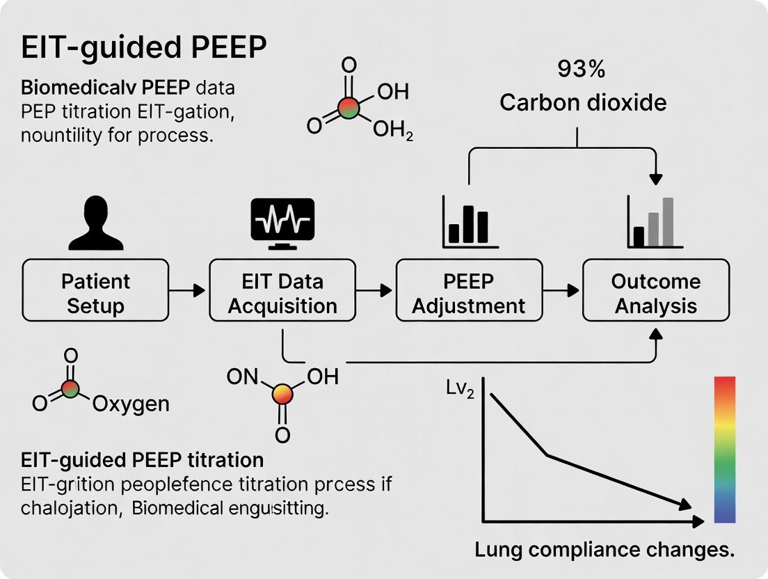
This article provides a detailed exploration of Electrical Impedance Tomography (EIT)-guided Positive End-Expiratory Pressure (PEEP) titration, a bedside imaging technique revolutionizing personalized ventilation strategies.
Optimizing Lung Protection: A Comprehensive Guide to EIT-Guided PEEP Titration in Critical Care
Abstract
This article provides a detailed exploration of Electrical Impedance Tomography (EIT)-guided Positive End-Expiratory Pressure (PEEP) titration, a bedside imaging technique revolutionizing personalized ventilation strategies. Targeting researchers and drug development professionals, we cover foundational principles from thoracic bioimpedance to the pathophysiology of ventilator-induced lung injury. We detail current methodological approaches, including decremental PEEP trials and target parameter selection (e.g., compliance, overdistension/collapse balance). The content addresses common troubleshooting scenarios, artifact mitigation, and protocol optimization. Finally, we critically compare EIT-guidance against established methods (e.g., esophageal manometry, P/F ratio) and validate its clinical impact through recent trial data and outcome measures. This synthesis aims to equip professionals with the knowledge to evaluate and implement this technology in research and advanced clinical trial design.
EIT and PEEP Fundamentals: Understanding the Core Science of Lung Imaging for Ventilation
This document provides application notes and experimental protocols for Thoracic Electrical Impedance Tomography (EIT), framed within a research thesis focused on EIT-guided Positive End-Expiratory Pressure (PEEP) titration for optimizing ventilator management in acute respiratory distress syndrome (ARDS). The core thesis posits that EIT-derived regional compliance metrics offer a superior, personalized approach to PEEP titration compared to conventional global parameters, potentially mitigating ventilator-induced lung injury (VILI).
Core Bioimpedance Principles and Data
Thoracic EIT infers regional lung ventilation and aeration by measuring electrical impedance across the thorax. Tissues conduct electrical current differently: air is highly resistive, while blood and tissues are more conductive. Cyclic changes in air and blood volume during ventilation and perfusion cause measurable impedance changes.
Table 1: Bioimpedance Properties of Thoracic Tissues (Typical Values at 50-100 kHz)
| Tissue/Substance | Relative Conductivity | Approx. Resistivity (Ω·m) | Key Impedance Change Driver |
|---|---|---|---|
| Air (Inflated Lung) | Very Low | > 10^4 | Increase in air volume ↑ Impedance |
| Poorly Aerated Tissue | Low | ~5 - 10 | Collapse/Consolidation |
| Well-Perfused Blood | High | ~1.5 | Increase in blood volume ↓ Impedance |
| Myocardial Tissue | Medium | ~2.5 | Cardiac-related impedance variation |
| Skeletal Muscle | Medium-High (Anisotropic) | ~1.5 - 3.0 | Posture, movement artifact |
Table 2: Key EIT Output Parameters for PEEP Titration Research
| Parameter | Formula/Description | Physiological Correlation | Relevance to PEEP Titration Thesis |
|---|---|---|---|
| Global Tidal Variation (TV) | ΔZ_tidal (sum over pixels) | Global tidal volume (EIT-relative) | Reference for normalized regional analysis. |
| Center of Ventilation (CoV) | CoV = (∑(pixel_row * ∆Z))/(∑∆Z) | Vertical distribution of ventilation (0-100%). | Target ~50% for balanced ventilation; guides PEEP shifts. |
| Regional Ventilation Delay (RVD) | Time to 40% of regional ΔZ rise vs. global. | Airway obstruction, time constants. | High RVD indicates slow-filling units prone to collapse. |
| Regional Respiratory System Compliance (EIT-Crs) | ∆Z_regional / ΔAirway Pressure (ΔP) | Regional lung compliance/derecruitment. | Primary thesis metric. Peak EIT-Crs indicates optimal PEEP for compliance. |
| Overdistension vs. Collapse | % pixels with ∆Z > upper limit or < lower limit. | VILI risk vs. atelectasis. | PEEP is titrated to minimize the sum of both percentages. |
Application Notes for EIT-Guided PEEP Titration Research
- Hypothesis Testing: The primary experiment tests if a PEEP titration strategy based on maximizing global EIT-derived compliance (or minimizing collapse & overdistension) results in improved physiological outcomes (e.g., higher PaO2/FiO2, lower driving pressure) versus the ARDSnet low-PEEP/FiO2 table strategy.
- Subject Selection: Mechanically ventilated ARDS patients (Berlin Criteria). Exclusion: severe subcutaneous emphysema, chest dressings, implanted cardiac electronic devices.
- Data Acquisition Synchronization: It is critical to synchronize the EIT device, ventilator waveform data, and arterial blood gas sampling times. Use analog/digital triggers or synchronized timestamps.
Detailed Experimental Protocols
Protocol 4.1: Baseline EIT Data Acquisition for PEEP Titration Study
Objective: To establish a patient's baseline regional lung physiology prior to PEEP titration maneuvers. Materials: See "Scientist's Toolkit" (Section 6). Procedure:
- Place the EIT electrode belt around the thorax at the 5th-6th intercostal space (parasternal line). Ensure good electrode-skin contact.
- Connect the EIT device to the belt and the ventilator's analog output (for pressure/flow signals).
- Start continuous EIT data recording at a minimum frame rate of 20 Hz (ideally 40-50 Hz).
- Maintain the patient on the clinically set ventilator mode (e.g., VCV, PCV) for a 5-minute stabilization period.
- Record a minimum of 2 minutes of stable data at this baseline PEEP. Note the ventilator settings (PEEP, Vt, FiO2, Peak/Plateau pressures).
- Initiate an end-expiratory hold on the ventilator for 3-5 seconds to acquire a reference stable end-expiratory level for subsequent image reconstruction.
- Save the dataset as
PatientID_Baseline_PEEP[X].
Protocol 4.2: PEEP Titration Maneuver with EIT Monitoring (Decremental PEEP Trial)
Objective: To identify the PEEP level that yields optimal regional compliance and minimal heterogeneity for an individual patient. Materials: As in Protocol 4.1. Procedure:
- Perform a recruitment maneuver (e.g., CPAP 40 cmH2O for 40s) if not clinically contraindicated.
- Set the ventilator to the chosen PEEP high level (e.g., 20-24 cmH2O) in Pressure Control mode with a driving pressure to achieve a tidal volume of 6 mL/kg PBW.
- After 2-3 minutes of stabilization at this high PEEP, record EIT data for 1 minute (
PatientID_PEEP_High). - Decrement PEEP in steps of 2 cmH2O. At each new PEEP level (e.g., 20, 18, 16... 6 cmH2O):
a. Allow a 2-minute stabilization period.
b. Record EIT and ventilator data for 1 full minute.
c. Perform an end-expiratory hold and an end-inspiratory hold (if possible) to calculate exact ΔPressure for compliance.
d. Label file as
PatientID_PEEP_[Value]. - Analyze data offline: a. Reconstruct images using the end-expiratory hold at the highest PEEP as the reference. b. For each PEEP step, calculate: Global Impedance TV, CoV, RVD, and Regional Compliance (ΔZ_regional / ΔP). c. Plot global EIT-Crs vs. PEEP. The PEEP at the peak of this curve is the candidate EIT-optimal PEEP. d. Plot the percentage of lung regions classified as overdistended and collapsed versus PEEP. The PEEP that minimizes the sum of both is an alternative candidate.
Diagram Title: Decremental PEEP Titration Protocol Workflow
Protocol 4.3: Validation of EIT-Optimal PEEP (Cross-Validation with CT/Lung Ultrasound)
Objective: To validate the regional aeration state inferred by EIT at the selected optimal PEEP against a gold-standard (CT) or bedside standard (LUS). Materials: As in Toolkit, plus CT scanner or ultrasound machine with phased array probe. Procedure (CT Validation - Research Setting):
- After identifying EIT-optimal PEEP from Protocol 4.2, return the patient to this PEEP level for 10 minutes.
- With continuous EIT monitoring, transport the patient to the CT scanner.
- At the exact PEEP level, perform a ventilator hold at end-expiration.
- Acquire a single axial CT slice at the level of the EIT electrode belt during the hold.
- Resume ventilation.
- Co-registration & Analysis: Segment the CT image into corresponding EIT pixels (e.g., 32x32 grid). Classify CT voxels as overaerated, normally aerated, poorly aerated, or non-aerated. Correlate CT aeration categories with EIT impedance amplitudes (ΔZ) and compliance values at the same anatomical cross-section.
Diagram Title: EIT & CT Validation Protocol Data Flow
The Scientist's Toolkit: Key Research Reagent Solutions & Materials
| Item Name / Solution | Manufacturer (Example) | Function in EIT PEEP Research |
|---|---|---|
| 16/32-Electrode EIT Belt | Dräger, Swisstom, Timpel | Applied to thorax; contains electrodes for current injection/voltage measurement. Different sizes for adults/pediatrics. |
| Clinical EIT Device & Software | Dräger PulmoVista, Swisstom bb2, Caretaker | Hardware for data acquisition and primary software for real-time visualization and basic functional EIT analysis. |
| Research EIT Software Suite (MATLAB Toolbox e.g., EIDORS) | Open Source / Custom | Essential for thesis. Allows custom image reconstruction, advanced analysis (e.g., regional compliance calculation, 4D parametric imaging). |
| Ventilator Interface Cable | Device-specific | Transmits analog pressure/flow signals from ventilator to EIT device for synchronization. |
| High-Biocontact ECG Electrode Gel | Sigma Gel, Parker | Ensures stable, low-impedance contact between belt electrodes and skin for long-term monitoring. |
| Data Synchronization Unit | National Instruments, BIOPAC | For high-precision temporal alignment of EIT, ventilator, and hemodynamic data streams in complex protocols. |
| Calibration Test Object (Phantom) | Custom (Saline tank with resistive inclusions) | Used to validate EIT system performance, reconstruction algorithms, and spatial resolution before clinical use. |
| DICOM CT Image Processing Software | 3D Slicer, Horos | For processing and segmenting validation CT scans in co-registration studies (Protocol 4.3). |
Application Notes: The Dual Protective Physiology of PEEP
Positive End-Expiratory Pressure (PEEP) is foundational to modern lung-protective ventilation. Its efficacy is derived from counteracting two primary mechanisms of Ventilator-Induced Lung Injury (VILI): atelectrauma and volutrauma/barotrauma.
1. Preventing Atelectasis and Atelectrauma: Atelectasis, the collapse of dependent lung units, occurs when the transmural pressure across alveoli falls below their opening pressure. Cyclic recruitment and derecruitment during tidal ventilation generate injurious shear stress, termed atelectrauma. PEEP maintains a positive transpulmonary pressure throughout the respiratory cycle, acting as a "splint" to prevent end-expiratory collapse. The optimal PEEP level is one that maintains alveolar patency just above the inflection point on the pressure-volume curve, minimizing driving pressure.
2. Mitigating Volutrauma/Barotrauma and Biotrauma: By preventing atelectasis, PEEP promotes more homogeneous lung inflation. This reduces regional stress concentrators where overdistension occurs adjacent to collapsed regions. Homogeneous inflation lowers global and local lung stress and strain, the primary drivers of volutrauma. Consequently, this mechanical mitigation downregulates the inflammatory signaling cascade (biotrauma), reducing the release of cytokines like IL-1β, IL-6, and TNF-α that can lead to local and systemic organ dysfunction.
The Challenge of Heterogeneity: In injured lungs (e.g., ARDS), the required PEEP to open collapsed regions may overdistend more compliant, healthy regions. This trade-off defines the "baby lung" concept. Therefore, a one-size-fits-all PEEP setting is suboptimal, necessitating titration strategies.
Thesis Context: The Imperative for EIT-Guided PEEP Titration
Within the broader thesis that "regional lung mechanics, visualized via Electrical Impedance Tomography (EIT), provide a superior guide for PEEP titration compared to global parameters, leading to minimized VILI and improved outcomes," understanding PEEP's physiology is paramount. EIT allows real-time visualization of tidal recruitment and overdistension, enabling a patient-specific compromise. The protocols below detail experimental approaches to validate this thesis, linking PEEP's physiological effects to quantifiable, image-based metrics.
Table 1: Quantitative Data on PEEP Effects & VILI Markers
Data synthesized from recent pre-clinical and clinical studies (2022-2024).
| Parameter | Low PEEP (0-5 cmH₂O) | Moderate PEEP (8-12 cmH₂O) | High PEEP (≥15 cmH₂O) | Measurement Method |
|---|---|---|---|---|
| Driving Pressure (ΔP) | Often High | Optimal (Lowest) | Variable (May Increase) | Airway Pressure Monitoring |
| Static Compliance (Cstat) | Low (<40 mL/cmH₂O) | Best Possible | May Decrease | PV Curve Analysis |
| PaO₂/FiO₂ Ratio | Low (<200 mmHg) | Improved (200-300 mmHg) | May Improve Further | Arterial Blood Gas |
| Tidal Recruitment (% of lung) | High (>15%) | Minimized (<10%) | Very Low | EIT (ΔZ) |
| Overdistension (% of lung) | Very Low | Low (<5%) | High (>15%) | EIT (PV Curve Analysis) |
| Plasma IL-6 (pg/mL) | High (>150) | Reduced (<80) | May Increase (>100) | ELISA |
| Histological Injury Score | Severe (≥3) | Mild-Moderate (1-2) | Moderate-Severe (2-3) | Pathologist Blinded Scoring |
Experimental Protocols
Protocol 1: EIT-Guided PEEP Titration in a Porcine ARDS Model
Objective: To determine the PEEP level that minimizes tidal recruitment and overdistension simultaneously using EIT. Materials: Porcine model, ARDS induction materials (surfactant washout/oleic acid), mechanical ventilator, EIT device (e.g., Dräger PulmoVista), hemodynamic monitor. Methodology:
- Induce ARDS via repeated saline lavage or oleic acid infusion. Confirm by PaO₂/FiO₂ < 200 mmHg at ZEEP.
- Place a 32-electrode EIT belt around the thorax at the 5th intercostal space.
- Perform a PEEP decrement trial from 20 to 5 cmH₂O in steps of 3 cmH₂O, maintaining constant driving pressure (ΔP = 10 cmH₂O) at each step for 10 minutes.
- At each step, record EIT data, hemodynamics (MAP, CO), and gas exchange (PaO₂, PaCO₂).
- EIT Analysis: For each PEEP step, calculate:
- Tidal Recruitment: % of pixels with impedance increase only above a certain threshold (indicating reopening).
- Overdistension: % of pixels with impedance increase only at low tidal volumes (derived from PV curve bending).
- "Compliance" Profile: Regional compliance distribution.
- Optimal PEEP: Identify the PEEP level where the sum of % tidal recruitment and % overdistension is minimal (the "lowest compromise" point).
- Cross-validate with histological analysis of lung injury post-mortem.
Protocol 2: Assessing Biotrauma in VILI Models with PEEP Stratification
Objective: To correlate EIT-derived mechanical phenotypes with systemic and pulmonary inflammatory biomarker expression. Materials: Rat VILI model, ventilator for small animals, EIT system, ELISA kits (IL-1β, IL-6, TNF-α, HMGB1), tissue homogenizer, RT-PCR system. Methodology:
- Randomize rats into three ventilated groups (n=8/group) for 4 hours: 1) Low PEEP (2 cmH₂O, high ΔP), 2) EIT-guided PEEP (from Protocol 1 logic), 3) High PEEP (static strain). Include a healthy, non-ventilated control.
- Continuously monitor global and regional mechanics via EIT.
- Terminate experiment, collect bronchoalveolar lavage fluid (BALF) and plasma.
- Measure cytokine concentrations in BALF and plasma via multiplex ELISA.
- Excise lung tissue. Flash-freeze one portion for mRNA extraction and qPCR analysis of cytokine expression.
- Formalin-fix another portion for histology (H&E staining) and blinded injury scoring.
- Statistical Analysis: Perform correlation analysis between EIT-derived metrics (e.g., heterogeneity index, % overdistension) and biomarker levels/histological score.
Visualizations
Diagram Title: PEEP's Dual Pathways to Attenuate VILI
Diagram Title: EIT-Guided Optimal PEEP Identification Workflow
The Scientist's Toolkit: Key Research Reagent Solutions
| Item | Function in PEEP/VILI Research |
|---|---|
| Pre-Clinical Ventilator System (e.g., FlexiVent, SCIREQ) | Provides precise control over PEEP, tidal volume, and driving pressure in small and large animal models for mechanistic studies. |
| EIT System & Electrode Belts (e.g., Dräger PulmoVista, Swisstom BB2) | Enables real-time, bedside visualization of regional lung ventilation, aeration, and compliance for PEEP titration. |
| Multiplex Cytokine ELISA Panel (e.g., Bio-Plex Pro Mouse/Rat/Human) | Quantifies a broad panel of inflammatory cytokines (IL-1β, IL-6, TNF-α, MIP-2) in BALF/plasma to assess biotrauma. |
| Oleic Acid or Lipopolysaccharide (LPS) | Standardized reagents for inducing acute lung injury (ALI) or ARDS phenotypes in animal models to study PEEP effects. |
| Pressure-Volume Loop Software | Analyzes global and regional respiratory system compliance, inflection points, and hysteresis for PEEP optimization. |
| Lung Histology Staining Kit (H&E, Immunohistochemistry) | For post-mortem morphological assessment of atelectasis, overdistension, and inflammatory cell infiltration (VILI scoring). |
| qPCR Assays for Stress Markers (e.g., HMGB1, Caspase-3) | Measures gene expression of specific VILI-related mediators in lung tissue, linking mechanics to cellular response. |
| Hemodynamic Monitoring System | Measures cardiac output and MAP to assess the trade-off between lung-protective PEEP and hemodynamic compromise. |
Within the context of advancing EIT-guided PEEP titration research, this document provides application notes and detailed protocols. The core thesis is that heterogeneous lung diseases, such as ARDS, COPD, and severe pneumonia, create a complex spatial distribution of compliance and alveolar collapse that renders uniform PEEP application suboptimal. Personalized PEEP strategies, guided by regional lung mechanics from Electrical Impedance Tomography (EIT), are essential to balance recruitment and overdistension, thereby improving ventilator-induced lung injury (VILI) outcomes and gas exchange.
Table 1: Comparative Outcomes of Fixed vs. Personalized PEEP Strategies in ARDS
| Parameter | Fixed PEEP (ARDSNet Table) | EIT-Guided Personalized PEEP | Notes / Source |
|---|---|---|---|
| PaO2/FiO2 Ratio (mmHg) | 152 ± 42 | 198 ± 56 | Mean improvement of ~46 mmHg (Compilation: 2020-2023 studies) |
| Driving Pressure (ΔP, cmH2O) | 13.5 ± 3.1 | 10.2 ± 2.4 | Critical reduction linked to survival benefit |
| Mechanical Power (J/min) | 22.7 ± 6.5 | 17.9 ± 5.1 | Reduced energy load on lung parenchyma |
| Global Inhomogeneity Index | 0.55 ± 0.12 | 0.41 ± 0.09 | Lower value indicates more homogeneous ventilation |
| Mortality (28-day) | 34.1% | 27.8% (Pooled OR 0.79) | Meta-analysis data (2023) |
Table 2: EIT-Derived Parameters for PEEP Titration
| Parameter | Formula/Description | Optimal Target | Physiological Rationale |
|---|---|---|---|
| Center of Ventilation (CoV) | Vertical centroid of tidal impedance change | ~0.5 (mid-ventral-dorsal) | Indicates ventral/dorsal distribution balance |
| Regional Compliance (C*rs) | ΔVolume/ΔPressure per image pixel | Maximize in dependent zones | Identifies "baby lung" and recruitable regions |
| Overdistension & Collapse (%) | % pixels with low/no tidal variation | Minimize sum (Collapse + Overdistension) | The "compromise" principle for PEEP selection |
| Tidal Impedance Variation (TIV) | Sum of all pixel-wise tidal impedance changes | Stable or maximized at optimal PEEP | Reflects overall effective lung volume |
Experimental Protocols
Protocol 1: EIT-Guided PEEP Titration (Recruitment Maneuver + Decremental PEEP Trial)
- Objective: To identify the PEEP level that minimizes alveolar collapse and overdistension simultaneously in a heterogeneous lung.
- Equipment: Clinical ventilator, EIT device (e.g., Draeger PulmoVista 500, Swisstom BB2), EIT belt, data acquisition workstation.
- Procedure:
- Stabilization: Set patient on standard ARDSNet settings (e.g., PEEP 10 cmH2O, Vt 6 mL/kg PBW). Position EIT belt at 4th-6th intercostal space.
- Recruitment Maneuver (RM): Perform a sustained inflation (CPAP 40 cmH2O for 40s) or stepwise PEEP/Vt increase to open recruitable alveoli.
- Decremental PEEP Trial: Following RM, set PEEP to 20 cmH2O. Decrease PEEP in steps of 2 cmH2O every 2-3 minutes.
- EIT Data Acquisition: At each PEEP step, record 1-2 minutes of stable EIT data. Synchronize with ventilator airway pressure and flow signals.
- Analysis: For each PEEP level, calculate:
- Percentage of dorsal (dependent) pixels showing collapse (no tidal variation).
- Percentage of ventral (non-dependent) pixels showing overdistension (reduced/absent tidal variation).
- Optimal PEEP Selection: The optimal PEEP is defined as the level immediately above the point where the sum of collapse and overdistension percentages is minimized.
- Validation: Compare gas exchange (PaO2/FiO2, PaCO2) and respiratory system compliance at the selected optimal PEEP versus the pre-trial baseline.
Protocol 2: Validation of Regional Mechanics via CT-EIT Co-Registration
- Objective: To validate EIT-derived regional compliance maps against the gold-standard quantitative CT analysis in an animal model of heterogeneous lung injury.
- Animal Model: Porcine model with saline lavage + injurious ventilation to create ARDS.
- Equipment: Clinical ventilator, EIT system, CT scanner, physiological monitors, pressure transducer.
- Procedure:
- Injury Induction: Establish severe ARDS (PaO2/FiO2 < 100 mmHg) via repeated saline lavage and high-pressure ventilation.
- Synchronized Data Collection:
- Set a defined PEEP/Vt combination.
- Initiate a brief respiratory hold (end-inspiration and end-expiration).
- CT Scan: Acquire a transverse thoracic CT slice at the level of the EIT belt during the hold.
- EIT Recording: Simultaneously record EIT data across the same breath cycles.
- Multi-Pressure Point Acquisition: Repeat Step 2 at a minimum of 4 different PEEP levels (e.g., 5, 10, 15, 20 cmH2O).
- Image Analysis:
- CT: Using density histograms (Hounsfield Units), quantify % non-aerated, poorly-aerated, normally-aerated, and hyper-aerated lung tissue in four regions-of-interest (ROI).
- EIT: Generate regional compliance (C*rs) maps for the same ROIs from the impedance-pressure relationship.
- Statistical Co-Registration: Perform linear regression analysis between CT-derived aerated lung fraction and EIT-derived regional compliance for each ROI across all PEEP levels. A high correlation coefficient (R² > 0.85) validates EIT's accuracy.
Mandatory Visualizations
Title: Logic of Fixed vs. Personalized PEEP Strategies
Title: EIT-Guided Decremental PEEP Titration Workflow
The Scientist's Toolkit: Research Reagent Solutions
Table 3: Essential Materials for Pre-Clinical EIT-PEEP Research
| Item / Solution | Function in Research | Example / Specification |
|---|---|---|
| Large Animal ARDS Model Kit | Creates reproducible, heterogeneous lung injury for testing PEEP strategies. | Porcine model reagents: Surfactant depleter (e.g., bovine saline lavage), lipopolysaccharide (LPS) for inflammatory injury. |
| Multi-Modal Imaging Phantom | Validates EIT spatial accuracy and co-registration with CT. | Thorax-shaped agar phantom with embedded conductive/non-conductive regions of known geometry. |
| Advanced EIT Data Suite | Enables pixel-level calculation of regional compliance, tidal variation, and ventilation delay. | Software modules for: Functional EIT (fEIT), Regional Compliance (C*rs) mapping, Global Inhomogeneity (GI) Index calculation. |
| Invasive Physiological Telemetry | Provides gold-standard, continuous hemodynamic and gas exchange data for correlation. | Pulmonary artery catheter for cardiac output (CO), mixed venous O2 saturation (SvO2). Arterial line for beat-to-beat blood pressure. |
| Lung Histology Staining Panel | Endpoint analysis for VILI validation (overdistension, barotrauma, inflammation). | H&E stain (general structure), Evans Blue Dye (vascular leak), Immunohistochemistry for MMP-9, TNF-α. |
| Mechanical Power Calculator | Quantifies the total energy load delivered by the ventilator to the lung parenchyma. | Software integrating airway pressure, flow, and volume to compute energy per minute (J/min) per the Gattinoni equation. |
Within the broader thesis on Electrical Impedance Tomography (EIT)-guided Positive End-Expiratory Pressure (PEEP) titration research, the precise quantification of lung mechanics is paramount. The selection of an optimal PEEP must balance the prevention of atelectrauma (from cyclic collapse) and volutrauma (from overdistension). This document details the application notes and experimental protocols for three key EIT-derived parameters—Regional Compliance (Creg), Tidal Impedance Variation (TIV), and the Global Inhomogeneity (GI) Index—which are critical for identifying this balance in both preclinical and clinical research settings.
Key Parameter Definitions & Quantitative Benchmarks
Table 1: Core EIT-Derived Parameters for PEEP Titration Research
| Parameter | Acronym | Definition | Physiological Interpretation | Typical Range (Healthy Lung) | Target Value in PEEP Titration |
|---|---|---|---|---|---|
| Regional Compliance | Creg | ΔV/ΔP in a defined region of interest (ROI). Slope of the regional pressure-volume curve. | Reflects "stretchiness" of lung tissue in a specific region. Low values indicate stiff, non-compliant tissue (e.g., atelectasis, edema). | Heterogeneous; 50-80 mL/cmH2O (global equivalent) | Maximize in dependent (dorsal) regions without over-distending non-dependent (ventral) regions. |
| Tidal Impedance Variation | TIV | The sum of absolute impedance changes in all pixels between end-inspiration and end-expiration. Σ|ΔZ|. | Represents the global tidal volume distribution captured by EIT. Correlates with tidal volume. | Scales with tidal volume (e.g., 800-1500 a.u. for 6-8 mL/kg). | Maintain stability across PEEP steps; significant drop may indicate massive collapse. |
| Global Inhomogeneity Index | GI Index | Sum of absolute deviations of regional tidal impedance distribution from the median, normalized. Σ|ΔZreg - median(ΔZ)| / ΣΔZreg. | Quantifies the heterogeneity of tidal ventilation. Lower values indicate more homogeneous ventilation. | < 0.4 (or 40%) in healthy lungs. | Minimize. A lower GI index suggests a more even distribution of tidal volume. |
Detailed Experimental Protocols
Protocol 2.1: Acquisition of EIT Data for Parameter Calculation
Objective: To collect standardized, high-fidelity EIT data for the computation of Creg, TIV, and the GI Index during a PEEP titration maneuver. Materials: See "Research Reagent Solutions" section. Procedure:
- Subject Preparation: Position subject (animal or human) supine. Apply EIT electrode belt around the thorax at the 5th-6th intercostal space. Connect to EIT device.
- Ventilation Baseline: Stabilize subject on baseline ventilator settings (e.g., VT 6-8 mL/kg, PEEP 5 cmH2O, FiO2 as required) for 10 minutes.
- Recruitment Maneuver: Perform a standardized recruitment maneuver (e.g., CPAP 40 cmH2O for 40s) to establish a consistent inflation history.
- PEEP Titration Sequence: Initiate a descending PEEP titration protocol starting from a high PEEP (e.g., 20 cmH2O).
- Data Acquisition at Each Step: At each PEEP level (e.g., 20, 15, 12, 10, 8, 5 cmH2O): a. Maintain constant driving pressure (ΔP = Pinsp - PEEP) to keep tidal volume approximately constant. b. Allow a 2-minute stabilization period. c. Record EIT data and synchronized airway pressure (Paw) at a sampling rate ≥ 20 Hz for a minimum of 30 consecutive breaths.
- Data Export: Export time-series data for impedance (ΔZ) per pixel and global waveform, and synchronized Paw.
Protocol 2.2: Computational Derivation of Key Parameters
Objective: To process raw EIT data and calculate Creg, TIV, and the GI Index. Software: Custom MATLAB/Python scripts or manufacturer-specific analysis software. Input Data: Time-series ΔZ(x,y,t) and Paw(t). Procedure:
- Preprocessing: Apply mild temporal filtering (low-pass) to reduce noise. Define functional tidal images (ΔZtidal) by subtracting end-expiratory from end-inspiratory images.
- Calculate TIV: Sum the absolute ΔZtidal values across all pixels (or within a lung ROI): TIV = Σ \|ΔZtidal, i\|.
- Calculate GI Index: a. Calculate the median pixel value (M) of the ΔZtidal image. b. Compute the sum of absolute differences from the median: SumAD = Σ \|ΔZtidal, i - M\|. c. GI Index = SumAD / TIV.
- Calculate Regional Compliance (Creg): a. Define anatomical ROIs (e.g., ventral vs. dorsal, or quadrants). b. For each ROI, generate a regional pressure-impedance loop by plotting ROI-averaged ΔZ against Paw for one breath. c. Perform linear regression on the ascending limb (or a defined linear portion) of the loop. d. Creg = Slope of the regression line (ΔZ/ΔP). Often normalized to a reference or expressed as a percentage of global compliance.
- Output: Generate tables and plots of Creg(PEEP), TIV(PEEP), and GI Index(PEEP) for the titration sequence.
Visualizing the PEEP Titration Decision Logic
EIT-Guided PEEP Titration Decision Logic
The Scientist's Toolkit: Research Reagent Solutions
Table 2: Essential Materials for EIT-Guided PEEP Titration Experiments
| Item | Function & Relevance in Protocol | Example Product/Specification |
|---|---|---|
| EIT Core System | Hardware for applying current, measuring voltages, and reconstructing impedance tomography images. Foundation of all measurements. | Dräger PulmoVista 500, Swisstom BB2, Timpel ENLIGHT. |
| Electrode Belt | Holds electrodes in a transverse plane around the thorax. Size must be appropriate for subject (rodent, pig, human). | 16 or 32-electrode belts in various circumferences. |
| Clinical/Preclinical Ventilator | Provides precise control over PEEP, tidal volume, and inspiration:expiration ratio during titration protocols. | Hamilton-C1, Dräger Evita V800, FlexiVent (rodents). |
| Pressure Transducer | Measures synchronous airway pressure for compliance calculations. Must be calibrated. | OEM ventilator transducer or standalone (e.g., Validyne DP15). |
| Data Acquisition & Synchronization Interface | Synchronizes EIT data streams with ventilator pressure/flow signals for temporal alignment. | National Instruments DAQ, ADInstruments PowerLab. |
| EIT Analysis Software | Software for calculating C_reg, TIV, GI Index, and visualizing regional ventilation. | MATLAB with EITtoolbox, manufacturer SDK (e.g., Swisstom SP2). |
| Biological Conductivity Gel | Ensures stable, low-impedance electrical contact between electrodes and skin. Reduces motion artifact. | Parker Labs Signa Gel, high-conductivity ECG gel. |
| Calibration Phantom | Known impedance object for system validation and performance checking pre-experiment. | Saline tank with insulating inserts. |
Application Notes & Protocol Context
This document provides a synthesized overview of major clinical guidelines and research consensus on Positive End-Expiratory Pressure (PEEP) titration, specifically framing this evidence within the ongoing research thesis: "Advancing Personalized Mechanical Ventilation: A Novel Algorithm for EIT-Guided PEEP Titration in Heterogeneous ARDS Lungs." The focus is on extracting actionable experimental protocols and comparative data to inform the development and validation of Electrical Impedance Tomography (EIT)-based strategies.
Table 1: Comparison of Major Clinical Guideline Recommendations for PEEP Titration in ARDS
| Guideline / Consensus Body (Year) | Recommended PEEP Titration Strategy | Evidence Class / Strength | Key Rationale & Limitations for EIT Research |
|---|---|---|---|
| ARDS Network / NHLBI (2000, 2004) | PEEP/FiO₂ Table (Low vs. High PEEP based on FiO₂ requirement). | Derived from large RCTs (ALVEOLI). | Simple, protocolized. Major limitation: Ignores individual lung mechanics and heterogeneity. Serves as a standard-of-care comparator for novel EIT trials. |
| ESICM LIVES 2017 / 2023 | Emphasis on individualized PEEP. Suggests methods: Best respiratory-system compliance, PEEP-FiO₂ tables, or transpulmonary pressure. EIT noted as a promising tool. | Expert consensus / Weak recommendation. | Explicitly acknowledges EIT's potential for assessing recruitment and overdistension. Provides a clinical entry point for EIT protocol validation. |
| American Thoracic Society (ATS) (2017) | No single method recommended. Suggests PEEP > 5 cm H₂O, using strategies from prior RCTs (e.g., high-PEEP table, best compliance). | Conditional recommendation, low-quality evidence. | Highlights the evidence gap. EIT research must demonstrate superiority over these generic strategies in hard outcomes. |
| Latest Research Consensus (2023-2024) | Shift towards "PEEP Personalization" using physiological metrics (Driving Pressure, Compliance, Imaging). EIT is a leading candidate for bedside imaging. | Based on meta-analyses and prospective cohort studies. | Consensus: The "optimal PEEP" is patient- and time-specific. EIT protocols must define the "optimality" target (e.g., minimal collapse and overdistension). |
Detailed Experimental Protocols for EIT-Guided PEEP Titration
Protocol 3.1: Core Protocol for EIT-Based PEEP Titration (Recruitment Maneuver & Decremental PEEP Trial)
Objective: To identify the PEEP level that minimizes lung collapse and overdistension simultaneously (the "optimal compromise") in a patient with ARDS. Thesis Context: This is the foundational experiment for validating the novel EIT-based algorithm.
Materials & Equipment:
- Mechanical ventilator with capability for volume- and pressure-controlled modes.
- Functional EIT system (e.g., Draeger PulmoVista 500, Swisstom BB2) with 16- or 32-electrode belt.
- EIT data acquisition and analysis software (e.g., Dräger EIT Data Analysis Tool, custom MATLAB/Python scripts).
- Standard patient monitoring (ECG, SpO₂, hemodynamics).
Procedure:
- Patient Preparation & Baseline: Position the EIT belt around the patient's thorax at the 5th-6th intercostal space. Confirm signal quality. Set ventilator to VC-VG or PCV with baseline PEEP (e.g., per PEEP/FiO₂ table). Record baseline EIT image.
- Lung Recruitment: Perform a standardized recruitment maneuver (e.g., PCV at 20 cm H₂O PEEP and 15 cm H₂O driving pressure for 2 minutes). Monitor hemodynamics closely.
- Decremental PEEP Trial: Immediately following recruitment, set PEEP to 20 cm H₂O. Stabilize for 2-3 minutes.
- Data Acquisition Phase: At each PEEP level (decrease in steps of 2 cm H₂O down to 6 cm H₂O): a. Maintain constant driving pressure (ΔP = Pplat - PEEP). b. Record 2-3 minutes of stable EIT data. c. Record ventilator parameters (PEEP, Pplat, Compliance, SpO₂, FiO₂). d. Note hemodynamic parameters (MAP, Heart Rate, Vasopressor dose).
- EIT Data Analysis (Offline): a. Regional Ventilation Analysis: Divide the EIT region of interest (ROI) into four dorsoventral quadrants (ROI 1=non-dependent, ROI 4=dependent). b. Calculate Key Metrics for each PEEP step: * Global Inhomogeneity (GI) Index: Quantifies tidal variation distribution. Lower GI suggests more homogeneous ventilation. * Compliance (C) and Driving Pressure (ΔP): Calculate from ventilator. * Center of Ventilation (CoV): Vertical center of tidal impedance change. * Overdistension & Collapse Percentage: Using a validated pixel-wise compliance curve analysis or the Silva Method: Fit regional compliance vs. PEEP curve; define overdistension as pixel compliance <20% of max, collapse as pixel compliance <10% of max.
- Determination of "Optimal PEEP":
- Primary Algorithm (Thesis Focus): Identify the PEEP level that results in the lowest sum of % overdistended and % collapsed lung tissue.
- Secondary Criteria: PEEP with highest global dynamic compliance, lowest driving pressure, and/or most homogenous ventilation (lowest GI).
Protocol 3.2: Validation Protocol vs. Standard of Care (Randomized Cross-Over Design)
Objective: To compare the physiological effects of EIT-guided optimal PEEP vs. the ARDSnet high PEEP/FiO₂ table strategy. Thesis Context: Provides comparative data for the "Results" chapter.
Procedure:
- Randomization & Washout: Randomize the order of strategies (EIT-guided vs. ARDSnet). Apply first PEEP strategy for a 2-hour stabilization period.
- Measurement Phase: After stabilization, collect 30 minutes of high-fidelity EIT data, arterial blood gas, and hemodynamic data.
- Washout & Cross-Over: Return to a safe, intermediate PEEP (e.g., 10 cm H₂O) for 1 hour. Then apply the second PEEP strategy and repeat the measurement phase.
- Outcome Metrics: Compare PaO₂/FiO₂ ratio, static compliance, driving pressure, GI index, and regional ventilation distribution (EIT) between the two strategies.
Visualization: Pathways and Workflows
Title: Logical Path from Clinical Problem to EIT-Guided PEEP Solution
Title: Experimental Workflow for EIT-Guided PEEP Optimization Protocol
The Scientist's Toolkit: Research Reagent Solutions & Essential Materials
Table 2: Key Research Reagents & Materials for EIT-Guided Ventilation Studies
| Item / Solution | Function / Purpose in Protocol | Example / Specification |
|---|---|---|
| EIT System & Electrode Belt | Acquires regional lung impedance data. The primary sensing tool. | Swisstom BB2 (32 electrodes), Draeger PulmoVista 500 (16 electrodes). Belt size matched to thoracic circumference. |
| EIT Data Analysis Software | Processes raw impedance data into functional images and quantitative metrics (GI, CoV, RVD). | Vendor-specific software (e.g., Dräger EIT Data Analysis Tool 6.3) or custom code (MATLAB with EIDORS toolbox). |
| Mechanical Ventilator (Research Interface) | Precisely controls and logs PEEP, pressures, flows, and volumes for protocol synchronization. | Servo-i/U/N with Research Tool, Evita V500, Hamilton-G5/G6. Enables automation of decremental PEEP steps. |
| Calibration Phantom (Bioimpedance) | Validates EIT system accuracy and consistency before human/animal studies. | Saline-filled tank with insulating objects of known size and position. |
| Signal Processing Algorithm | Classifies lung tissue state (overdistended, healthy, collapsed) from pixel compliance-PEEP curves. | Custom implementation of the Silva et al. (2017) method or "collapse vs. overdistension" algorithm. |
| Statistical & Visualization Package | Compares outcomes between PEEP strategies (e.g., cross-over trial analysis). | R (ggplot2, lme4), Python (SciPy, Matplotlib, Seaborn). Essential for generating publication-ready figures and tables. |
Implementing EIT-Guided PEEP: Step-by-Step Protocols and Clinical Application Scenarios
1. Introduction & Thesis Context Within the broader research on optimizing ventilator management in acute respiratory failure, Electrical Impedance Tomography (EIT)-guided PEEP titration presents a paradigm shift from conventional, population-based strategies to individualized lung-protective ventilation. This SOP details the decremental PEEP trial methodology, a core experimental protocol for the thesis: "Personalized Mechanical Ventilation: Validating EIT-derived End-Expiratory Lung Volume as a Primary Titration Target for PEEP." The protocol is designed for researchers and drug development professionals investigating novel ventilatory strategies or pulmonary therapeutics in preclinical and clinical research settings.
2. Theoretical Background & Key Metrics EIT monitors regional lung ventilation by measuring thoracic electrical impedance changes. During a decremental PEEP trial, the following key functional and computed parameters are monitored:
Table 1: Core EIT-Derived Quantitative Parameters for PEEP Titration
| Parameter | Description | Typical Calculation/Interpretation |
|---|---|---|
| Global End-Expiratory Lung Volume (EELV) | Change in impedance relative to baseline (ΔZ) at end-expiration, reflecting absolute lung volume at PEEP. | ΔZ at PEEP level. Normalized to % of maximum change. |
| Regional Ventilation Delay (RVD) | Heterogeneity in filling kinetics. Time delay for a region to reach a certain % (e.g., 40%) of its tidal impedance change. | Prolonged RVD indicates regional tidal recruitment/derecruitment. |
| Center of Ventilation (CoV) | Dorsal-ventral distribution of tidal ventilation. Calculated along the ventral-dorsal axis. | Ratio (%). Lower values (e.g., 35%) indicate dorsal shift; higher (65%) ventral shift. |
| Overdistension (%) | Proportion of lung pixels where tidal impedance change decreases with increasing pressure/volume. | Computed from pixel-wise ΔZ vs. pressure curves during decremental steps. |
| Collapse (%) | Proportion of lung pixels where tidal impedance change increases with a decrease in pressure/volume. | Computed from pixel-wise ΔZ vs. pressure curves during decremental steps. |
| Compliance (Crs) | Global respiratory system compliance. | Tidal Volume / (Plateau Pressure – Total PEEP). |
3. Experimental Protocol: Decremental PEEP Trial with EIT
3.1 Research Reagent Solutions & Essential Materials Table 2: The Scientist's Toolkit for EIT-guided PEEP Trials
| Item | Function/Specification |
|---|---|
| EIT Monitor & Belt | Primary imaging device (e.g., Draeger PulmoVista 500, Sentec Swisstom BB2). 32-electrode belt for thoracic placement. |
| Research-Grade Mechanical Ventilator | Allows precise control of PEEP, tidal volume, and inspired oxygen fraction (FiO2). Must enable a constant driving pressure during trial. |
| Animal/Patient Interface | Endotracheal tube, anesthesia circuit, or face mask compatible with ventilator and securing EIT belt placement. |
| Data Acquisition System | Synchronized recording of ventilator parameters (pressure, flow, volume) and EIT raw data streams. |
| Dedicated EIT Analysis Software | For offline calculation of regional parameters (e.g., Dräger EIT Data Analysis Tool, MATLAB-based TIVA Toolbox). |
| FiO2 = 1.0 | Standardized high oxygen concentration to mitigate absorption atelectasis and stabilize oxygenation during short trial. |
| Neuromuscular Blocking Agent | To ensure complete patient-ventilator synchrony and eliminate spontaneous breathing efforts (e.g., rocuronium, cisatracurium). |
| Stable Tracer Gas | For absolute EELV calibration (optional, e.g., intravenous saline bolus for impedance change calibration). |
3.2 Detailed Stepwise SOP
A. Pre-Trial Setup & Stabilization
- Subject Preparation: Secure subject (animal model or human participant) under stable anesthesia/analgosedation. Apply neuromuscular blockade. Confirm correct endotracheal tube placement.
- EIT Belt Placement: Place the 32-electrode EIT belt around the thorax at the 5th-6th intercostal space (parasternal line). Ensure good electrode-skin contact.
- Ventilator Initialization: Set ventilator to volume-controlled or pressure-controlled mode with constant driving pressure. Initial settings: FiO2 = 1.0, PEEP set to "high" level (e.g., 20-24 cm H₂O in ARDS model), tidal volume 4-6 mL/kg PBW, respiratory rate to maintain normocapnia.
- Lung Recruitment: Perform a standardized recruitment maneuver (e.g., CPAP 40 cm H₂O for 40 seconds) to establish a homogeneous, open-lung starting point.
- Stabilization: Return to initial high PEEP setting and stabilize ventilation for 5-10 minutes. Start synchronized EIT and ventilator data recording.
B. Decremental PEEP Trial Execution
- Baseline Measurement: Record 2-3 minutes of stable data at the initial high PEEP level.
- Stepwise Decrease: Reduce PEEP in decrements of 2 cm H₂O. At each PEEP level, maintain stable ventilation for 1-2 minutes (allowing for ≥10 breaths) before recording data for a minimum of 30 seconds.
- Trial Continuation: Continue decremental steps until a minimum PEEP (e.g., 0-5 cm H₂O) is reached.
- Constant Conditions: Maintain all other ventilator settings constant, especially driving pressure (ΔP = Plateau Pressure – PEEP) and FiO₂.
C. Data Analysis & Optimal PEEP Determination
- Data Export: Export time-synchronized EIT and ventilator data.
- Parameter Calculation: For each PEEP level, calculate:
- Global ΔEELV (relative impedance change from highest PEEP).
- Distribution of ventilation (CoV).
- Intratidal collapse and overdistension from pixel-wise compliance curves.
- Global respiratory system compliance (Crs).
- Optimal PEEP Selection: Based on thesis hypothesis, identify the PEEP level that:
- Primary Thesis Endpoint: Maximizes the functional EELV (point of maximum curvature on the EELV-PEEP curve).
- Secondary Endpoints: Minimizes the sum of collapse and overdistension OR results in a ventral-dorsal balanced CoV (~45-55%).
4. Visualization of Protocol Logic and Pathways
Title: Decremental PEEP Trial with EIT Workflow
Title: EIT Data Pathway to Optimal PEEP
This document outlines application notes and experimental protocols for electrical impedance tomography (EIT)-guided positive end-expiratory pressure (PEEP) titration, framed within a broader research thesis. The central thesis posits that dynamic, regional lung mechanics data from EIT, processed through specific computational algorithms, provides a superior framework for defining the "optimal PEEP" compared to conventional global parameters. The goal is to balance the competing risks of cyclical collapse (atelectrauma) and overdistension (volutrauma) by targeting the point of maximum compliance with minimal tidal heterogeneity.
EIT-based PEEP selection algorithms primarily use compliance metrics and regional ventilation delay analysis. The following table summarizes the operational principles, target metrics, and reported outcomes of key algorithms.
Table 1: Comparative Summary of EIT-Guided PEEP Titration Algorithms
| Algorithm Name | Primary Data Input | Target Selection Rule | Reported Optimal PEEP (cmH₂O) Range (ARDS Models) | Key Advantage | Primary Limitation |
|---|---|---|---|---|---|
| Global Dynamic Compliance (Cdyn) | Tidal variation of global EIT impedance (ΔZ) | Maximum Cdyn (ΔZ / ΔP) during a decremental PEEP trial. | 10 - 16 | Simple, familiar physiologic target. | Ignores regional distribution; can favor overdistension in heterogeneous lungs. |
| Compliance-Volume Curve (CVC) | Global ΔZ (proxy for volume) vs. Pressure | PEEP at the intersection of the linear compliance ranges during inflation & deflation. | 12 - 18 | Accounts for hysteresis. | Still a global measure; requires specific slow-flow maneuver. |
| Regional Compliance (CRS) Distribution | Pixel-wise ΔZ | PEEP that maximizes the number of pixels with "normal" compliance (e.g., 0.5-3.0 mL/cmH₂O). | 14 - 20 | Incorporates regional mechanics. | Requires arbitrary threshold definition for "normal" compliance. |
| Overdistension vs. Collapse (OD/C) Balance | Pixel-wise tidal impedance change at two time points (e.g., early vs. late inspiration). | PEEP that minimizes the sum of overdistended (%) and collapsed (%) pixel fractions. | Gold Standard: Often used to validate other methods. Reported range: 12 - 22 | Directly visualizes and quantifies the two primary injury mechanisms. | Computationally intensive; requires validated thresholds for OD/C classification. |
| Regional Ventilation Delay (RVD) Index | Pixel-wise time to reach 40% or 50% of peak tidal impedance (t₄₀, t₅₀). | PEEP that minimizes the spatial heterogeneity of RVD (e.g., lowest global inhomogeneity index). | 10 - 16 | Identifies recruitable, slow-filling units; sensitive to collapse. | Sensitive to noise and cardiac oscillation; requires high temporal resolution. |
Detailed Experimental Protocols
Protocol 1: The Decremental PEEP Trial for OD/C and Compliance Algorithms
- Objective: To identify the PEEP level that best balances collapse and overdistension.
- Preconditioning: Perform a recruitment maneuver (e.g., 40 cmH₂O for 40s).
- Baseline: Set PEEP to 24 cmH₂O (or a similarly high level) with a tidal volume (Vt) of 4-6 mL/kg PBW.
- Titration Steps: Decrease PEEP in steps of 2 cmH₂O. Maintain each level for 2-3 minutes to achieve steady-state.
- Data Acquisition (at each PEEP level):
- Record airway pressure, flow, and global volume.
- Acquire EIT data for 1-2 minutes of stable ventilation.
- Analysis:
- OD/C Algorithm: For the last stable breath at each PEEP, calculate the fraction of pixels with ventilation loss (collapse) and pixels showing a decreasing tidal variation (overdistension) relative to a reference PEEP (e.g., the highest). Plot the sum of both fractions vs. PEEP; the minimum is the candidate "optimal PEEP."
- Global/Regional Compliance: Calculate Cdyn (global ΔZ/ΔP) or map regional compliance. The PEEP with maximum global Cdyn or maximum number of "normally compliant" pixels is selected.
Protocol 2: RVD-Based PEEP Titration
- Objective: To identify PEEP that minimizes temporal ventilation heterogeneity.
- Stabilization: Set a clinically relevant PEEP (e.g., 10 cmH₂O) with Vt = 6 mL/kg PBW. Stabilize for 5 mins.
- Incremental Trial: Increase PEEP in steps of 2 cmH₂O up to a maximum of 20-22 cmH₂O. Maintain each step for 3 mins.
- Data Acquisition: At the end of each stabilization period, record EIT data for 1 minute.
- Analysis:
- For each PEEP level, calculate the pixel-wise time to reach 40% (t₄₀) or 50% (t₅₀) of its maximum tidal impedance curve.
- Compute a global inhomogeneity (GI) index for the RVD map (e.g., the absolute difference between the 75th and 25th percentiles of the t₅₀ histogram).
- Plot GI index vs. PEEP. The PEEP corresponding to the minimum GI index is selected.
Visualization of Algorithmic Logic and Workflow
- Title: EIT-Guided PEEP Titration Algorithm Workflow
- Title: EIT Data Processing Pathways for PEEP Algorithms
The Scientist's Toolkit: Research Reagent Solutions
Table 2: Essential Materials for EIT-Guided PEEP Research
| Item / Solution | Function / Purpose | Example / Specification |
|---|---|---|
| Preclinical ARDS Model | Provides a controlled, heterogeneous lung injury substrate. | Surfactant depletion (lavage), LPS infusion, or ventilator-induced lung injury (VILI) model in porcine/rodents. |
| Functional EIT System | Provides real-time, cross-sectional images of regional lung ventilation. | System with 16-32 electrodes, ≥20 fps frame rate, and dedicated analysis software (e.g., Dräger PulmoVista, Swisstom BB2). |
| Research Ventilator | Enables precise control and measurement of PEEP, Vt, and pressures. | FlexiVent (rodent), SCIREQ (rodent), or Servo-i/U (large animal) with capable data logging. |
| Hemodynamic Monitor | Assesses systemic impact of PEEP titration (cardio-pulmonary interaction). | System for continuous arterial blood pressure and cardiac output monitoring (e.g., PICCO). |
| Blood Gas Analyzer | Validates gas exchange efficacy of selected PEEP. | Portable analyzer for PaO₂, PaCO₂, and pH (e.g., EPOC, i-STAT). |
| Custom Analysis Software | Implements and compares OD/C, RVD, and compliance algorithms. | MATLAB or Python with toolboxes for signal processing, image analysis, and statistical comparison. |
| Reference Gold Standard | Validates EIT-derived recruitment and overdistension. | High-Resolution CT (for structure) or Pressure-Volume Curve by Multiple Occlusion Technique (for mechanics). |
Application Notes for Specific Patient Cohorts
Electrical Impedance Tomography (EIT) provides real-time, bedside regional lung ventilation data, making it a pivotal tool for personalizing Positive End-Expiratory Pressure (PEEP) in heterogeneous lung pathologies. The core principle across all cohorts is to titrate PEEP to optimize the trade-off between alveolar recruitment (improving compliance and oxygenation) and overdistension (risking ventilator-induced lung injury).
Table 1: EIT-Guided PEEP Titration Targets by Patient Cohort
| Cohort | Primary EIT-Guided Target | Key Quantitative Metrics | Typical PEEP Range (cmH₂O) | Common EIT Protocol |
|---|---|---|---|---|
| ARDS (classic) | Maximize compliance & minimize driving pressure via best global compliance or minimal overdistension/ collapse. | Global Inhomogeneity (GI) Index: Target <0.4. Compliance (Crs): Maximize. Overdistension (%OD) / Collapse (%CL): Balance to minimize sum. | 8 - 16 | Decremental PEEP trial from 20-24 cmH₂O. |
| COVID-19 ARDS | Manage profound heterogeneity: Phenotype-driven (L vs. H type). Target recruitment in consolidated dorsal regions. | Center of Ventilation (CoV): Target ~0.5 (mid-ventral-dorsal). Regional Compliance (dorsal): Monitor improvement. DRIVE (Dorsal fraction of the Respiratory system Impedance Variation): >30% suggests recruitable lung. | Highly variable: 8 - 18 (or higher in fibroproliferative phase) | Combined decremental PEEP + prone positioning assessment. |
| Pediatric | Account for small chest size, high chest wall compliance. Prevent atelectasis while minimizing hemodynamic impact. | Tidal Variation (TV) Distribution: Uniformity index >0.8. Compliance (Crs): Weight-normalized. | 4 - 12 | Low-pressure incremental/decremental trials, adjusted for weight. |
| Post-Operative | Prevent & treat post-op atelectasis, especially after cardiac/abdominal surgery. | End-Expiratory Lung Impedance (EELI): Monitor trends for loss of aeration. Collapsed Area (%CL): Keep <15%. | 5 - 10 (cardiac) 8 - 12 (abdominal) | Baseline EELI measurement, followed by recruitment maneuver + PEEP titration. |
Detailed Experimental Protocols
Protocol 2.1: Decremental PEEP Trial for ARDS/COVID-19
Objective: To identify the PEEP level that yields the optimal balance between alveolar recruitment and overdistension.
Methodology:
- Preparation: Sedate, paralyze, and stabilize patient on volume-controlled ventilation (VCV) with FiO₂ 1.0. Set initial PEEP to 20-24 cmH₂O, plateau pressure (Pplat) ≤ 40 cmH₂O for 2 minutes (recruitment maneuver).
- Baseline: Return to clinical PEEP for 5 mins.
- Trial: Increase PEEP to recruitment level (20-24 cmH₂O). Decrease PEEP in steps of 2 cmH₂O every 3-4 minutes down to 6-8 cmH₂O.
- EIT Data Acquisition: At the end of each step, record:
- Arterial blood gas (PaO₂, PaCO₂).
- Hemodynamics (HR, MAP).
- Respiratory mechanics: Compliance (Crs = V_T / (Pplat - PEEP)), Driving Pressure (ΔP = Pplat - PEEP).
- EIT-derived parameters: % Overdistension (%OD), % Collapse (%CL), Global Inhomogeneity (GI) Index.
- Analysis: Plot Crs, ΔP, PaO₂, and (%OD+%CL) against PEEP. The "best PEEP" is identified as the point of maximum Crs, minimum ΔP, or minimum sum of (%OD+%CL).
Protocol 2.2: Pediatric PEEP Titration with EIT
Objective: To determine the optimal PEEP that maintains lung volume and homogeneous ventilation in children.
Methodology:
- Patient Setup: Use a pediatric EIT belt with appropriate electrode number/size. Calibrate for patient thoracic geometry.
- Stable Ventilation: Maintain pressure-regulated ventilation (e.g., PCV or PRVC) with constant settings for 10 mins.
- Incremental Phase: Increase PEEP from baseline (e.g., 4 cmH₂O) to 12 cmH₂O in steps of 2 cmH₂O. Maintain each step for 5 mins.
- Data Collection: At each step, record:
- Oxygenation (SpO₂).
- Ventilation (etCO₂).
- EIT: Tidal variation distribution (image), uniformity index (UI), and regional time constants.
- Hemodynamics (HR, NIBP).
- Determination: Optimal PEEP is selected at the level just before a significant drop in UI or a rise in HR, indicating inhomogeneity or hemodynamic compromise.
The Scientist's Toolkit: Research Reagent Solutions
Table 2: Essential Materials for EIT-Guided PEEP Research
| Item / Reagent | Function / Application in Research |
|---|---|
| Clinical EIT Device (e.g., Draeger PulmoVista 500, Swisstom Swisstone) | Core hardware for acquiring cross-sectional impedance data. Must be certified for clinical use. |
| EIT Electrode Belts (Multiple Sizes) | Contains electrode array; size must match patient cohort (adult, pediatric, neonatal). |
| EIT Data Analysis Software (e.g., Dräger EIT Data Analysis Tool, custom MATLAB/Python toolkits) | For offline calculation of GI Index, CoV, %OD, %CL, DRIVE, and generation of functional EIT images. |
| Research Ventilator | Precisely controls PEEP, tidal volume, and modes. Enables automated PEEP titration protocols. |
| Cardio-Respiratory Simulator & Thorax Phantom | Validates EIT device performance, tests new algorithms under controlled, reproducible conditions. |
| High-Fidelity Hemodynamic Monitor | Synchronously records MAP, CVP, cardiac output (if available) with EIT data to assess cardiopulmonary interactions. |
| Blood Gas Analyzer & Cartridge Reagents | Provides gold-standard PaO₂, PaCO₂, and lactate data to correlate with EIT-derived parameters. |
| Data Synchronization Interface | Hardware/software to temporally align EIT, ventilator, and hemodynamic data streams for multimodal analysis. |
Diagrams
EIT-Guided Decremental PEEP Trial Protocol
Logical Flow of EIT-Guided PEEP Research
Within the context of a broader thesis on EIT-guided PEEP titration research, this document outlines detailed application notes and protocols for integrating Electrical Impedance Tomography (EIT) into both clinical and research workflows. This integration is pivotal for studies aimed at optimizing Positive End-Expiratory Pressure (PEEP) to improve ventilation-perfusion matching and minimize ventilator-induced lung injury (VILI). The process encompasses device setup, standardized data acquisition, and frameworks for real-time interpretation, essential for generating reproducible data in clinical trials and preclinical drug development studies.
Device Setup and Calibration Protocol
A standardized setup is critical for ensuring data fidelity and comparability across multi-center trials.
2.1 Equipment and Materials
- EIT Device: A commercially available, medically certified thoracic EIT monitor (e.g., Draeger PulmoVista 500, Sentec SenTemple).
- Electrode Belt: A 16- or 32-electrode textile belt appropriate for the subject's thoracic circumference.
- Reference Electrodes: Disposable adhesive electrodes for ECG/EIT reference.
- Coupling Gel: High-impedance electrode gel.
- Data Interface: Secure digital connection (e.g., isolated USB, Ethernet) to a dedicated research computer running acquisition software.
- Calibration Phantom: A saline-filled tank with known, stable impedance distribution for periodic system validation.
2.2 Step-by-Step Setup Procedure
- System Check: Power on the EIT device and connected computer. Verify system self-tests pass.
- Belt Preparation: Select the correct electrode belt size. Align belt markers with the subject's sternum and spine at the 5th-6th intercostal space (for standard thoracic imaging). Apply a thin layer of electrode gel to each electrode segment.
- Subject/Belt Placement: Position the belt snugly around the subject's thorax. Ensure even skin contact without excessive pressure. Attach reference electrodes as per manufacturer instructions.
- Impedance Check: Initiate the electrode-skin impedance test via the EIT device interface. Acceptable impedance is typically <5 kΩ. Re-prepare skin or adjust belt if impedances are high or uneven.
- Signal Calibration: Perform the device's internal calibration routine against its internal reference impedance. This establishes a baseline for relative impedance changes (∆Z).
- System Validation (Weekly/Pre-Study): Connect the electrode belt to the calibration phantom filled with 0.9% NaCl solution at room temperature. Acquire a 5-minute recording. The measured impedance distribution should be stable with a coefficient of variation (CV) < 2% across all image pixels, confirming system stability.
Table 1: Acceptable Ranges for EIT System Setup Parameters
| Parameter | Target Range | Corrective Action if Out of Range |
|---|---|---|
| Electrode-Skin Impedance | < 5 kΩ, balanced across channels | Re-prep skin with alcohol/gel; adjust belt tension |
| Baseline Signal Noise (RMS) | < 1% of Ventilation Signal | Check connections; ensure subject is still |
| Calibration Phantom CV | < 2% | Service device; ensure phantom temperature stability |
| Frame Rate | 40-50 Hz (adult human studies) | Adjust device settings as per study protocol |
Data Acquisition Protocol for PEEP Titration Studies
This protocol is designed for a quasi-static PEEP titration maneuver to identify the optimal PEEP based on EIT-derived parameters.
3.1 Pre-Acquisition Configuration
- Ventilator Synchronization: Synchronize the EIT device's clock with the ventilator's output signal (e.g., via analog pressure feed or digital trigger) to tag each EIT frame with the corresponding respiratory phase.
- Data Acquisition Software: Configure software to record:
- Raw EIT data (voltages/reconstructed images).
- Ventilator parameters: PEEP, tidal volume (Vt), airway pressure (Paw), FiO2.
- Physiological monitors: ECG, SpO2, blood pressure (if available).
- File Naming Convention: Use a structured format:
[StudyID]_[SubjectID]_[Date]_[PEEPLevel].eit
3.2 The PEEP Titration Maneuver Protocol
- Stabilization: Ventilate the subject at baseline PEEP (e.g., 5 cm H₂O) for 5-10 minutes. Record a 2-minute baseline EIT recording.
- Recruitment Maneuver (Optional, as per study design): Perform a standardized recruitment maneuver (e.g., CPAP 40 cm H₂O for 40s).
- Stepwise PEEP Titration: Increase PEEP in steps of 2-3 cm H₂O. At each PEEP level:
- Maintain ventilation for 3-5 minutes to reach a steady state.
- Record the last 60 stable breaths at that PEEP level.
- Decremental Phase: Following the highest PEEP (e.g., 20-24 cm H₂O), reduce PEEP in steps of 2-3 cm H₂O back to baseline.
- At each step, wait 3-5 minutes and record 60 breaths.
- Return to Baseline: Return to the baseline PEEP setting and record a final 2-minute segment.
Table 2: Key EIT-Derived Metrics for PEEP Optimization Analysis
| Metric | Calculation | Physiological Relevance | Target for Optimization |
|---|---|---|---|
| Global Inhomogeneity (GI) Index | Sum of absolute deviation of pixel ΔZ from median, divided by sum of all ΔZ. | Quantifies global tidal volume distribution heterogeneity. Lower = more homogeneous. | Minimize |
| Center of Ventilation (CoV) | Vertical centroid of tidal impedance change distribution. | Indicates ventral-dorsal distribution of ventilation. | Trend monitoring during titration. |
| Compliance (EIT-derived) | ΔGlobal Impedance (proxy for Vt) / ΔDriving Pressure. | Regional/global lung mechanics. | Maximize (often at "best PEEP") |
| Overdistension & Collapse | % of pixels with ΔZ > upper threshold or < lower threshold. | Estimates non-functional lung (overdistended or collapsed). | Balance to minimize sum. |
| Regional Ventilation Delay (RVD) | Time delay for regional curve to reach 40% of peak vs. global signal. | Identifies slow-filling, potentially recruitable units. | Minimize at optimal PEEP. |
Framework for Real-Time Interpretation
Real-time interpretation enables immediate feedback during interventional studies or clinical applications.
4.1 Data Processing Pipeline Raw voltages → Image Reconstruction (e.g., GREIT algorithm) → Functional Image Calculation (e.g., tidal variation, impedance change) → Parameter Extraction (GI, CoV, etc.) → Visualization/Alert.
EIT Real-Time Data Processing Pipeline
4.2 Interpretation Logic for PEEP Titration The core logic for real-time PEEP guidance involves balancing recruitment and overdistention.
Logic for Real-Time PEEP Titration Guidance
The Scientist's Toolkit: Essential Research Reagents & Materials
Table 3: Key Reagents and Materials for EIT-Guided Research
| Item | Function/Application | Example/Notes |
|---|---|---|
| Thoracic EIT Monitor & Belt | Core device for non-invasive, radiation-free imaging of regional lung ventilation. | Draeger PulmoVista 500, Sentec SenTemple. Must have research data export capabilities. |
| Calibration Phantom | Validates system stability and performance over time, ensuring longitudinal data integrity. | Saline tank with precise conductivity; essential for multi-center trial protocol adherence. |
| High-Impedance Electrode Gel | Ensures stable electrical contact between skin and electrodes, minimizing signal drift. | Spectra 360, Parker Labs. Reduces skin-electrode impedance. |
| Research Data Acquisition Suite | Software for synchronized recording of EIT, ventilator, and hemodynamic data streams. | Custom LabVIEW, BioBench, or manufacturer-specific research software (e.g., Draeger EIT Data Review Tool). |
| Image Reconstruction & Analysis Software | Converts raw data into functional images and calculates quantitative parameters (GI, CoV). | MATLAB with EIT toolkit (EIDORS), custom Python scripts using sci-kit learn, or proprietary software. |
| Mechanical Ventilator with Research Interface | Precisely controls and logs PEEP, tidal volume, and pressures for synchronized protocols. | Maquet Servo-i, Hamilton G5. Requires digital/analog output for trigger signals. |
| Lung Simulation/Test System | For pre-study protocol validation and device testing under controlled conditions. | ASL 5000 Breathing Simulator with variable compliance/resistance. |
Application Notes & Quantitative Data
Within the broader thesis on EIT-guided PEEP optimization, these advanced applications demonstrate EIT's role in dynamic physiological assessment and protocol guidance. Key metrics are summarized below.
Table 1: EIT Metrics for Advanced Clinical Applications
| Application | Primary EIT Metric | Typical Quantitative Change / Target | Clinical/Research Significance |
|---|---|---|---|
| Recruitment Maneuver (RM) Guidance | Global Inhomogeneity (GI) Index | Decrease of 10-15% post-RM indicates successful homogenization. | Objective endpoint for RM; prevents over-distension by identifying compliance plateau. |
| Regional Ventilation Delay (RVD) | Reduction in pendelluft fraction (<5%) and RVD time constant. | Quantifies temporal heterogeneity and gas redistribution during RM. | |
| Prone Positioning Assessment | Center of Ventilation (CoV) in dorsoventral axis | Ventral shift of CoV by >5% total thoracic height indicates favorable redistribution. | Confirms physiological effect of proning; guides optimal PEEP re-titration in new posture. |
| Dorsal fraction of tidal variation (ΔZ) | Increase from <20% (supine) to >30% (prone) in ARDS. | Direct measure of recruitment in dependent lung regions. | |
| Spontaneous Breathing Effort Assessment | Regional Tidal Variation (ΔZ) & ΔEELI | Paradoxical ΔZ in dorsal regions during inspiration indicates intense effort/pendelluft. | Detects injurious spontaneous effort and patient-ventilator asynchrony. |
| Global ΔEELI (end-expiratory lung impedance) | Negative global ΔEELI signifies expiratory muscle activity (auto-PEEP generation). | Identifies occult expiratory effort and dynamic hyperinflation risk. |
Detailed Experimental Protocols
Protocol 1: EIT-Guided Recruitment Maneuver in ARDS Model
Objective: To perform and evaluate a staircase RM, using EIT to identify optimal recruitment and avoid over-distension. Materials: See Scientist's Toolkit.
- Baseline: Stabilize patient on VC-VI (Vt 6 mL/kg PBW, PEEP per prior EIT titration). Acquire 2-min EIT baseline.
- RM Execution: Switch to PC-VI. Increase PEEP in steps of 5 cm H₂O every 2 minutes (e.g., 15 → 20 → 25 → 30 cm H₂O). Maintain constant driving pressure (ΔP = 10-15 cm H₂O).
- EIT Monitoring: Continuously record. Calculate Global Inhomogeneity (GI) Index and plot regional compliance curves in real-time.
- Endpoint Determination: The RM is terminated when: a) GI index plateaus or increases, OR b) Compliance of the non-dependent lung region decreases, indicating over-distension.
- Decremental PEEP Titration: Immediately after RM peak, conduct a standard decremental PEEP trial (EIT-guided) to identify the new optimal PEEP.
Protocol 2: EIT Assessment of Prone Positioning Efficacy
Objective: To quantify the regional ventilation redistribution before and after prone positioning. Materials: See Scientist's Toolkit.
- Supine Baseline: Record 5-min stable EIT data under current ventilator settings. Note CoV and dorsal ΔZ fraction.
- Prone Transition: Carefully reposition patient. Re-stabilize ventilator (often requires increased PEEP by 2-5 cm H₂O initially).
- Prone Assessment: After 15-30 min, record 5-min EIT data at the same settings. Re-calculate CoV and dorsal ΔZ fraction.
- Analysis: Compare maps. A successful response is defined as a ventral shift of CoV >5% and a >10% absolute increase in dorsal ΔZ fraction. If no change, consider further PEEP adjustment or evaluate for non-recruitable lung.
Protocol 3: EIT for Spontaneous Breathing Effort & Asynchrony Detection
Objective: To detect and quantify injurious spontaneous breathing efforts during assisted ventilation modes. Materials: See Scientist's Toolkit.
- Data Acquisition: In PSV or NAVA mode, record high-temporal-resolution EIT (>40 Hz) for at least 10 minutes.
- Regional Tidal Impedance Analysis: Generate waveforms of ΔZ for dependent (dorsal) and non-dependent (ventral) ROIs.
- Pendelluft Detection: Identify paradoxical ventilation (dorsal ΔZ decreases during inspiratory trigger before ventilator support is delivered).
- Expiratory Effort Analysis: Calculate ΔEELI on a breath-by-breath basis. A negative global ΔEELI indicates active expiration.
- Quantification: Calculate the pendelluft volume (integral of paradoxical ΔZ) and the magnitude of global ΔEELI drop to grade effort intensity.
Visualizations
EIT-Guided Recruitment Maneuver Workflow
Pathway of Pendelluft During Spontaneous Effort
The Scientist's Toolkit: Key Research Reagents & Materials
Table 2: Essential Materials for Advanced EIT Research Applications
| Item / Solution | Function & Specification | Example Vendor/Model |
|---|---|---|
| 16/32-Electrode EIT Belt & Data Acquisition System | Captures cross-sectional thoracic impedance data at high temporal resolution (≥40 Hz). Core hardware. | Dräger PulmoVista 500, Swisstom BB2, Timpel ENLIGHT |
| EIT Analysis Software Suite | For calculating GI Index, CoV, RVD, ΔEELI, and generating regional time-curves & compliance profiles. | Manufacturer-specific (e.g., Dräger EIT Data Review Tool) or custom MATLAB/Python toolkits. |
| Research Ventilator with Full Waveform Export | Precisely delivers RM protocols and provides synchronized pressure/flow data for EIT correlation. | Hamilton-G5, Servo-u, MAQUET FLOW-i |
| Animal ARDS Model Reagents | For preclinical validation (e.g., Porcine Oleic Acid Model). Oleic Acid, LPS, saline lavage kit. | Sigma-Aldrich (O1008), E. coli O55:B5 LPS (L5418) |
| Medical-Grade Electrode Gel | Ensures stable, low-impedance contact between electrodes and skin for signal fidelity. | Parker Laboratories Signa Gel |
| Synchronization Hardware (DAQ Device) | Aligns EIT data stream with ventilator timestamps and other physiological signals (e.g., Paw, Flow). | National Instruments USB-6008, ADInstruments PowerLab |
Troubleshooting EIT-Guided PEEP: Artifacts, Pitfalls, and Protocol Optimization Strategies
Within the framework of a broader thesis on EIT-guided PEEP titration research, achieving reliable, reproducible data is paramount. Electrical Impedance Tomography (EIT) is a sensitive, bedside imaging modality for monitoring regional lung ventilation and aeration. However, its signal fidelity is highly susceptible to technical artifacts that can confound the interpretation of regional compliance curves and impedance trends critical for optimal PEEP selection. This document details the identification and mitigation strategies for three pervasive artifacts: Electrode Contact Impedance Variability, Whole-Body Position Shifts, and Cardiac-Related Electrical Interference. Effective management of these artifacts is a prerequisite for validating any EIT-derived index for PEEP titration in clinical research.
Artifact Identification & Quantitative Impact
Table 1: Characteristics and Impact of Common EIT Artifacts in PEEP Titration Research
| Artifact Type | Primary Cause | Typical Signal Manifestation | Quantitative Impact on Global Impedance (ΔZ) | Risk to PEEP Titration Protocol |
|---|---|---|---|---|
| Poor Electrode Contact | High skin-electrode impedance, uneven gel, loose strap. | Step changes, increased noise, non-physiological regional patterns. | Up to ±30% baseline drift. | Misleading compliance calculation; erroneous identification of recruitment/collapse. |
| Body Position Shift | Patient movement (e.g., supine to lateral), bed angle adjustment. | Global impedance drift, slow baseline wander, altered ventral-dorsal gradient. | Drift of 5-15% over 1-5 minutes. | Obscures true PEEP-induced impedance change; corrupts trend analysis. |
| Cardiac Interference | Pulsatile blood volume changes in thorax. | Periodic, high-frequency oscillations superimposed on ventilation waveform. | Amplitude ~5-10% of tidal ΔZ. | Contaminates tidal variation measurements; affects ROI analysis near heart. |
Detailed Experimental Protocols for Artifact Management
Protocol 3.1: Pre-Experimental Electrode Contact Optimization & Validation
Objective: To establish a stable, low-impedance electrode-skin interface prior to PEEP titration sequences. Materials: See Scientist's Toolkit. Procedure:
- Skin Preparation: Shave if necessary. Clean skin with 70% alcohol gauze. Lightly abrade with Nuprep gel or equivalent using a lint-free applicator until slight erythema appears. Wipe clean.
- Electrode Application: Apply conductive hydrogel electrodes pre-filled with adhesive gel. Place 16 electrodes equidistantly around the 5th/6th intercostal space using a dedicated electrode belt. Apply firm, uniform pressure.
- Contact Impedance Test: Using the EIT device's impedance check function, measure and record the contact impedance (Zc) for each electrode. Acceptance Criterion: Zc < 3 kΩ and variation across all electrodes < 1 kΩ.
- Stabilization Period: Allow a 5-minute stabilization period with the patient at baseline ventilator settings before initiating protocol.
Protocol 3.2: Continuous Monitoring & Rejection of Position Shift Artifacts
Objective: To detect and segment data corrupted by whole-body movement during prolonged PEEP steps. Procedure:
- Reference Data Acquisition: At the start of each PEEP level (after a 2-minute stabilization period), acquire 30 seconds of stable EIT data as a positional reference frame.
- Drift Monitoring: Calculate the moving average of global end-expiratory impedance (EEI) over 30-second epochs.
- Rejection Threshold: If the EEI drift for a given epoch exceeds ±5% from the reference frame's EEI, flag the data.
- Protocol Action: If flagged, pause PEEP step progression. Instruct the patient to relax back to original position. If drift persists, re-establish reference frame. Exclude flagged epochs from final analysis.
Protocol 3.3: Gating and Filtering of Cardiac Interference
Objective: To separate cardiac-induced impedance changes from ventilation signals. Procedure:
- Synchronous ECG Acquisition: Acquire a synchronized ECG signal (Lead II) simultaneously with EIT data.
- Cardiac Gating: Use the R-peak of the ECG as a trigger to average EIT frames over multiple cardiac cycles. This creates a "cardiac template" for each pixel.
- Template Subtraction: For each new EIT image sequence, subtract the corresponding pixel-specific cardiac template synchronized to the concurrent ECG.
- Alternative - High-Pass Filtering: If ECG is unavailable, apply a digital high-pass filter with a cutoff frequency of 1.5 Hz (90 breaths/min) to remove slower cardiac oscillations. Note: This may also attenuate low-frequency ventilation trends.
Visualization of Workflows and Relationships
Diagram Title: EIT PEEP Titration Artifact Management Workflow
Diagram Title: EIT Signal Decomposition Path for PEEP Analysis
The Scientist's Toolkit: Key Research Reagent Solutions
Table 2: Essential Materials for EIT PEEP Titration Research
| Item | Specification / Example | Primary Function in Artifact Management |
|---|---|---|
| Electrode Gel | Adhesive hydrogel, Ag/AgCl, high conductivity (e.g., Parker Signa Gel). | Ensures stable, low-impedance electrical contact between skin and electrode. Reduces contact noise. |
| Skin Prep Abrasive Gel | Slightly abrasive, low-residue gel (e.g., Nuprep). | Removes stratum corneum, lowering baseline skin impedance for improved signal quality. |
| Disposable ECG Electrodes | Wet-gel, foam, or cloth-based Ag/AgCl electrodes. | Provides synchronized ECG signal for cardiac gating (Protocol 3.3). |
| Structured Electrode Belt | Elastic belt with predefined, equidistant electrode positions (e.g., Draeger EIT belt). | Standardizes electrode placement, minimizes position-related geometry errors. |
| Impedance Check Device | Integrated in EIT hardware (e.g., Swisstom BB2, Dräger PulmoVista). | Quantifies electrode-skin contact impedance pre-experiment (Protocol 3.1). |
| Digital High-Pass Filter Software | MATLAB highpass, Python scipy.signal.butter. |
Removes slow drift from position shifts post-hoc (if gating fails). |
| Data Annotation Log | Digital or paper form synchronized to EIT recording clock. | Records timing of patient movement, nursing events, or interventions for artifact correlation. |
1. Introduction & Thesis Context Within the broader research thesis on optimizing positive end-expiratory pressure (PEEP) titration using Electrical Impedance Tomography (EIT), a critical challenge lies in the accurate interpretation of EIT images under pathological lung conditions. Severe asymmetry, pneumothorax, and subcutaneous emphysema introduce artifacts and pathophysiological changes that can mislead algorithms designed for homogeneous ARDS lungs. Misinterpretation can lead to erroneous PEEP recommendations, invalidating study outcomes and posing risks in translational research. This document outlines application notes and protocols to identify, mitigate, and account for these pitfalls in EIT-guided research.
2. Quantitative Data Summary: Impact of Pathologies on EIT Parameters
Table 1: EIT Parameter Deviations in Pathological States vs. Homogeneous ARDS
| Pathological State | Global Inhomogeneity Index (GI) | Center of Ventilation (CoV) | Tidal Impedance Variation (ΔZ) | Regional Compliance Curve Morphology |
|---|---|---|---|---|
| Severe Asymmetry (e.g., unilateral consolidation) | Markedly Increased (>0.6) | Lateralized (>65% to affected side) | Reduced in affected region, increased in contralateral | Biphasic/broadened; distinct curves per region |
| Pneumothorax | Sharply Increased (>0.8) | Shifted away from affected hemithorax | Near-zero in affected region | Flattened/uninterpretable in affected region |
| Subcutaneous Emphysema | Artificially Increased | Unreliable (signal attenuation) | Globally reduced (dampened signal) | Poor signal-to-noise ratio; erratic |
| "Ideal" Homogeneous ARDS (for reference) | 0.4 - 0.6 | Centered (45-55%) | Symmetrical distribution | Uniform, identifiable inflection point |
Table 2: Recommended Actions for Suspected Pathology in EIT-Guided PEEP Trials
| EIT Alert Signal | Confirmatory Diagnostic (Gold Standard) | Protocol Action in PEEP Titration Study |
|---|---|---|
| Sudden GI increase >0.3 + CoV shift | Bedside ultrasound / Chest X-ray | Pause titration protocol. Exclude subject if pneumothorax confirmed. |
| Persistent unilateral ΔZ <15% of contralateral | CT scan (if available) / Clinical exam | Flag data. Use separate region-of-interest (ROI) analysis. Do not use global EIT parameters. |
| Global ΔZ drop >30% with stable mechanics | Physical exam (crepitus) / X-ray | Note as confounding factor. Data may be unsuitable for primary endpoint analysis. |
3. Experimental Protocols
Protocol 3.1: Detection and Validation of Pneumothorax in an EIT Study Arm Objective: To systematically identify and confirm pneumothorax during an EIT-guided PEEP titration study. Materials: EIT monitor with belt, mechanical ventilator, bedside ultrasound, standardized data acquisition software. Procedure:
- Continuous EIT Monitoring: During recruitment maneuvers or PEEP steps, monitor real-time EIT functional images.
- Trigger Criteria: If a region shows a sudden, persistent drop in ΔZ to near-zero, accompanied by a shift of ventilation to the contralateral side.
- Study Pause: Immediately pause the PEEP titration protocol. Maintain protective ventilation settings.
- Ultrasound Confirmation: Perform a lung ultrasound (LUS) following the BLUE protocol. The presence of a lung point confirms pneumothorax.
- Documentation: Document the time, ventilator settings, and EIT parameters at the event.
- Subject Management: Clinically manage the subject per standard care. Their data must be excluded from the primary analysis of the PEEP optimization algorithm but reported as an adverse event.
Protocol 3.2: Managing Severe Asymmetry in Regional Compliance Analysis Objective: To derive separate regional PEEP-compliance curves for severely asymmetric lungs. Materials: EIT device, ventilator with PEEP titration capability, data analysis software with ROI segmentation. Procedure:
- Pre-Trial Identification: Use baseline CT or EIT to define left and right ROI (or more diseased vs. less diseased).
- Titration Protocol: Execute a standardized PEEP titration (e.g., from 20 to 5 cm H₂O in steps of 2 cm H₂O).
- Dual-ROI Data Acquisition: At each PEEP step, record global and separate ROI data for: ΔZ (tidal variation), end-expiratory lung impedance (EELI), and driving pressure.
- Regional Compliance Calculation: For each ROI, calculate dynamic compliance as Cdyn(ROI) = ΔZ(ROI) / (Plateau Pressure - PEEP).
- Curve Generation: Plot Cdyn(ROI) vs. PEEP for each ROI. Identify the PEEP for optimal compliance per region.
- Algorithm Input: The research algorithm must process these curves separately and may output a compromised PEEP or suggest a differential lung strategy.
4. Visualization: EIT Pitfall Identification Workflow
EIT Pitfall ID and Action Workflow
5. The Scientist's Toolkit: Key Research Reagent Solutions
Table 3: Essential Materials for EIT Pitfall Research
| Item | Function in Research | Example/Specification |
|---|---|---|
| High-Fidelity EIT System | Primary data acquisition for regional lung impedance. Must have high temporal resolution. | Dräger PulmoVista 500, Swisstom BB2 |
| Programmable Research Ventilator | Precisely execute PEEP titration protocols and pressure waveforms. | Hamilton-C6, SERVO-i with research software |
| Animal/Phantom Lung Models | Simulate pathologies (pneumothorax, asymmetry) for controlled algorithm validation. | Porcine ARDS model, asymmetric compliance test lung |
| Bedside Ultrasound System | Gold-standard confirmatory imaging for pneumothorax and consolidation within the lab. | Portable LUS device with linear/phased array probes |
| ROI Segmentation Software | Custom analysis of defined lung regions in asymmetric cases. | MATLAB EIT Toolkit, custom Python scripts using pyEIT |
| Data Synchronization Hub | Time-sync EIT, ventilator, and hemodynamic data for event correlation. | Biopac MP160, custom LabVIEW interface |
| Signal Processing Library | Filter subcutaneous emphysema artifacts, calculate GI, CoV. | SciPy (Python), Wavelet Toolbox (MATLAB) |
Within the broader research on Electrical Impedance Tomography (EIT)-guided Positive End-Expiratory Pressure (PEEP) titration, optimal signal acquisition is foundational. The fidelity of regional lung compliance and ventilation data, critical for identifying the "optimal PEEP" point, is directly contingent upon precise electrode placement, secure belt positioning, and meticulous patient preparation. This protocol details standardized procedures to maximize signal-to-noise ratio and ensure reproducible, high-quality EIT data collection for research and drug development studies in critical care and pulmonary medicine.
Patient Preparation Protocol
Objective: To establish a stable, low-impedance interface between the patient's skin and the electrodes, minimizing motion artifact and baseline drift.
Detailed Methodology:
- Site Selection: Identify the thoracic transverse plane at the 5th-6th intercostal space (parasternal line). This corresponds to the axial plane with the largest lung cross-section. Mark the circumference.
- Skin Abrasion and Cleansing:
- Shave excess hair if present.
- Gently abrade the skin at each marked electrode site using fine-grit abrasive paper (e.g., NuPrep) until slight erythema is observed.
- Thoroughly cleanse the abraded sites with 70% isopropyl alcohol swabs. Allow to air dry completely.
- Skin Impedance Check: Use a multimeter or dedicated impedance checker. Target skin-electrode interface impedance below 5 kΩ. Re-prepare sites exceeding this threshold.
Electrode Placement Protocol
Objective: To apply electrodes in a consistent, equidistant configuration for accurate anatomical reconstruction and tidal impedance variation measurement.
Detailed Methodology:
- Electrode Type: Use self-adhesive, pre-gelled Ag/AgCl electrodes, typically 16 or 32 electrodes for modern EIT systems.
- Placement Pattern: Adhere electrodes equidistantly around the marked thoracic circumference. For a 16-electrode array, place electrodes at 22.5° intervals.
- Reference Electrode: Place one additional reference/ground electrode on the patient's abdomen, away from the belt area.
- Verification: Visually confirm uniform electrode-skin contact with no folds or air gaps.
Belt Positioning and Securement Protocol
Objective: To fix the electrode belt in a stable, reproducible position that prevents slippage during patient movement or mechanical ventilation cycles.
Detailed Methodology:
- Belt Application: Thread the electrode leads through the dedicated EIT belt. Center the belt on the marked plane.
- Tensioning: Fasten the belt snugly using the Velcro or clip mechanism. Ensure it is tight enough to prevent slippage but does not restrict natural thoracic expansion or cause patient discomfort. A single finger should fit comfortably under the belt.
- Secondary Securement: Apply a loop of hypoallergenic medical tape over the belt at the sternum and spine to provide additional anti-rotation stability.
- Lead Management: Secure excess lead wires to the bed linen with tape to reduce cable tugging artifacts.
Table 1: Target Parameters for Optimal EIT Signal Acquisition
| Parameter | Target Value / Standard | Measurement Method | Rationale |
|---|---|---|---|
| Skin-Electrode Impedance | < 5 kΩ | Multimeter / System Check | Minimizes injected current loss and thermal noise. |
| Electrode Spacing | Equidistant (e.g., 22.5° for 16-electrode) | Protractor / Placement Template | Ensures uniform spatial resolution in image reconstruction. |
| Belt Plane (Adults) | 5th-6th intercostal space | Anatomical palpation / ultrasound | Captures largest cross-section of lung tissue. |
| Belt Tightness | Snug, 1-finger comfortable fit | Clinical assessment | Prevents slippage while avoiding ventilation restriction. |
| Signal-to-Noise Ratio (SNR) | > 30 dB | EIT system software analysis | Ensures tidal variation is distinguishable from system noise. |
Experimental Protocol for Signal Quality Validation in PEEP Titration Studies
Title: Protocol for Baseline Signal Integrity Check in EIT-guided PEEP Trials.
Purpose: To establish a baseline of valid EIT data acquisition before commencing a PEEP titration maneuver (e.g., decremental PEEP trial).
Procedure:
- Prepare, place, and secure electrodes and belt as per Sections 1-3.
- Connect the EIT device to the patient monitor to synchronize with ventilator timing.
- Initiate EIT data recording for a 2-minute stabilization period with the patient on baseline ventilator settings.
- Instruct the patient to remain passive (if sedated) or to breathe normally without speaking or moving.
- Perform a "tidal challenge": Visually inspect the real-time global impedance waveform. It must show a clean, regular, and synchronous waveform with the ventilator's inspiratory phase.
- Quality Check: Calculate the Global Inhomogeneity (GI) Index or similar metric over 10 consecutive stable breaths.
- Acceptance Criterion: The coefficient of variation for the GI index across these breaths should be < 10%. If exceeded, re-check electrode contact, belt tightness, and patient conditions.
- Only upon passing this check, commence the formal PEEP titration protocol.
Visualization: EIT Signal Optimization Workflow
Title: Workflow for EIT Signal Acquisition Optimization
The Scientist's Toolkit: Research Reagent & Essential Materials
Table 2: Essential Materials for EIT Signal Quality Research
| Item / Reagent Solution | Function / Purpose |
|---|---|
| Ag/AgCl Electrodes (Self-adhesive, Pre-gelled) | Provides stable, low-noise electrical interface with skin; pre-gel ensures consistent impedance. |
| Skin Abrasion Gel (e.g., NuPrep) | Removes dead skin cells and oils to dramatically reduce skin-electrode impedance. |
| 70% Isopropyl Alcohol Wipes | Cleanses skin, removes residual abrasive gel, and degreases for optimal adhesive contact. |
| Disposable Measuring Tape & Skin Marker | Ensures reproducible belt placement at the correct intercostal space plane. |
| Multi-Electrode EIT Belt (16 or 32 channel) | Holds electrodes in fixed, equidistant geometry; critical for accurate image reconstruction. |
| Hypoallergenic Medical Tape (e.g., Micropore) | Provides secondary belt securement to prevent rotation and slippage during long trials. |
| Impedance Check Meter | Quantifies skin-electrode interface resistance to verify preparation quality (<5 kΩ target). |
| Phantom Test Object (Saline/ Agar) | Validates EIT system function and baseline image reconstruction pre-patient use. |
| EIT Data Acquisition System with Synchronization | Hardware/software for data collection, often with ventilator sync input for temporal alignment. |
| Global Inhomogeneity (GI) Index Analysis Script | Software tool to quantify signal stability and ventilation homogeneity for quality control. |
This application note details the computational and software protocols essential for reliable Electrical Impedance Tomography (EIT) image analysis within a broader thesis on EIT-guided Positive End-Expiratory Pressure (PEEP) Titration. Accurate reconstruction and interpretation of thoracic EIT data are critical for identifying the optimal PEEP that balances alveolar recruitment with overdistension. The inherent noisiness of bioimpedance signals necessitates rigorous algorithmic post-processing to extract clinically valid parameters for ventilation heterogeneity, compliance, and tidal impedance variation. This document provides standardized protocols for sensitivity adjustment, signal filtering, and Region-of-Interest (ROI) analysis to ensure reproducible and physiologically plausible results.
Core Algorithmic Adjustments and Quantitative Settings
Table 1: Standardized Software Settings for EIT-Guided PEEP Titration Analysis
| Parameter Category | Specific Setting | Recommended Value/Range | Rationale & Physiological Correlation |
|---|---|---|---|
| Image Reconstruction | Reconstruction Algorithm | GREIT (Graz consensus) or Gauss-Newton with Tikhonov prior | Standardized, reproducible image generation. |
| Regularization Strength (λ) | 0.1 - 0.3 (subject to L-curve analysis) | Balances image fidelity against noise amplification. | |
| Sensitivity & Calibration | Reference Frame Selection | End-expiration of a stable breath cycle pre-intervention | Establishes baseline impedance (ΔZ = 0). |
| Drift Compensation | Linear or adaptive filter (see Section 3.1) | Mitigates long-term impedance drift from electrode contact or perfusion. | |
| Temporal Filtering | High-Pass Filter (for cardiac artifact) | Cut-off: 0.5 - 1.0 Hz (Butterworth, 3rd order) | Removes cardiogenic impedance oscillations. |
| Low-Pass Filter (for noise smoothing) | Cut-off: 10 - 15 Hz (Butterworth, 3rd order) | Attenuates high-frequency measurement noise. | |
| Spatial Filtering | Median Filter (kernel size) | 3x3 pixels | Reduces salt-and-pepper noise in reconstructed images. |
| ROI Definition | Ventral & Dorsal ROI Split | Typically at 50% of ventral-dorsal image height | Enables calculation of dorsal ventilation share and recruitment. |
| Global & Regional Impedance Calculation | Sum of ΔZ pixels within ROI, normalized to global sum. | Quantifies distribution of tidal ventilation. |
Detailed Experimental Protocols
Protocol 3.1: Adaptive Drift Compensation for Long-Term Measurements Objective: To remove low-frequency impedance drift not associated with ventilation. Procedure:
- Acquire a 5-minute baseline EIT recording at constant ventilator settings before PEEP titration.
- Export the global impedance waveform (Z_global(t)).
- Apply a least-squares linear fit to Z_global(t) over the baseline period.
- Subtract this linear trend line from the entire recording (baseline + titration phases).
- For non-linear drift, implement a moving-average filter with a window length of 10-15 breath cycles to define the dynamic baseline, then subtract.
Protocol 3.2: Definition of Functional ROIs for Ventilation Analysis Objective: To define consistent, non-anatomical ROIs for calculating regional ventilation parameters. Procedure:
- Reconstruct EIT images using the settings in Table 1.
- For each image pixel, calculate the standard deviation (SD) of the tidal impedance change (ΔZ) over a representative 1-minute stable period.
- Generate a "ventilation map" based on pixel-wise SD.
- Apply a threshold: pixels with SD < 30% of the maximum SD in the image are excluded as poorly ventilated.
- Define the functional lung ROI as all pixels above this threshold.
- Split this functional ROI horizontally (ventral-dorsal) at the defined percentage (e.g., 50%). The centroid of the functional ROI can guide the split level.
Protocol 3.3: Calculation of Key PEEP Titration Metrics Objective: To compute quantitative indices for identifying optimal PEEP. Procedure:
- Global Inhomogeneity (GI) Index: At each PEEP level, calculate the sum of absolute deviations of pixel ΔZ values from the median ΔZ within the functional ROI, normalized by the sum of all ΔZ. Lower GI indicates more homogeneous ventilation.
- Dorsal Ventilation Share (DVS): (Sum of ΔZ in dorsal ROI) / (Sum of ΔZ in global functional ROI). Monitors recruitment of dependent lung regions.
- Regional Compliance (Cregion): For each ROI, divide the change in ΔZ between end-inspiration and end-expiration by the corresponding driving pressure (Plateau Pressure - PEEP). Plot Cregion vs. PEEP to identify the PEEP maximizing dorsal compliance without reducing ventral compliance.
The Scientist's Toolkit: Research Reagent Solutions
Table 2: Essential Materials for EIT-Guided PEEP Research
| Item | Function & Application Note |
|---|---|
| 32-Electrode EIT Belt (Active/Passive) | Sensor array for thoracic impedance measurement. Choose size appropriate for subject/patient chest circumference to ensure consistent electrode contact. |
| Reference Saline Solution (0.9% NaCl) | Used to moisten electrode contacts, ensuring stable skin-electrode impedance. Must be applied consistently across all electrodes. |
| Biocompatible Adhesive Tape & Spacers | Secures belt position and prevents electrode slippage. Spacers maintain consistent belt tension. Critical for reproducible ROI alignment. |
| Digital Phantom (EIDORS/Sim4Life) | Software-based calibrated impedance model. Used for pre-experimental algorithm validation, tuning reconstruction parameters, and troubleshooting. |
| Calibration Resistor Network | Precision electrical circuit mimicking known thoracic impedances. Used for system calibration and performance verification pre- and post-measurement. |
Visualized Workflows and Pathways
Diagram 1: EIT Data Processing Pipeline for PEEP Titration
Diagram 2: Decision Logic for Identifying Optimal PEEP
Electrical Impedance Tomography (EIT)-guided Positive End-Expiratory Pressure (PEEP) titration is a dynamic, bedside method for personalizing ventilator settings in acute respiratory failure. This protocol refinement document, situated within a broader thesis on optimizing lung-protective ventilation, addresses three critical operational challenges: the criteria for repeating titration procedures, the management of rapidly changing lung physiology, and the integration of EIT data into weaning decisions. The goal is to translate research findings into robust, repeatable application notes for clinical scientists.
When to Repeat EIT-Guided PEEP Titration: Criteria and Triggers
PEEP titration is not a one-time event. Lung mechanics and morphology can change rapidly due to disease progression, therapeutic intervention, or patient effort. The following table synthesizes current evidence on triggers mandating repetition of an EIT-guided PEEP titration procedure.
Table 1: Triggers for Repeating EIT-Guided PEEP Titration
| Trigger Category | Specific Indicator | Proposed Action | Supporting Rationale / Threshold |
|---|---|---|---|
| Clinical Change | ≥20% change in PaO₂/FiO₂ ratio | Repeat full titration protocol | Indicates significant change in gas exchange efficiency. |
| Change in ventilatory mode (e.g., PCV to VCV) | Re-assess PEEP via EIT post-switch | Mechanics and distribution may alter with mode change. | |
| Significant change in hemodynamics (e.g., MAP change >15%) | Consider re-titration, balancing lung & circulation | Optimal PEEP for lung may compromise preload/afterload. | |
| EIT Parameter Drift | Change in global end-expiratory lung impedance (EELI) >10% from baseline | Repeat titration | Suggerts substantial change in lung volume or aeration. |
| Shift in Center of Ventilation (CoV) >10% cranio-caudally | Repeat regional compliance analysis | Indicates gravitational redistribution of ventilation. | |
| Therapeutic Milestone | After recruitment maneuver (RM) | Mandatory re-titration post-RM | RM alters recruitment state; previous PEEP may be suboptimal. |
| Change in patient position (prone to supine) | Mandatory re-titration in new position | Gravitational forces and regional compliance are reset. | |
| Scheduled Re-assessment | Time-based protocol (e.g., every 24h) | Scheduled re-titration | Catches slow, cumulative changes not captured by acute triggers. |
Handling Dynamic Lung Conditions
Lung conditions such as pneumothorax, progressive consolidation, or increasing pleural effusion dynamically alter EIT images and data interpretation.
Protocol for Differentiating Artifact from Pathology
Objective: To distinguish between a true decrease in regional ventilation (e.g., consolidation) and an artifact caused by external factors (e.g., electrode dislodgement, pleural air). Method:
- Immediate Check: Visually inspect electrode belt position and skin contact.
- Waveform Analysis: Examine the regional impedance-time curves. A true pathological decrease shows dampened but present waveforms. Sudden loss of signal with flatline curves suggests electrode artifact.
- Global Impedance: Check global tidal variation. A sudden drop may indicate pneumothorax (often focal) or disconnection (global).
- Cross-Validation: Use bedside lung ultrasound (if available) to confirm suspected pathology (consolidation, effusion) identified by EIT.
- Action: If artifact is confirmed, correct setup and re-acquire baseline. If pathology is confirmed, note the region as "non-recruitable" for subsequent PEEP titration analysis.
Adaptive PEEP Titration in Rapidly Changing Compliance
Objective: To adjust the PEEP titration protocol when overall respiratory system compliance (Crs) is changing rapidly (>10% between PEEP steps). Revised Experimental Protocol:
- Perform a standard decremental PEEP trial from a recruitment level (e.g., 20 cm H₂O) down to 5 cm H₂O in steps of 2-3 cm H₂O.
- At each step: Hold for 1-2 minutes, record EIT data (tidal impedance variation, compliance) and Crs from the ventilator.
- Real-time Analysis: If Crs changes by >10% from the previous step before the 1-minute mark, shorten the hold time at subsequent steps to 30-45 breaths to complete the titration sweep before physiology drifts significantly.
- Post-hoc Analysis: Use the Stress Index from airway pressure waveforms (if available in volume control) or regional compliance-time profiles from EIT to identify continuing recruitment or overdistension at each step, despite the short hold.
Diagram 1: Workflow for Dynamic Condition Assessment
Weaning Considerations Informed by EIT
EIT provides unique regional data to guide the transition from controlled to assisted ventilation and eventual liberation.
Protocol for Assessing Readiness for Spontaneous Breathing Trials (SBT)
Objective: To use EIT parameters as predictors of SBT success or failure, complementing standard clinical criteria. Experimental Protocol:
- Baseline Measurement: Under stable, controlled ventilation, record 5-minute EIT baseline. Key metrics: global inhomogeneity index (GI), regional ventilation delay (RVD), and Center of Ventilation (CoV).
- Initiate SBT: Switch to low-pressure support (e.g., 5-7 cm H₂O over PEEP) or T-piece.
- EIT Monitoring during SBT: Continuously monitor for:
- Increased Inhomogeneity: A rise in GI index >15% from baseline.
- Ventilation Redistribution: A cranial shift of CoV >10%, indicating increased inspiratory effort and potential diaphragm fatigue.
- Development of Pendelluft: Use pixel-level waveform analysis to detect paradoxical inflation of dependent lung regions during early inspiration (a sign of severe failure).
- Decision Point: If EIT shows marked deterioration in homogeneity or significant pendelluft within the first 5-10 minutes of SBT, consider aborting the trial even if global parameters (SpO₂, RR) remain acceptable, as this indicates high regional stress.
Guiding PEEP Reduction During Weaning
Objective: To systematically reduce PEEP while preventing derecruitment, using EIT as a safety monitor. Method:
- Reduce PEEP in steps of 2 cm H₂O.
- After each reduction, monitor for 5-10 minutes.
- Primary EIT Stop Signal: A decrease in end-expiratory lung impedance (EELI) by >10% in dependent lung regions, indicating derecruitment.
- Secondary Stop Signal: A significant increase in ventilation inhomogeneity (GI index).
- Return to the previous PEEP level if a stop signal is triggered.
Table 2: EIT Parameters for Weaning Prognostication
| EIT Parameter | Trend Predicting SBT Success | Trend Predicting SBT Failure | Proposed Threshold for Concern |
|---|---|---|---|
| Global Inhomogeneity (GI) Index | Stable or decreases | Increases markedly | Increase >15-20% from pre-SBT baseline |
| Regional Ventilation Delay (RVD) | Remains stable or improves (decreases) | Worsens (increases) in >30% of lung regions | Significant increase in dependent zones |
| Center of Ventilation (CoV) | Stable along cranio-caudal axis | Shifts cranially (ventral) | Cranial shift >10% of thorax height |
| Tidal Impedance Variation | Stable or increases with effort | Decreases globally or in dependent regions | Decrease >20% in dependent EELI |
The Scientist's Toolkit: Key Research Reagent Solutions
Table 3: Essential Materials for EIT-Guided PEEP Research
| Item / Solution | Function in Protocol | Example & Notes |
|---|---|---|
| EIT Device & Electrode Belt | Data acquisition. Creates impedance image of transverse lung slice. | Example: Draeger PulmoVista 500, Sentec SDC Electric. Note: Belt size must match patient thoracic circumference. |
| Calibration Phantom / Test Load | Validates EIT system function pre-study. Ensures signal accuracy. | Resistive test load mimicking human thoracic impedance. Used for daily quality checks. |
| EIT Data Analysis Software | Processes raw impedance data into physiological parameters. | Vendor-specific software (e.g., Draeger EIT Data Analysis Tool) or open-source platforms (e.g., MATLAB EIT toolkit). |
| Dedicated Research Ventilator | Allows precise, reproducible control of PEEP steps and modes. | Enables automated PEEP titration protocols with exact timing. |
| Lung Phantom (Advanced) | For method validation and simulation of pathologies. | Anatomical torso phantom with variable saline-filled compartments to simulate consolidation, effusion, pneumothorax. |
| Time-Sync Interface | Synchronizes EIT data with ventilator data streams. | Critical for correlating PEEP steps, pressure, and flow with impedance changes. |
| Standardized Data Export Format | Enables pooled analysis and sharing. | Adherence to formats like the TRIPOD+EIT statement recommendations facilitates meta-analysis. |
Diagram 2: Logical Structure of Protocol Refinement
EIT vs. Alternatives: Validating Efficacy and Comparing Outcomes in Clinical Research
Benchmarking Against the Gold Standard? Comparing EIT to Esophageal Pressure (Pes) and CT-Derived PEEP
Application Notes
Within the thesis exploring EIT-guided PEEP titration, benchmarking against established "gold standard" methods is a critical step in validating EIT's clinical utility. This document outlines the comparative landscape between Electrical Impedance Tomography (EIT), esophageal manometry (Pes), and Computed Tomography (CT)-derived metrics for determining optimal PEEP.
Core Conceptual Comparison:
- EIT: Provides dynamic, bedside regional ventilation data. Its PEEP titration strategies (e.g., global inhomogeneity index, compliance-based) aim to balance collapse and overdistension without direct pressure or anatomical measurement.
- Esophageal Pressure (Pes): Serves as a surrogate for pleural pressure, allowing calculation of transpulmonary pressure (PL = Paw - Pes). PEEP titration based on PL aims to maintain positive end-expiratory PL, theoretically preventing atelectasis.
- CT-Derived PEEP: Provides anatomical "ground truth" for lung aeration states (hyperinflated, normally aerated, poorly aerated, non-aerated). The "PEEP of best compromise" minimizes non-aerated and hyperinflated compartments. It is a static, snapshot assessment.
The central hypothesis for the thesis is that EIT-derived PEEP will show strong concordance with Pes- and CT-derived optimal PEEP in moderate to severe ARDS, while offering superior temporal resolution and eliminating the disadvantages of invasiveness (Pes) or radiation/transport (CT).
Table 1: Comparative Analysis of PEEP Titration Modalities
| Feature / Metric | Electrical Impedance Tomography (EIT) | Esophageal Pressure (Pes) | CT-Derived Analysis |
|---|---|---|---|
| Primary Measured Variable | Relative impedance change (ΔZ) | Esophageal pressure swing | X-ray attenuation (Hounsfield Units) |
| Key Derived Parameter for PEEP | Global Inhomogeneity (GI) Index, Compliance (C({}_{\text{dyn}})), Center of Ventilation (CoV) | End-expiratory transpulmonary pressure (PL_ee) | Distribution of lung aeration compartments |
| Typical Optimal PEEP Target | Minimize GI OR maximize C({}_{\text{dyn}}) | PL_ee = 0 to +2 cm H₂O | Maximize "normally aerated" lung, minimize "non-aerated" & "hyperinflated" |
| Spatial Resolution | Fair (∼ region of interest, slice) | None (global estimate) | Excellent (voxel-level) |
| Temporal Resolution | High (∼20-50 Hz) | High (∼100 Hz) | Very Low (single snapshot) |
| Bedside Capability | Yes | Yes | No |
| Invasiveness / Risk | Non-invasive | Minimally invasive (catheter) | Radiation exposure, requires transport |
| Representative Concordance with CT (from literature) | 75-85% (for identifying collapse/overdistention trends) | 70-80% (for PEEP to maintain positive PL) | Gold Standard (anatomical) |
| Major Limitation | Relative measures, 2D slice, influenced by chest geometry | Assumes representative pleural pressure, positioning sensitive | Static, no dynamics, radiation dose |
Table 2: Example Experimental Results from a Comparative Study (Composite Data)
| PEEP (cm H₂O) | EIT: GI Index (a.u.) | Pes: PL_ee (cm H₂O) | CT: Non-aerated Tissue (%)* | CT: Hyperinflated Tissue (%)* |
|---|---|---|---|---|
| 5 | 0.52 | -3.2 | 45 | 1 |
| 8 | 0.48 | -1.1 | 38 | 2 |
| 10 | 0.41 | +0.5 | 28 | 5 |
| 12 | 0.39 | +1.8 | 22 | 9 |
| 14 | 0.44 | +3.5 | 20 | 15 |
| 16 | 0.55 | +5.0 | 18 | 28 |
*Hypothetical CT data at corresponding PEEP level. Bold indicates suggested optimal PEEP from each method's primary criterion (min GI, PL~0-2, best compromise).
Experimental Protocols
Protocol 1: Simultaneous EIT and Pes Measurement for PEEP Titration
Objective: To compare EIT-derived and transpulmonary pressure-derived optimal PEEP levels at the bedside.
Materials: See "Scientist's Toolkit" below. Procedure:
- Patient Setup & Calibration:
- Place EIT belt around the patient's thorax at the 5th-6th intercostal space. Connect to EIT monitor.
- Insert esophageal balloon catheter per standard protocol (validate occlusion test). Connect to pressure transducer and ventilator/physiologic monitor.
- Ensure ventilator is in a volume-controlled mode with constant inspiratory flow for compliance calculations.
Data Acquisition Sequence:
- Set ventilator to a "high" PEEP level (e.g., 20 cm H₂O) for a recruitment maneuver (30-40 seconds).
- Implement a descending PEEP titration protocol (e.g., 16, 14, 12, 10, 8, 5 cm H₂O).
- Maintain each PEEP level for 3-5 minutes to achieve steady-state ventilation.
- During the last 1-2 minutes at each step, record: a. EIT data: Capture continuous impedance data. b. Pes data: Record end-expiratory (Pes_ee) and end-inspiratory (Pes_ei) hold values. c. Airway pressure (Paw) and flow data from the ventilator.
Data Processing:
- EIT: Reconstruct images. Calculate Global Inhomogeneity (GI) Index and dynamic respiratory system compliance (C_{\text{dyn,rs}}) for each PEEP step.
- Pes: Calculate transpulmonary pressure at end-expiration (PL_ee = PEEP - Pes_ee) and end-inspiration.
- Determine Optimal PEEP:
- EIT-PEEP: The PEEP level resulting in the minimum GI index OR the highest C_{\text{dyn,rs}}.
- Pes-PEEP: The PEEP level resulting in PL_ee between 0 and +2 cm H₂O.
Protocol 2: EIT vs. CT for Regional Aeration Analysis
Objective: To validate EIT-derived ventilation distribution against the anatomical gold standard (CT) at different PEEPs.
Materials: Adds CT scanner, controlled ventilator for transport. Procedure:
- Pre-CT Baseline: In the ICU, perform the descending PEEP titration as in Protocol 1, recording EIT data at each step. Identify the EIT-derived optimal PEEP (PEEP_EIT).
- CT Imaging Protocol:
- Stabilize patient at a defined PEEP (start with PEEP_EIT).
- Transport to CT with portable ventilator and monitor (including EIT).
- At the scanner, during an end-expiratory hold, acquire a single whole-lung CT scan.
- Adjust PEEP to the next predefined level (e.g., PEEP_EIT ± 4 cm H₂O, ± 8 cm H₂O). Allow 5-10 minutes for stabilization before the next end-expiratory CT scan.
- Repeat for 3-4 distinct PEEP levels.
- Co-registration & Analysis:
- Correlate the axial CT slice location with the position of the EIT belt.
- CT Analysis: Segment the lungs. Classify each voxel based on Hounsfield Units into non-aerated, poorly aerated, normally aerated, and hyperinflated tissue. Calculate percentages for each compartment per PEEP.
- EIT Analysis: For the EIT data at the precise PEEP levels used for CT, generate functional tidal variation images. Divide the lung region into dependent and non-dependent regions of interest (ROIs). Calculate the ventilation distribution ratio (e.g., dependent/non-dependent tidal impedance variation).
- Correlation: Compare the trend of EIT-based ventilation shift with the change in CT-based non-aerated tissue in the dependent region across PEEP levels.
Diagrams
PEEP Method Comparison Workflow
Pes Physiology Pathway
The Scientist's Toolkit
Table 3: Key Research Reagent Solutions & Materials
| Item | Function in Experiment | Critical Notes |
|---|---|---|
| EIT Device & Belt (e.g., Draeger PulmoVista, Swisstom BB2) | Generates safe alternating current, measures surface voltages, reconstructs impedance distribution images. | Ensure correct belt size and placement (sternum to spine). Electrode contact quality is paramount. |
| Esophageal Balloon Catheter Kit (e.g., CooperSurgical, SmartCath) | Measures pressure fluctuations in the lower esophagus as a surrogate for pleural pressure. | Correct placement (validated by occlusion test) and proper balloon inflation volume (0.5-1.0 ml air) are essential. |
| Dual-Pressure Transducer & Amplifier | Converts Pes and Paw signals for simultaneous digital recording. | Required for accurate, synchronous PL calculation. Must be calibrated before each study. |
| Research Ventilator or Interface (e.g., Evita V800, Hamilton G5 w/ RS232) | Allows precise, programmable PEEP titration protocols and digital data export (Paw, flow). | Protocol standardization depends on reproducible ventilator settings. |
| CT Scanner & Analysis Software (e.g., 128-slice CT, OsiriX, 3D Slicer) | Provides high-resolution anatomical reference for lung aeration. | Requires radiation dose management plan. Software must allow HU-based tissue classification. |
| Data Synchronization System (e.g., LabChart, VitalRecorder, custom daq) | Timestamps and aligns EIT, Pes, Paw, and ventilator data streams. | Synchronization accuracy (<100 ms) is critical for comparing dynamic parameters. |
| Custom Analysis Scripts (Python/MATLAB) | For calculating GI index, PL, compliance, and generating correlation statistics (Bland-Altman). | Enables standardized, reproducible analysis of complex multimodal data. |
Application Notes: Clinical Outcomes in EIT-Guided PEEP Titration
Electrical Impedance Tomography (EIT)-guided positive end-expiratory pressure (PEEP) titration is a dynamic, bedside strategy to personalize lung-protective ventilation. This approach aims to optimize the trade-off between alveolar collapse (atelectrauma) and alveolar overdistension by identifying the PEEP level associated with best regional compliance or lowest driving pressure. The ultimate validation of this physiological rationale depends on its impact on patient-centered clinical outcomes and correlated biomarkers. The following notes synthesize evidence from recent randomized controlled trials (RCTs) and meta-analyses.
Key Findings:
- Mortality: Current evidence does not demonstrate a statistically significant reduction in overall mortality with EIT-guided PEEP. However, signals of benefit exist in specific, high-risk subgroups, such as patients with moderate-to-severe ARDS.
- Ventilator-Free Days (VFDs): This composite outcome (encompassing mortality and duration of mechanical ventilation) is the most consistently improved endpoint in RCTs. EIT-guided strategies significantly increase VFDs at day 28, indicating a faster liberation from mechanical ventilation among survivors.
- Biomarkers: EIT-guided PEEP titration is associated with favorable trends in physiological (driving pressure, compliance) and inflammatory (IL-6) biomarkers, suggesting a reduction in ventilator-induced lung injury (VILI) burden.
Table 1: Key RCTs on EIT-Guided PEEP Titration and Clinical Outcomes
| Study (Year) | Population (n) | Primary Outcome | Mortality (EIT vs. Control) | VFDs at Day 28 (EIT vs. Control) | Key Biomarker Findings |
|---|---|---|---|---|---|
| Zhao et al. (2020) | ARDS (86) | Compliance at 24h | 20.9% vs. 34.9% (p=0.16) | 12 [0-18] vs. 5 [0-15] (p=0.04) | Lower driving pressure, higher compliance at 24h. |
| Perier et al. (2021) | Moderate-severe ARDS (50) | VFDs at day 28 | 28% vs. 44% (p=0.25) | 12 [0-22] vs. 0 [0-17] (p=0.04) | Trend toward lower IL-6 levels at day 3. |
| Sella et al. (2022) | Moderate-severe ARDS (78) | VFDs at day 28 | 30.8% vs. 43.6% (p=0.24) | 14.5 [0-22] vs. 5 [0-20.5] (p=0.045) | Lower driving pressure and mechanical power. |
| Hsu et al. (2023) | ICU patients (350) | VFDs at day 28 | 22.4% vs. 23.4% (p=0.84) | 22 [0-26] vs. 20 [0-25] (p=0.46) | No significant biomarker differences. |
Table 2: Meta-Analysis Findings (Selected)
| Meta-Analysis (Year) | Included Studies (n) | Pooled Mortality (RR, 95% CI) | Pooled VFDs (MD, 95% CI) | Conclusion |
|---|---|---|---|---|
| Wang et al. (2022) | 5 RCTs (589) | 0.84 [0.65, 1.09] | +3.05 days [1.07, 5.03] | EIT guidance increases VFDs without significant mortality reduction. |
| Zhang et al. (2023) | 7 RCTs (784) | 0.91 [0.73, 1.14] | +2.89 days [1.33, 4.45] | Significant improvement in VFDs and pulmonary compliance. |
Detailed Experimental Protocols
Protocol 1: Core EIT-Guided PEEP Titration RCT Protocol (e.g., for VFDs Primary Outcome)
Objective: To compare the effect of an EIT-guided PEEP titration strategy versus an empirical high-PEEP/FiO2 table strategy on the number of ventilator-free days (VFDs) at day 28 in patients with moderate to severe ARDS.
Population:
- Inclusion: Intubated adults (≥18 yrs), within 48h of ARDS diagnosis (Berlin criteria: PaO2/FiO2 ≤200 mmHg with PEEP ≥5 cmH2O).
- Exclusion: Pregnancy, severe chronic respiratory disease, contraindication for EIT belt, expected withdrawal of life support within 72h.
Intervention (EIT-Guided Arm):
- Place a 16-electrode EIT belt around the patient's chest at the 5th-6th intercostal space.
- Perform a PEEP Titration Maneuver upon enrollment and at least every 24h: a. Set ventilator to volume-controlled mode, FiO2 to achieve SpO2 92-95%. b. Increase PEEP to 20 cmH2O for 1 minute (recruitment). c. Decrease PEEP in steps of 2 cmH2O every 30-60 seconds down to 5 cmH2O. d. EIT device continuously calculates regional compliance and tidal impedance variation.
- Determine Optimal PEEP: Identify the PEEP level yielding the lowest global overdistension-to-collapse ratio or the highest global compliance during the decremental phase.
- Set the ventilator to this calculated PEEP.
Control Arm: PEEP set according to the ARDSNet high-PEEP/FiO2 table.
Common Care: Both arms use low tidal volume ventilation (4-8 mL/kg PBW), plateau pressure ≤30 cmH2O. Sedation, weaning, and other care per ICU protocol.
Primary Outcome Measurement (VFDs):
- Define: VFDs = 0 for patients who die before day 28. For survivors, VFDs = (28 - days of invasive mechanical ventilation within the first 28 days).
- Extubation Readiness: Daily spontaneous breathing trial (SBT) when: FiO2 ≤0.5, PEEP ≤8 cmH2O, no vasopressors, RASS ≥-2.
- Success: SBT success (30-120 min) leads to extubation. If reintubation occurs ≤48h, sum ventilator days.
- Data Lock: Calculate VFDs at day 28 post-enrollment.
Protocol 2: Serial Biomarker Assessment Protocol (e.g., for Plasma IL-6)
Objective: To assess the trajectory of systemic inflammation in response to ventilation strategy.
Sampling:
- Timepoints: Enrollment (T0), 24h (T1), 48h (T2), 72h (T3).
- Method: Collect 5 mL venous blood in EDTA tubes. Centrifuge at 1500-2000 x g for 15 min at 4°C within 30 min. Aliquot plasma into cryovials. Store at -80°C until batch analysis.
Analysis (ELISA for IL-6):
- Reagent: Commercial human IL-6 ELISA kit.
- Procedure: Thaw samples on ice. Follow kit instructions: coat plate with capture antibody, block, add standards and samples in duplicate, incubate. Add detection antibody, then streptavidin-HRP. Develop with TMB substrate, stop with sulfuric acid.
- Quantification: Read absorbance at 450 nm (correction 570 nm). Generate standard curve (4-parameter logistic fit). Inter- and intra-assay CVs should be <10%.
Diagrams & Visualizations
Title: RCT Workflow for EIT vs. Control PEEP Titration
Title: Proposed Pathway from EIT PEEP to Improved Outcomes
The Scientist's Toolkit: Research Reagent Solutions
| Item | Function in EIT PEEP Research |
|---|---|
| EIT Device & Electrode Belt | Core hardware for bedside, real-time imaging of regional lung ventilation and compliance. Enables the titration maneuver. |
| Mechanical Ventilator with Research Mode | Allows precise control and adjustment of PEEP, tidal volume, and mode necessary for protocolized titration maneuvers. |
| EDTA Blood Collection Tubes | Preserves plasma samples for subsequent biomarker analysis (e.g., cytokines like IL-6) by inhibiting coagulation. |
| Human IL-6 ELISA Kit | Validated immunoassay for quantitatively measuring interleukin-6 concentrations in plasma/serum, a key inflammatory biomarker. |
| Clinical Data Capture (EDC) System | Secure, compliant platform for recording and managing patient demographics, daily ventilator settings, and outcome data (VFDs, mortality). |
| -80°C Ultra-Low Freezer | For long-term, stable storage of biological samples (plasma) prior to batch analysis, ensuring biomarker integrity. |
| Statistical Software (R, SAS) | Essential for performing intention-to-treat analysis, calculating VFDs, and conducting survival & repeated measures analyses. |
Application Notes
This analysis provides a framework for comparing Electrical Impedance Tomography (EIT) against established bedside methods for Positive End-Expiratory Pressure (PEEP) titration within a research thesis context. The objective is to position EIT not as a replacement but as a complementary, physiologically granular tool that validates and refines insights from global parameters.
- P/F Ratio (PaO₂/FiO₂): A cornerstone for defining Acute Respiratory Distress Syndrome (ARDS) severity, the P/F ratio offers a simple, continuous global assessment of gas exchange efficiency. However, it is a nonspecific integration of shunt, V/Q mismatch, and diffusion limitations. Its primary limitation for PEEP titration is its delayed response to recruitment and its inability to inform on regional overdistension, creating risk for ventilator-induced lung injury (VILI). In research, it serves as an essential secondary endpoint to correlate EIT-derived optimal PEEP with systemic oxygenation.
- Stress Index (SI): Derived from the pressure-time curve during constant-flow volume-controlled ventilation, the SI identifies non-linear respiratory system mechanics. A SI ~1 suggests linear compliance, <1 suggests tidal recruitment (potential for higher PEEP), and >1 suggests tidal overdistension (need for lower PEEP). It is a dynamic, breath-by-breath global indicator. Its research utility lies in its real-time feedback on the trend of mechanical strain, providing a time-series correlate to EIT's spatial analysis of compliance.
- Pressure-Volume (P-V) Curve Mechanics: The quasi-static P-V curve, derived via a low-flow or supersyringe technique, is the classical method for defining the lower and upper inflection points (LIP, UIP), representing global recruitment and overdistension thresholds, respectively. It is considered a gold standard for defining the "mechanical window" of the lung. In modern EIT research, the P-V curve provides the foundational mechanical context. EIT can be seen as providing a regional decomposition of this global curve, identifying which lung units contribute to the LIP/UIP.
Table 1: Quantitative Comparison of Bedside PEEP Titration Methods
| Feature | P/F Ratio | Stress Index (SI) | Quasi-Static P-V Curve | EIT (Reference) |
|---|---|---|---|---|
| Primary Measured | Gas Exchange (Oxygenation) | Dynamic Respiratory System Compliance | Static Respiratory System Compliance | Regional Ventilation & Aeration |
| Output Parameter | Ratio (mmHg, unitless) | Dimensionless Index | Curve with LIP/UIP (cmH₂O, mL) | Global Inhomogeneity Index, RVD, CoV (%) |
| Spatial Resolution | None (Global) | None (Global) | None (Global) | High (Regional, ~32 regions) |
| Temporal Resolution | Intermittent (blood gas) | Breath-by-Breath | Very Slow (Snapshot, minutes) | Real-time (Breath-by-breath) |
| Key Limitation | Nonspecific, delayed to change | Assumes constant flow; affected by chest wall | Requires sedation/paralysis, disconnection | Relative, not absolute measures; belt placement |
| Research Role | Essential clinical correlate/endpoint | Dynamic trend analysis for strain | Defining global mechanical boundaries | Spatial validation & titration target |
Experimental Protocols
Protocol 1: Synchronized Multi-Modal Data Acquisition for PEEP Titration Study Objective: To concurrently collect data from P/F ratio, SI, P-V curve, and EIT during a standardized PEEP titration maneuver.
- Subject Preparation: Intubated, deeply sedated, and paralyzed subjects (animal model or human) under volume-controlled ventilation (VCV) with constant flow.
- Baseline Stabilization: Set initial PEEP per ARDSNet low-PEEP/FiO₂ table. Stabilize for 20 minutes.
- Instrumentation & Calibration:
- Insert arterial line for blood gas sampling.
- Connect ventilator to research-grade data acquisition system to record airway pressure (Paw), flow, and volume.
- Place EIT belt at the 5th-6th intercostal space per consensus and calibrate.
- PEEP Titration Sequence: Perform decremental PEEP titration from 20 cmH₂O to 5 cmH₂O in steps of 3 cmH₂O.
- At each PEEP step: Allow 5-10 minutes for stabilization.
- EIT Data: Record 2-minute stable segment.
- SI Data: Automatically calculate from last 10 breaths of the segment (Paw-time curve fit).
- P/F Data: Draw arterial blood gas at the end of the segment.
- P-V Curve Acquisition: At the conclusion of the titration, return to a predefined recruiting PEEP (e.g., 20 cmH₂O for 30s), then perform a low-flow (<10 L/min) inflation from 0 to 35 cmH₂O (or safety limit) followed by deflation. Record Paw and volume.
- Data Synchronization: All devices synchronized to a common master clock (e.g., via analog/digital trigger pulses).
Protocol 2: Analysis of Method Concordance for Optimal PEEP Objective: To determine the PEEP level identified as "optimal" by each method and analyze discordance.
- P/F-Defined Optimal PEEP: The PEEP yielding the highest P/F ratio.
- SI-Defined Optimal PEEP: The PEEP level where SI is closest to 1.0 (linear compliance).
- P-V Curve-Defined Optimal PEEP:
- LIP Method: PEEP set 2 cmH₂O above the lower inflection point of the inflation limb.
- Compliance Method: PEEP at the point of maximum static respiratory system compliance (Crs) on the deflation limb.
- EIT-Defined Optimal PEEP:
- Global Inhomogeneity (GI) Index: The PEEP with the minimum GI index, indicating most homogeneous ventilation distribution.
- Compliance (Crs) Cross-Check: Confirm that EIT-derived optimal PEEP aligns with the region of highest global dynamic Crs from ventilator data.
- Concordance Analysis: Tabulate optimal PEEP from all methods for each subject. Define concordance as values within ±2 cmH₂O. Calculate Cohen's kappa for agreement.
Visualizations
Title: Multi-Method PEEP Study Workflow
Title: Method Interdependence Logic Model
The Scientist's Toolkit: Research Reagent Solutions
Table 2: Essential Materials for Multi-Modal PEEP Titration Research
| Item | Function in Research |
|---|---|
| Research-Grade EIT System (e.g., Draeger PulmoVista, Swisstom BB2) | Core device for acquiring regional impedance data. Must allow raw data export and have software development kits (SDKs) for custom analysis. |
| Animal/Ventilator Interface (e.g., flexiVent, Servo-i with research mode) | Provides precise control over ventilation modes (e.g., low-flow P-V maneuver) and high-fidelity digital output of pressure, flow, and volume signals. |
| Physiological Data Acquisitor (e.g., ADInstruments PowerLab, BIOPAC MP160) | Hardware/software system to synchronously record analog/digital signals from ventilator, blood pressure, and trigger events into a single time-aligned file. |
| Arterial Blood Gas Analyzer & Consumables | Essential for obtaining absolute PaO₂ values to calculate the P/F ratio, serving as the gold-standard clinical oxygenation endpoint. |
| EIT Electrode Belt & Contact Gel | Subject-specific hardware. Multiple sizes are needed for different species/thoracic diameters. Hypoallergenic gel ensures stable electrode-skin contact impedance. |
| Custom Data Fusion Software (e.g., MATLAB, Python with SciPy) | Critical for in-house analysis. Used to calculate derived indices (GI, SI, LIP), synchronize datasets, generate composite visualizations, and perform statistical tests. |
| Lung Phantom/Test Object | A calibrated resistive-capacitive object or saline-filled balloon used for pre-study validation of EIT system function and multi-device signal synchronization. |
Application Notes
Electrical Impedance Tomography (EIT) is a non-invasive, radiation-free imaging modality for monitoring regional lung ventilation and aeration. Its application in PEEP titration to mitigate ventilator-induced lung injury (VILI) is a core focus of modern critical care research. The feasibility and cost-benefit ratio of implementing EIT-guided protocols vary dramatically between high-intensity research settings (e.g., university hospitals, pharmaceutical R&D) and resource-limited environments (e.g., low- and middle-income country (LMIC) research clinics, small academic labs). This analysis provides a framework for deploying EIT within a broader thesis on personalized PEEP titration.
Key Cost-Benefit Considerations:
- Capital & Operational Costs: High-intensity settings can absorb the high initial cost of commercial EIT systems (>€50,000) and dedicated personnel. Resource-limited settings may rely on emerging lower-cost hardware (<€15,000) or open-source EIT initiatives, trading some performance for accessibility.
- Protocol Complexity vs. Clinical Utility: High-intensity research can employ complex, multi-parameter EIT protocols (e.g., combined ventilation/perfusion, silent spaces titration). In resource-limited settings, simplified, robust protocols focusing on key metrics like global inhomogeneity index or center of ventilation are more feasible and still provide significant benefit over standard care.
- Data Analysis & Expertise: Advanced settings utilize proprietary software and custom algorithms for biomechanical modeling. Resource-limited research can leverage validated, open-access analysis software (e.g., EITdiag, MATLAB toolboxes) to reduce barriers.
Table 1: Cost & Infrastructure Comparison for EIT-Guided Research
| Component | High-Intensity Research Setting | Resource-Limited Research Setting | Notes |
|---|---|---|---|
| System Cost | €50,000 - €120,000 | €10,000 - €30,000 (or DIY/Open Source) | RL cost based on emerging compact systems & educational kits. |
| Software | Proprietary, with advanced analytics. | Open-source (e.g., EIDORS, pyEIT), limited freeware. | Open-source requires technical expertise for setup. |
| Personnel | Dedicated biomedical engineer/data scientist. | Clinician-researcher with dual role. | Major driver of operational feasibility. |
| Key Performance Metric | High spatial-temporal resolution, multi-frequency. | Adequate resolution for core metrics (tidal variation). | RL systems may sacrifice some image fidelity. |
| Protocol Integration | Fully integrated with research ventilator, ECG. | Stand-alone use, manual data syncing. | Integration complexity increases cost. |
| Regulatory Path | FDA/CE-marked for clinical use, GCP compliance. | Often used under investigational device exemption. | Impacts patient recruitment and trial design. |
Table 2: Benefit Indicators in PEEP Titration Research (Hypothetical Study Data)
| Outcome Metric | Standard Care (CXR/Blood Gas) | EIT-Guided Protocol | Relative Benefit |
|---|---|---|---|
| Optimal PEEP Identification Rate | 65% (estimated) | 89% (Pulm. Ther. 2023) | +37% improvement |
| Time to Stabilize Oxygenation | 180 ± 45 min | 115 ± 30 min | ~1 hour faster |
| Incidence of Over-Distension | 28% | 11% (Crit Care 2022) | ~60% reduction |
| Daily Research Data Points | 2-4 (intermittent) | 500-1000 (continuous) | Orders of magnitude increase |
| Patient Recruitment Appeal | Baseline | High (novel monitoring) | Enhances trial feasibility. |
Experimental Protocols
Protocol A: High-Intensity Setting – Comprehensive EIT-guided PEEP Titration with Silent Spaces Mapping Objective: To determine the PEEP level that minimizes lung inhomogeneity and cyclic atelectasis while preventing over-distension in an ARDS model. Materials: Commercial EIT system (e.g., Draeger PulmoVista 500, Swisstom BB2), mechanical research ventilator, large animal model, integrated data acquisition suite. Methodology:
- Setup: Place a 16- or 32-electrode EIT belt around the subject's thorax at the 5th-6th intercostal space. Connect to EIT device and research ventilator via digital interface.
- Initialization: Acquire a 5-minute baseline EIT recording at standard clinical PEEP. Perform a reference measurement (e.g., saline injection or deep inspiration) if using absolute impedance.
- PEEP Titration Maneuver:
- Perform a recruitment maneuver (e.g., PEEP incremental to 30 cmH₂O for 30s).
- Decrement PEEP in steps of 2 cmH₂O every 3-5 minutes from 20 cmH₂O to a minimum of 5 cmH₂O.
- At each step, record EIT data continuously, along with hemodynamics and blood gases.
- Data Analysis (Proprietary Software):
- Calculate Global Inhomogeneity (GI) Index and Compliance for each PEEP step.
- Generate regional tidal variation (TV) and time-constant maps.
- Identify the "silent spaces" (non-ventilated areas) and "over-distended" areas from the PV curves.
- Optimal PEEP Selection: Choose the PEEP level that yields the lowest GI index, provided it corresponds with the peak respiratory system compliance and minimizes cyclic atelectasis without >10% over-distension.
Protocol B: Resource-Limited Setting – Simplified EIT-guided PEEP Trial Using Open-Source Tools Objective: To identify a PEEP level that improves and homogenizes lung ventilation compared to a fixed standard PEEP. Materials: Lower-cost or older-generation EIT system, standard ICU ventilator, laptop with open-source analysis software (EIDORS, MATLAB runtime). Methodology:
- Setup: Place EIT electrode belt. Connect EIT to laptop via USB. Manually note ventilator settings and time.
- Calibration: Record 2-minute baseline at standard-of-care PEEP.
- PEEP Testing Protocol:
- Test 3-4 distinct PEEP levels (e.g., Low: 5, Medium: 10, High: 15 cmH₂O) in a random or stepwise order.
- Maintain each PEEP for 10 minutes to achieve stability.
- Record EIT data for the final 3 minutes at each level.
- Data Analysis (Open-Source Pipeline):
- Export raw EIT data.
- Use a pre-configured EIDORS script to reconstruct images and calculate Center of Ventilation (CoV) along the ventral-dorsal axis and Tidal Impedance Variation (TIV) for the entire lung and quadrants.
- The primary outcome is the PEEP level resulting in the most dorsal CoV (improved dorsal recruitment) without a significant drop in global TIV.
- Decision Rule: Select the PEEP that shifts the CoV >5% dorsally from baseline without reducing global TIV by >15%. This simplified protocol focuses on a key, interpretable EIT metric.
Visualizations
Title: Research Strategy Flow for Two Settings
Title: Generic EIT PEEP Titration Workflow
The Scientist's Toolkit: Key Research Reagent Solutions
Table 3: Essential Materials for EIT-Guided PEEP Research
| Item / Reagent | Function in Research | Example / Specification |
|---|---|---|
| EIT Hardware System | Acquires trans thoracic voltage data to reconstruct impedance images. | Draeger PulmoVista 500, Swisstom BB2, Timpel ENLIGHT. |
| Electrode Belt | Holds electrodes in stable, reproducible positions on the thorax. | 16- or 32-electrode array, multiple sizes for subject fit. |
| Electrode Gel | Ensures stable, low-impedance electrical contact with skin. | High-conductivity, non-irritating ECG/US gel. |
| Research Ventilator | Precisely controls and logs PEEP, tidal volume, and other parameters. | FlexiVent, Servo-i (Research Mode). |
| Data Sync Interface | Synchronizes EIT and ventilator data timestamps for accurate analysis. | Digital trigger box or software-based (e.g., LabChart). |
| Image Reconstruction Software | Converts raw voltage data into 2D/3D impedance distribution images. | Manufacturer software, EIDORS, pyEIT. |
| Analysis Software Suite | Extracts quantitative metrics (GI, CoV, TV) from EIT images. | MATLAB with custom scripts, Python (SciPy, NumPy). |
| Calibration Phantom | Validates system performance and image reconstruction algorithms. | Saline tank with known inclusion objects. |
| Animal Disease Model | Provides a controlled ARDS/ALI substrate for PEEP titration studies. | Porcine model with lavage or oleic acid injury. |
Application Notes on EIT-Guided PEEP Titration in Drug Development Context
Electrical Impedance Tomography (EIT) provides dynamic, bedside imaging of regional lung ventilation. Within drug development, particularly for novel therapeutics in Acute Respiratory Distress Syndrome (ARDS) or severe pneumonia, EIT-guided Positive End-Expiratory Pressure (PEEP) titration offers a paradigm for assessing drug efficacy on lung mechanics and heterogeneity. These application notes detail its integration into preclinical and clinical validation strategies.
- EIT as a Functional Pharmacodynamic Biomarker: Drug candidates aiming to improve lung compliance, reduce edema, or modulate inflammation should demonstrably improve EIT-derived metrics (e.g., global inhomogeneity index, compliance). This provides direct, regionalized evidence of physiological effect beyond systemic biomarkers.
- Standardization Gap: The lack of consensus on optimal EIT-driven PEEP titration algorithms (e.g., targeting best compliance vs. minimizing overdistension and collapse) introduces variability. This hampers cross-trial comparisons and meta-analyses of drug effects.
- Correlation with Traditional Biomarkers: A critical validation need is establishing robust correlations between EIT metrics (functional imaging) and established/novel molecular biomarkers (e.g., plasma IL-6, SP-D, receptor occupancy). This bridges organ-level function with molecular mechanism of action (MoA).
Table 1: Key EIT-Derived Metrics for Drug Efficacy Assessment
| Metric | Description | Relevance to Drug Development |
|---|---|---|
| Global Inhomogeneity (GI) Index | Quantifies tidal variation distribution; lower = more homogeneous ventilation. | Primary endpoint for drugs aiming to reduce regional strain heterogeneity. |
| Compliance (Cdyn) | Dynamic compliance calculated from EIT-derived tidal volume and airway pressure. | Measures improvement in overall lung mechanics post-therapeutic intervention. |
| Overdistension & Collapse (%) | Percentage of lung pixels indicating non-ventilated or hyperinflated areas. | Safety & efficacy: drugs should ideally reduce both collapsed and overdistended regions. |
| Center of Ventilation (CoV) | Vertical spatial center of tidal impedance change. | Tracks gravity-dependent shifts in ventilation, indicating recruitment. |
Detailed Experimental Protocols
Protocol A: Preclinical Validation of a Lung-Protective Therapeutic Using EIT in a Porcine ARDS Model
Objective: To evaluate the efficacy of Drug Candidate X on lung recruitment and homogeneity using EIT-guided PEEP titration.
Materials: Porcine model (n=8/group), ARDS induction materials (e.g., saline lavage, oleic acid), mechanical ventilator with EIT capability (e.g., Dräger PulmoVista 500), Drug Candidate X/Placebo, invasive hemodynamic monitoring.
Procedure:
- Baseline Phase: Anesthetize, instrument, and stabilize subject. Acquire 5-minute baseline EIT data at standardized ventilator settings (PEEP 5 cm H₂O, FiO₂ 0.5).
- ARDS Induction: Induce lung injury via repeated saline lavage until PaO₂/FiO₂ ratio < 150 mmHg. Confirm increased lung heterogeneity via EIT (calculate baseline GI index).
- Pre-Treatment EIT PEEP Titration: Perform a decremental PEEP trial (from 20 to 5 cm H₂O in steps of 3 cm H₂O). At each step, after 5-min stabilization, record EIT data, hemodynamics, and blood gas.
- Intervention: Randomize to receive intravenous infusion of Drug Candidate X or matching placebo.
- Post-Treatment EIT PEEP Titration (90-min post-dose): Repeat the identical decremental PEEP trial.
- Data Analysis: For each subject and PEEP step, calculate GI Index, Cdyn, % overdistension/collapse. Determine "best PEEP" as the point of minimum GI. Compare the shift in "best PEEP" and the values of all metrics at that PEEP between treatment groups.
Protocol B: Clinical Study Protocol for Correlating EIT Metrics with Serum Biomarkers in a Phase IIa ARDS Drug Trial
Objective: To establish correlations between EIT-derived functional improvements and systemic pharmacodynamic biomarkers.
Materials: Approved clinical EIT device, ventilator, serum collection tubes, validated ELISA/multiplex assay kits for target biomarkers (e.g., inflammatory cytokines, epithelial injury markers).
Procedure:
- Patient Enrollment & Baseline: Enroll moderate-severe ARDS patients. Within 12h of diagnosis, collect baseline blood sample (Biomarker Timepoint B1) and perform a brief EIT recording at clinically set PEEP (EIT Timepoint T1).
- EIT-Guided PEEP Optimization & Dosing: Perform a standardized EIT-based PEEP titration (e.g., compliance-guided) to determine patient-specific optimal PEEP. Administer study drug/placebo.
- Serial Paired Measurements: At 24h and 48h post-dose:
- Obtain blood sample (B2, B3) for biomarker analysis.
- With ventilator set at the determined optimal PEEP, perform a 10-minute EIT recording (T2, T3) under steady-state conditions.
- Correlative Analysis: For each patient, plot longitudinal changes (Δ) in key EIT metrics (e.g., ΔGI, ΔCompliance) against Δ in key serum biomarker concentrations. Perform linear mixed-model regression to assess significance of correlations.
Diagrams
Dot Script for Signaling Pathways & Validation Logic
Diagram 1: MoA to Outcome Validation Pathway (94 chars)
Dot Script for EIT PEEP Titration Workflow
Diagram 2: EIT PEEP Titration Experimental Workflow (85 chars)
The Scientist's Toolkit: Essential Research Reagent Solutions
Table 2: Key Materials for EIT-Guided Pharmacological Studies
| Item | Function in Context | Example/Supplier Note |
|---|---|---|
| Preclinical ARDS Model Kit | Standardized induction of lung injury for therapeutic testing. | Porcine Oleic Acid/Lipopolysaccharide (LPS) models; murine ventilator-induced lung injury (VILI) setups. |
| Clinical & Preclinical EIT System | Non-invasive, real-time imaging of regional lung ventilation and compliance. | Dräger PulmoVista 500 (clinical), Sentec/BBT research systems (preclinical). |
| Multiplex Cytokine Assay Panel | Quantify a broad panel of inflammatory mediators from serum/BALF to correlate with EIT findings. | Meso Scale Discovery (MSD) U-Plex, Luminex Human Cytokine Panels. |
| Lung Epithelial Injury Marker ELISA Kits | Measure specific biomarkers of alveolar-capillary barrier damage (e.g., Surfactant Protein-D, RAGE). | Quantikine ELISA Kits (R&D Systems). |
| Mechanical Ventilator with Research Interface | Allows precise control and data logging of PEEP, tidal volume, and pressures during titration protocols. | Servo-i/Servo-u (Getinge), Evita V800 (Dräger) with research software. |
| Data Analysis Software for EIT | Processes raw impedance data to calculate metrics like GI index, compliance, and tidal recruitment. | MATLAB with EIT toolkit, vendor-specific analysis suites (e.g., Dräger EIT Data Analysis Tool). |
Conclusion
EIT-guided PEEP titration represents a paradigm shift towards physiologically-informed, personalized mechanical ventilation. This review synthesizes evidence that moving beyond generic settings to strategies informed by real-time regional lung mechanics—specifically balancing collapse and overdistension—holds significant promise for mitigating VILI and improving outcomes. For researchers and drug development professionals, EIT is not just a clinical tool but a critical research technology. It provides a dynamic, quantifiable endpoint for assessing novel therapeutics (e.g., surfactants, anti-inflammatories) aimed at modulating lung recruitability and homogeneity. Future directions must focus on large-scale, multicenter validation with hard clinical endpoints, the development of standardized, automated algorithms, and the integration of EIT data with multi-omics biomarkers to unlock truly precision-based critical care. Its role in defining phenotypically homogenous patient cohorts for clinical trials is particularly compelling for the next generation of pulmonary and critical care research.